. 2016 Apr 18:7:11311.
doi: 10.1038/ncomms11311.

Induction of IL-25 secretion from tumour-associated fibroblasts suppresses mammary tumour metastasis

Affiliations

Induction of IL-25 secretion from tumour-associated fibroblasts suppresses mammary tumour metastasis

Shu-Yi Yin et al. Nat Commun. .

Erratum in

Abstract

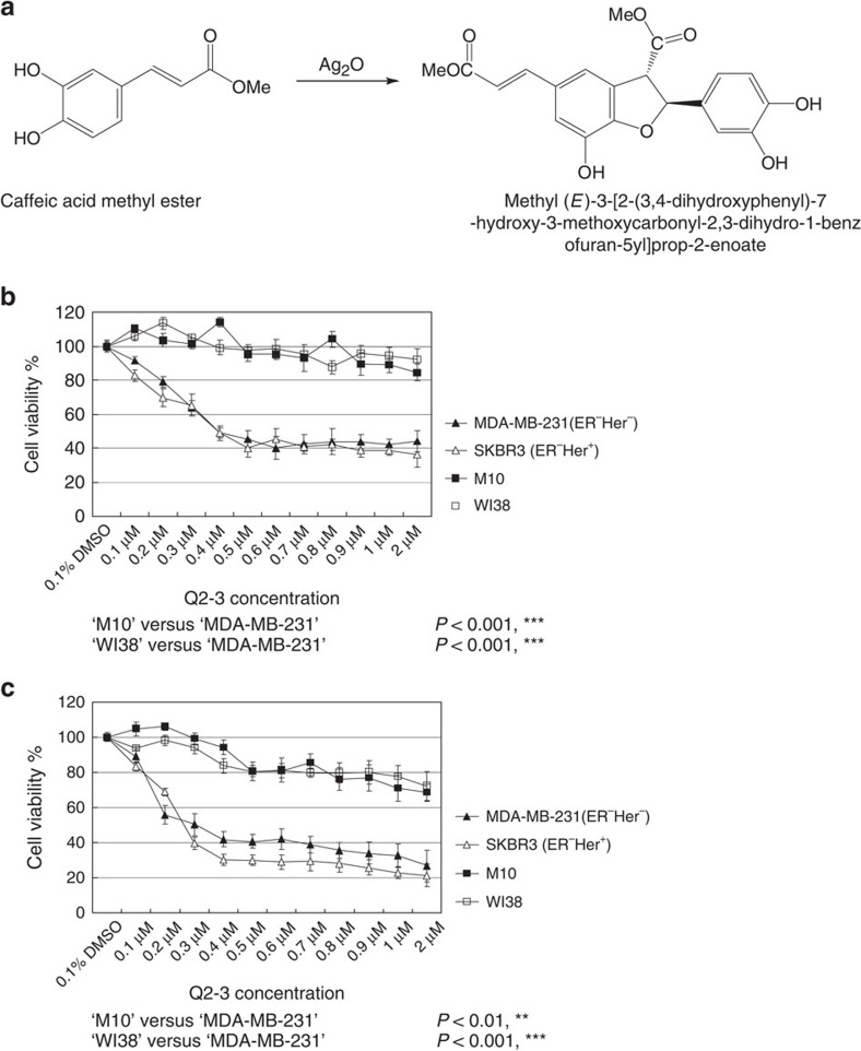
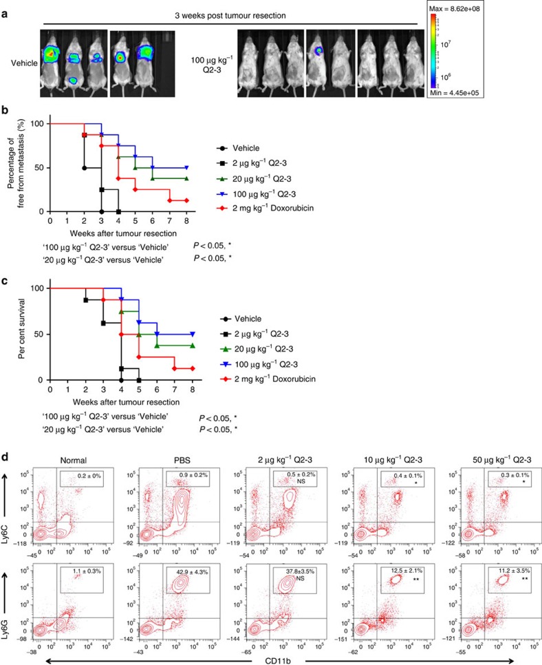
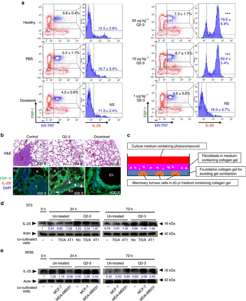
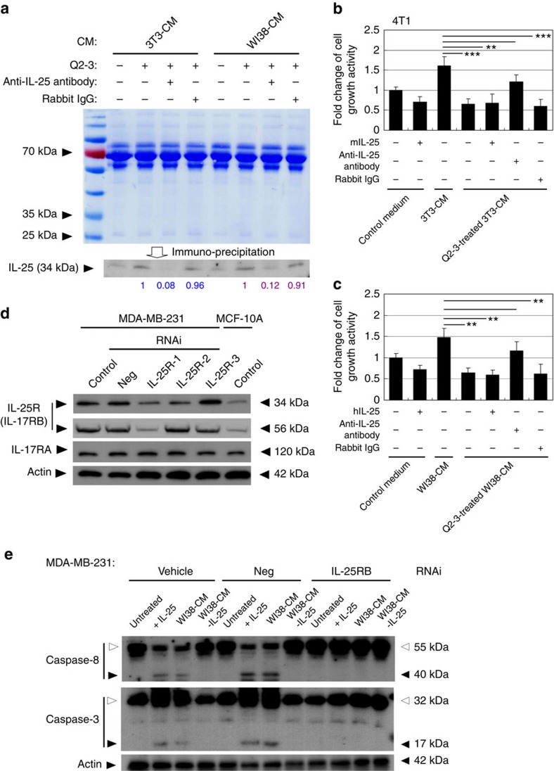
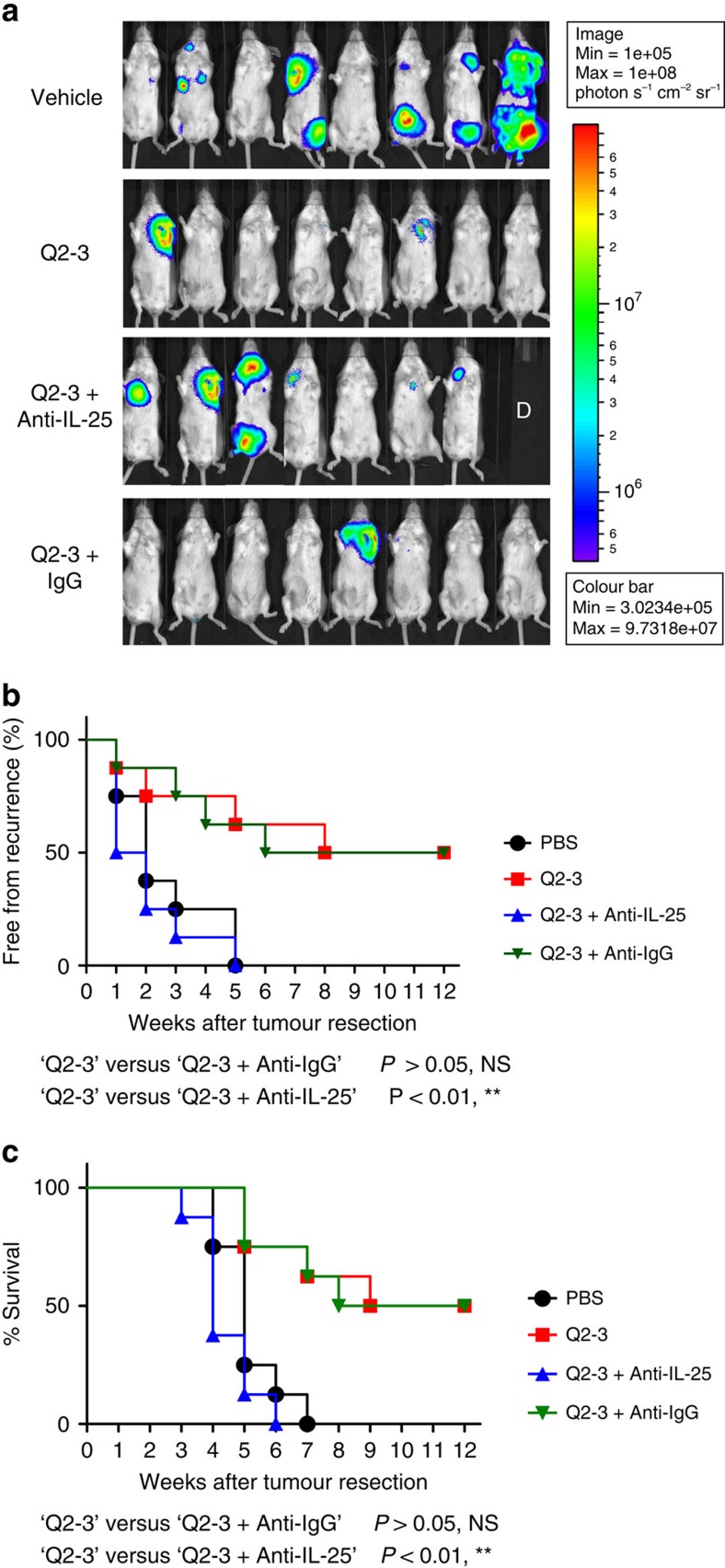
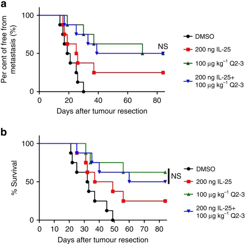
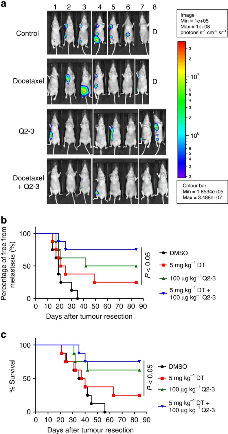
Tumour-associated fibroblasts (TAFs), as a functionally supportive microenvironment, play an essential role in tumour progression. Here we investigate the role of IL-25, an endogenous anticancer factor secreted from TAFs, in suppression of mouse 4T1 mammary tumour metastasis. We show that a synthetic dihydrobenzofuran lignan (Q2-3), the dimerization product of plant caffeic acid methyl ester, suppresses 4T1 metastasis by increasing fibroblastic IL-25 activity. The secretion of IL-25 from treated human or mouse fibroblasts is enhanced in vitro, and this activity confers a strong suppressive effect on growth activity of test carcinoma cells. Subsequent in vivo experiments showed that the anti-metastatic effects of Q2-3 on 4T1 and human MDA-MD-231 tumour cells are additive when employed in combination with the clinically used drug, docetaxel. Altogether, our findings reveal that the release of IL-25 from TAFs may serve as a check point for control of mammary tumour metastasis and that phytochemical Q2-3 can efficiently promote such anticancer activities.


Figures

Figure 1
Figure 1. Dihydrobenzofuran lignan (Q2-3) exhibits a specific cytotoxic effect on breast tumour cells.
(a) Chemical reaction formula for synthesis of Q2-3 compound (methyl (E)-3-[2-(3,4-dihydroxyphenyl)-7-hydroxy-3-methoxycarbonyl-2,3-dihydro-1-benzofuran-5yl]prop-2-enoate) from two caffeic acid methyl ester molecules. (b,c) MTT assays for cytotoxic effect of Q2-3 on SKBR3 (ERHer+ human breast cancer cell line), MDA-MB-231 (ERHer human breast cancer cell line), M10 (human normal epithelial cell line) and WI38 (human lung fibroblast cell line). 2 × 10⁴ cells were seeded and compared for their growth activity after treatment with Q2-3 for 24 or 72 h. The beginning cytotoxic dosage of Q2-3 was detected at 0.3 μM, with >50% kill rate on SKBR3 and MDA-MB-231 cells. Data are reported as mean±s.d. (n=4). **P<0.01, ***P<0.001 (Two-tailed t-test). Similar results were obtained from three independent experiments.
Figure 2
Figure 2. In vivo treatment of Q2-3 significantly suppresses the metastasis of mouse (4T1) mammary tumour cells after a tumour resection process.
(a) Representative bioluminescent images of tumour-bearing mice (n=8 per group) after in vivo treatment with PBS (0.1% DMSO in saline), doxorubicin (2 mg kg−1) or different dosages of Q2-3, after resection of the orthotopic primary tumours. In the PBS-treated group (vehicle), three mice died before 3 weeks post tumour resection. The red signal represents the highest level on the colorimetric scale. (b) Quantification of tumour metastasis by measuring luciferase activity in photons s−1 cm−2 sr−1 in mice revealed along the indicated time course (n=8 per group). (c) Survival of test mice after different treatments. P<0.05, were obtained between the control (DMSO) and Q2-3-treated (20 or 100 μg kg−1) groups (Kaplan–Meier results were analysed by log-rank test). (d) Effect of Q2-3 (2, 20 or 50 μg kg−1) on the population change of monocytic (CD11b+Ly6C+) and granulocytic (CD11b+Ly6G+) MDSCs in lung tissues of test mice were quantified by flow cytometry analysis, and were compared with MDSC populations in PBS group. Data are reported as mean±s.d. (n=3). *P<0.05; **P<0.01; NS, not significant (two-tailed t-test). Similar results were obtained from three independent experiments.
Figure 3
Figure 3. Q2-3 treatment significantly upregulates IL-25 expression in tumour-associated fibroblasts, as evaluated by 3D cell co-culture system.
(a) Effect of Q2-3 on population change of FSP-1+ER-TR7+ cells and their IL-25 expression level in lungs of test mice, quantified using flow cytometry. The percentage of IL-25+ fibroblasts in gated FSP-1+ER-TR7+ cells were compared with corresponding IL-25+ fibroblast population in PBS group. Data are reported as mean±s.d. (n=3). *P<0.05; ***P<0.001; NS, not significant (two-tailed t-test). FSP-1, fibroblast specific protein-1; ER-TR7, Erasmus University Rotterdam-thymic reticulum-7.IF staining; lung tissues of control (0.1% DMSO in PBS), Q2-3-treated and docetaxel-treated mice were collected at 21 days post tumour resection and stained for the presence of IL-25-expressing cells (red and indicated with arrowheads), FSP-1 (green) and DAPI (blue). T, tumour; A, alveoli. (b) IF staining, lung tissues of control (0.1% DMSO in PBS), Q2-3-treated and docetaxel-treated mice were collected at 21 days post tumour resection and stained for the presence of IL-25-expressing cells (red and indicated with arrowheads), FSP-1 (green) and DAPI (blue). T, tumour; A, alveoli. (c) Construction of 3D cell co-culture system for mammary tumour cells and tumour-associated fibroblasts. Western blot analyses of the expression of (d) mouse IL-25 in 3T3 fibroblasts and (e) human IL-25 in WI38 fibroblasts, in response to Q2-3 treatments in 3D culture. Some fibroblasts in the upper layer were co-cultured with mammary tumour cells as indicated. The expression level of IL-25 in 3T3 and WI38 fibroblasts were quantified and normalized using ImageJ software. Fold changes of IL-25 expression level in test samples were normalized with the value in fibroblasts of the 0 h group and were indicated by the number labelled in blue. β-Actin served as an internal control. Similar results were obtained from three independent experiments.
Figure 4
Figure 4. IL-25 secreted by Q2-3-treated fibroblasts suppresses the growth of mammary tumour cells.
(a) Western blot analysis of the IL-25 secretion activity of mouse (3T3) and human (WI38) fibroblasts in response to Q2-3 treatment. Different fibroblast-conditioned media (CM), including 3T3-CM and WI38-CM, were collected from the 3D cultures and were stained with coomassie blue, revealing that the total protein level in tested CM was normalized. Aliquots of 3T3-CM and WI38-CM were immunodepleted for IL-25. Rabbit IgG (isotype control) was used as a negative control. Amounts of IL-25 (relative staining intensity) were further normalized with the value detected for Q2-3-treated 3T3-CM (blue) or Q2-3-treated WI38-CM samples (purple). (b) Reduction in cytotoxicity of 3T3-CM on 4T1 cells after immunodepletion of IL-25. The control (fresh) medium, 3T3-CM, Q2-3-treated 3T3-CM, Q2-3-treated 3T3-CM with added IL-25 protein, Q2-3-treated 3T3-CM with the immunodepletion of IL-25 and Q2-3-treated 3T3-CM with control IgG-mediated immunodepletion, were compared for their suppressive effect on growth of 4T1 cells. (c) Reduction in cytotoxicity of WI38-CM on MDA-MB-231 after immunodepletion of IL-25. Similarly, WI38-CM with added IL-25 protein, WI38-CM with the immunodepletion of IL-25, or Q2-3-treated WI38-CM with control IgG-mediated immunodepletion, were compared for their effect on growth of MDA-MB-231 cells. The growth activity of 4T1 cells or MDA-MB-231 cells was determined using MTT assay at 5 days post cultivation, and was normalized to the control group (with fresh medium only). Data are reported as mean±s.d. (n=3). *P<0.05; **P<0.01; ***P<0.001 (two-tailed Student's t-test). (d) Western blot analysis of IL-17RA and IL-17RB (IL-25R) expression in MDA-MB-231 and MCF-10A cells. Knock down of human IL-25R (both 56 and 33 kDa molecules) expression in MDA-MB-231 cells was performed with three designed siRNAi treatments. Neg, negative control RNAi. (e) Western blot analysis of the cleavage of caspases 8 and 3 in MDA-MB-231 cells. MDA-MB-231 cells were treated by control-, Neg RNAi- or IL-25R RNAi, for 48 h. Some cells in each test group were cultivated with recombinant human IL-25 (200 ng ml−1), WI38-CM, or WI38-CM with the immunodepletion of IL-25, for another 24 h. White arrowheads, cleaved protein. Black arrowheads, uncleaved protein. β-Actin served as an internal control. Similar results were obtained from three independent experiments.
Figure 5
Figure 5. In vivo neutralization of IL-25 activity can effectively invalidate the anti-metastatic activity of Q2-3.
(a) Representative bioluminescent images of tumour-resected mice (n=8 per group) after in vivo treatment with PBS (0.1% DMSO in saline), Q2-3 (100 μg kg−1; 3 injections per week), anti-mouse IL-25 Ab (100 μg per mice; 2 injections per week), Q2-3+IL-25 Ab, or Q2-3+isotype IgG, at 3 weeks post tumour resection. The label ‘D' in the photograph denotes the mice died before 3 weeks post tumour resection. (b) Quantification of tumour metastasis by measuring luciferase activity photons s−1 cm−2 sr−1 in mice, as revealed along the indicated time course (12 weeks). (c) Survival of test mice after different treatments. NS, no significant difference between the ‘Q2-3' and ‘Q2-3+Anti-IgG' groups. **P<0.01, was obtained between the ‘Q2-3' and ‘Q2-3+Anti-IL-25' groups (Kaplan–Meier results were analysed by log-rank test). Similar results were obtained from two independent experiments.
Figure 6
Figure 6. In vivo treatment of Q2-3 confers comparable anti-metastatic activity with IL-25 administration.
(a) Tumour-resected mice (n=8 per group) were treated with PBS (0.1% DMSO in saline), IL-25 (200 ng per mice), Q2-3 (100 μg kg−1) or co-treated with IL-25 and Q2-3 for 3 weeks. Quantification of tumour metastasis by measuring luciferase activity in photons s−1 cm−2 sr−1 in mice revealed along the indicated time course. (b) Survival of test mice after different treatments. NS, no significant difference between the Q2-3 and co-treatment groups (Kaplan–Meier results were analysed by log-rank test). Similar results were obtained from three independent experiments.
Figure 7
Figure 7. Q2-3 administration can confer a complementary anti-metastatic effect on human cancer cells (MDA-MB-231), when used in combination with docetaxel.
(a) Representative bioluminescent images of MDA-MB-231 tumour-bearing nude mice (n=8 per group) after in vivo treatment with PBS (0.1% DMSO in saline), Q2-3 (100 μg kg−1), docetaxel (5 mg kg−1) or co-treatment with docetaxel and Q2-3 for 3 weeks, after resection of the orthotopic primary tumours. In PBS-treated (control) and docetaxel-treated groups, one mice was died before 3 weeks post tumour resection. (b) Quantification of tumour metastasis by measuring luciferase activity in photons s−1 cm−2 sr−1 in mice revealed along the indicated time course. (c) Survival of test mice after different treatments. P<0.05, were obtained between the docetaxel- and co-treated mice (Kaplan–Meier results were analysed by log-rank test). Similar results were obtained from two or three independent experiments.

Similar articles

Cited by

References

    1. Herbst R. S. et al. Clinical Cancer Advances 2005: major research advances in cancer treatment, prevention, and screening--a report from the American Society of Clinical Oncology. J. Clin. Oncol. 24, 190–205 (2006). - PubMed
    1. Weigelt B., Peterse J. L. & van 't Veer L. J. Breast cancer metastasis: markers and models. Nat. Rev. Cancer 5, 591–602 (2005). - PubMed
    1. Joyce J. A. & Pollard J. W. Microenvironmental regulation of metastasis. Nat. Rev. Cancer 9, 239–252 (2009). - PMC - PubMed
    1. Oskarsson T. et al. Breast cancer cells produce tenascin C as a metastatic niche component to colonize the lungs. Nat. Med. 17, 867–874 (2011). - PMC - PubMed
    1. Psaila B. & Lyden D. The metastatic niche: adapting the foreign soil. Nat. Rev. Cancer 9, 285–293 (2009). - PMC - PubMed

Publication types

MeSH terms