. 2016 Apr 19:6:24687.
doi: 10.1038/srep24687.

Olfactory epithelium changes in germfree mice

Affiliations

Olfactory epithelium changes in germfree mice

Adrien François et al. Sci Rep. .

Abstract

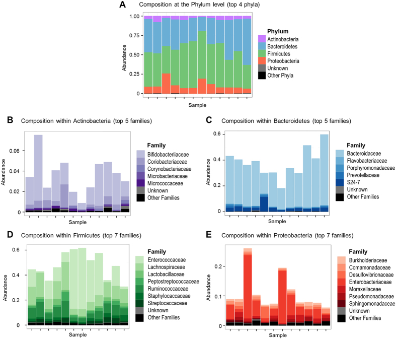
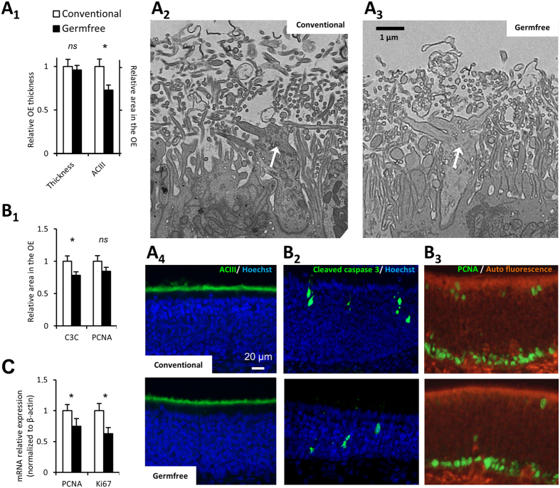
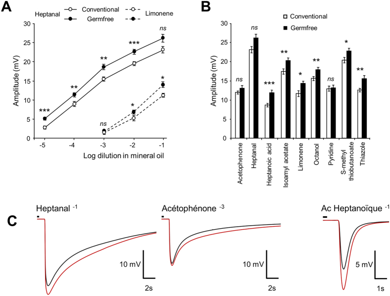
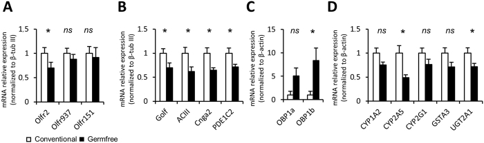
Intestinal epithelium development is dramatically impaired in germfree rodents, but the consequences of the absence of microbiota have been overlooked in other epithelia. In the present study, we present the first description of the bacterial communities associated with the olfactory epithelium and explored differences in olfactory epithelium characteristics between germfree and conventional, specific pathogen-free, mice. While the anatomy of the olfactory epithelium was not significantly different, we observed a thinner olfactory cilia layer along with a decreased cellular turn-over in germfree mice. Using electro-olfactogram, we recorded the responses of olfactory sensitive neuronal populations to various odorant stimulations. We observed a global increase in the amplitude of responses to odorants in germfree mice as well as altered responses kinetics. These changes were associated with a decreased transcription of most olfactory transduction actors and of olfactory xenobiotic metabolising enzymes. Overall, we present here the first evidence that the microbiota modulates the physiology of olfactory epithelium. As olfaction is a major sensory modality for most animal species, the microbiota may have an important impact on animal physiology and behaviour through olfaction alteration.


Figures

Figure 1
Figure 1. Taxonomic diversity of the microbiota associated with the mouse olfactory epithelium.
Analysis was based on 16S rDNA sequencing. Bar graphs show the relative distribution of phyla (A) and of families within the most abundant phyla (B–E) in the olfactory epithelium samples collected from conventional, specific-pathogen free, mice (n = 12).
Figure 2
Figure 2. Thinner cilia layer and reduced turn-over in the OE of germfree animals.
(A1) The OE thickness was compared between conventional and germfree animals. The cilia layer thickness was evaluated based on adenylate cyclase III (ACIII) staining, whose expression is restricted to olfactory cilia. Results were expressed as mean of the OE thickness and mean of the ACIII signal area in the OE normalized to conventional animals ± SEM (n = 8). (A2,3) Representative transmission electron images of olfactory dendritic knob (white arrow) and associated cilia layer (x2000). (A4) Representative image of ACIII staining in the OE. (B1) The cellular turn-over of the OE was evaluated by quantifying the areas with cleaved caspase 3 (C3C) and PCNA stainings, taken as indices of apoptosis and proliferation levels, respectively. Results were expressed as mean of the C3C and PCNA signal area in the OE normalized to conventional animals ± SEM (n = 19). Representative image of (B2) C3C staining with OSN ongoing apoptosis and (B3) PCNA staining mainly present in basal cells same after (B2). (C) The level of proliferation was further evaluated by quantification of ki67 and PCNA expression on cDNAs from olfactory mucosa. Their expression levels were normalized to that of β-actin and are given as mean ± SEM (n = 7). (*) P < 0.05.
Figure 3
Figure 3. Global increase of responses to odorants recorded by EOG in germfree animals.
(A,B) EOG responses to various odorants in conventional and germfree animals. Values represent the mean of peak amplitudes ± SEM (n = 12) (*P < 0.05; **P < 0.01; ***P < 0.001). (C) Average traces for 3 odorants from conventional (black) and germfree (red) animals. Small black line on top of recordings indicates odorant stimulation.
Figure 4
Figure 4. Modulation of the expression of genes related to odorant detection in germfree animals.
Quantitative PCR (qPCR) analysis of cDNAs from olfactory mucosa. Genes are grouped by functions; (A) olfactory receptors sensitive to odorants tested in the EOG experiment; (B) main olfactory transduction pathway components; (C) olfactory binding proteins; (D) detoxifying enzymes. Their expression levels were normalized to that of β-tubIII (A,B) or β-actin (C,D) and are given as mean ± SEM (n = 7). (*) P < 0.05.

References

    1. Gustafsson B. E., Daft F. S., Mc D. E., Smith J. C. & Fitzgerald R. J. Effects of vitamin K-active compounds and intestinal microorganisms in vitamin K-deficient germfree rats. J Nutr 78, 461–468 (1962). - PubMed
    1. Hsiao E. Y. et al. Microbiota modulate behavioral and physiological abnormalities associated with neurodevelopmental disorders. Cell 155, 1451–1463 (2013). - PMC - PubMed
    1. Smith K., McCoy K. D. & Macpherson A. J. Use of axenic animals in studying the adaptation of mammals to their commensal intestinal microbiota. Seminars in immunology 19, 59–69 (2007). - PubMed
    1. Lozupone C. A., Stombaugh J. I., Gordon J. I., Jansson J. K. & Knight R. Diversity, stability and resilience of the human gut microbiota. Nature 489, 220–230 (2012). - PMC - PubMed
    1. Yan M. et al. Nasal microenvironments and interspecific interactions influence nasal microbiota complexity and S. aureus carriage. Cell host & microbe 14, 631–640 (2013). - PMC - PubMed

Publication types

Substances

LinkOut - more resources