. 2017 Jan;91(1):481-494.
doi: 10.1007/s00204-016-1697-8. Epub 2016 Apr 18.

Role of AMP-activated protein kinase α1 in 17α-ethinylestradiol-induced cholestasis in rats

Affiliations

Role of AMP-activated protein kinase α1 in 17α-ethinylestradiol-induced cholestasis in rats

Xiaojiaoyang Li et al. Arch Toxicol. 2017 Jan.

Abstract

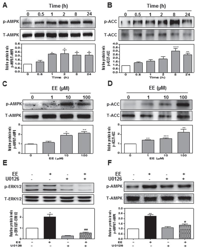
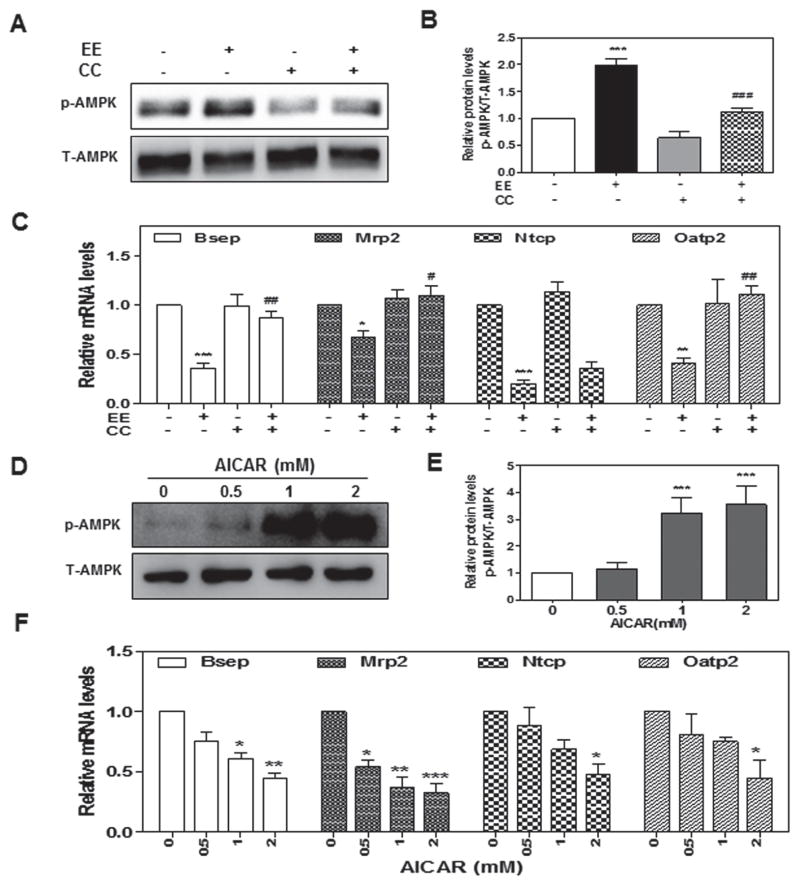
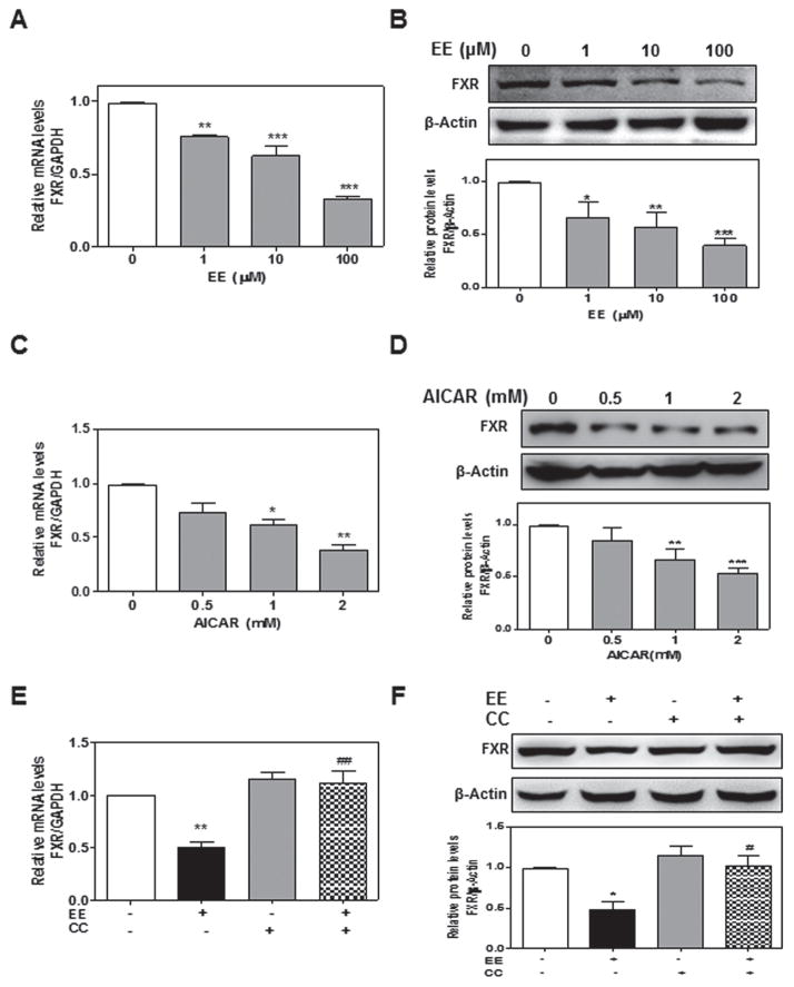
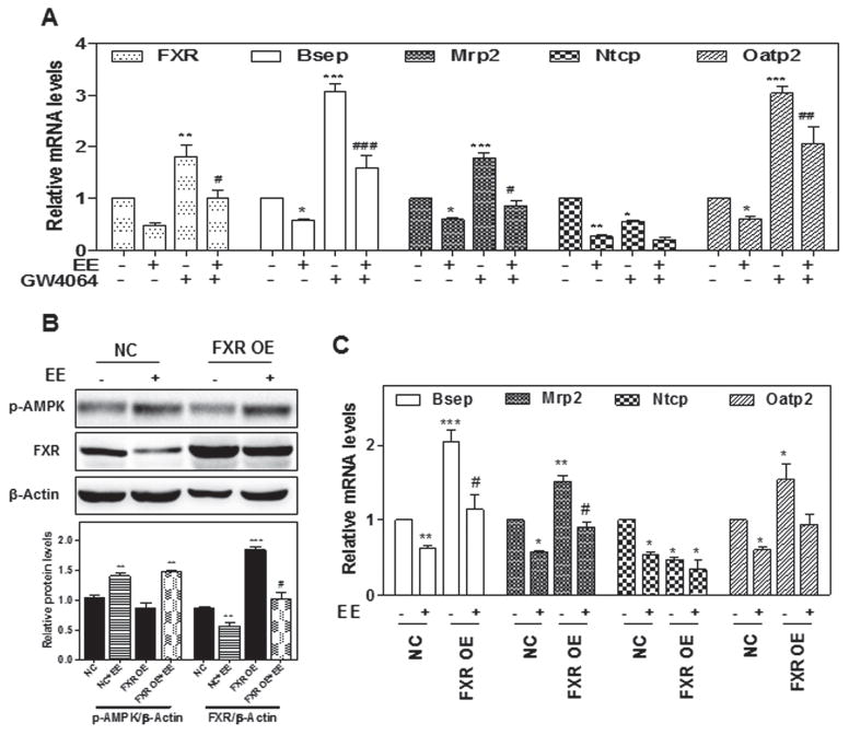
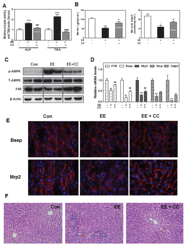
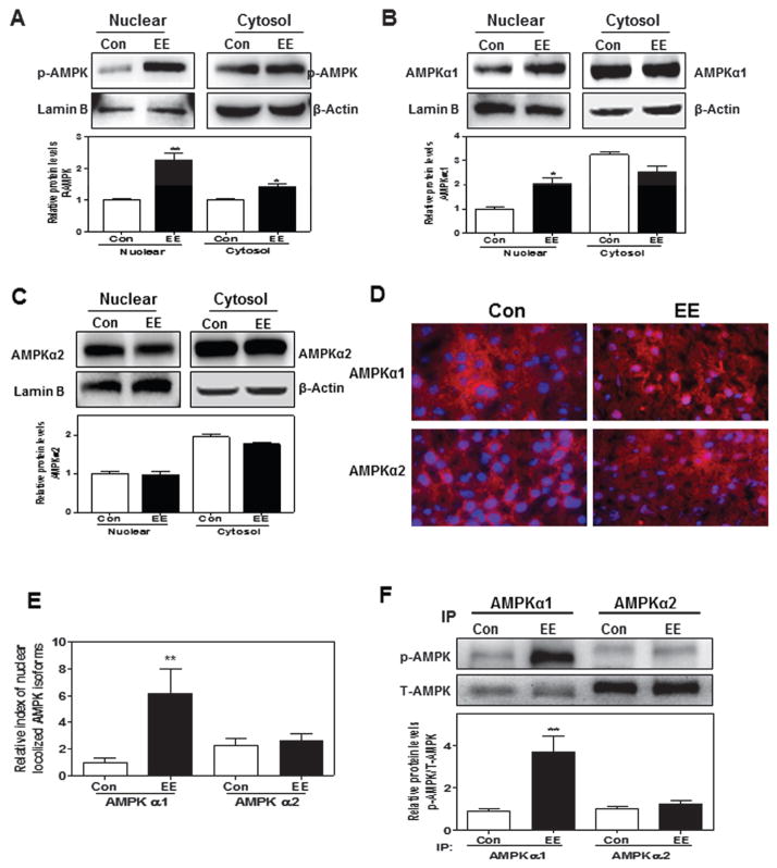
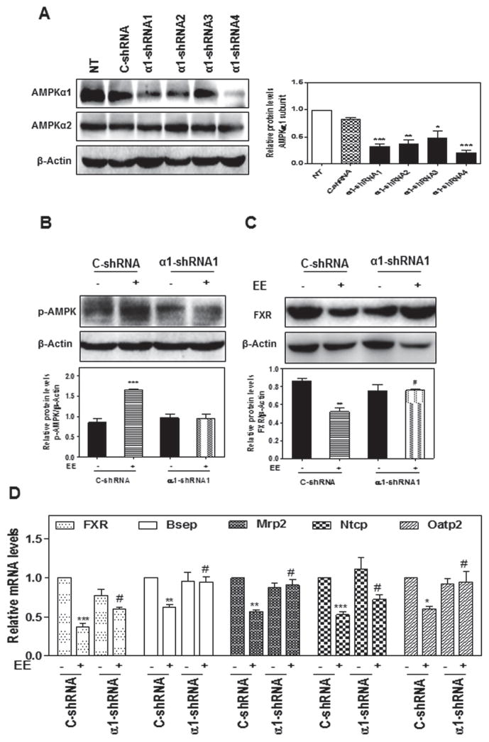
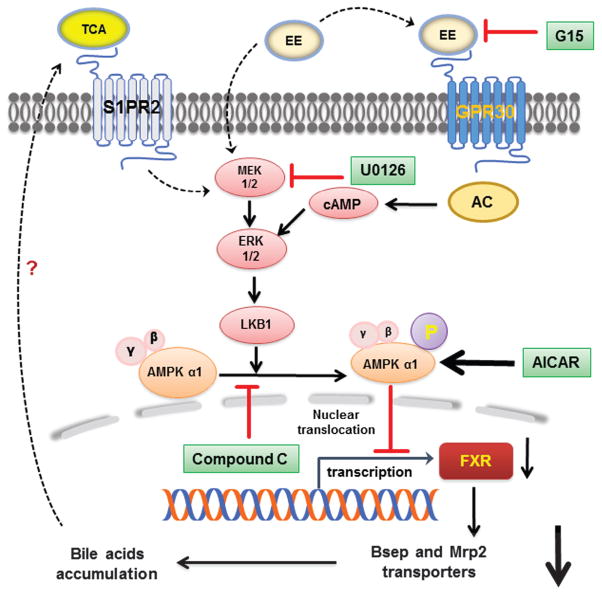
Estrogen-induced cholestasis occurs in many women who are susceptible due to pregnancy or hormone replacement therapy for postmenopausal syndrome. 17α-Ethinylestradiol (EE), as a synthetic estrogen, has been widely used to study the underlying mechanisms of estrogen-induced cholestasis. Recent studies have also reported that liver kinase B1 (LKB1)-mediated activation of AMP-activated protein kinase (AMPK) plays a critical role in the regulation of canalicular network formation. However, the role of AMPK in EE-induced cholestasis remains to be determined. In this study, the effects of EE (1-100 µM) on AMPK activation and the expression of farnesoid X receptor (FXR) and hepatic bile acid transporters were examined in in vitro using 3D-cultured rat primary hepatocytes and in in vivo using rat cholestasis models. We also used specific chemical agonist and antagonist of AMPK, AMPK subunit-specific antibodies and lentiviral shRNAs for AMPKα1 and AMPKα2 to delineate the role of AMPK in EE-induced cholestasis and potential cellular mechanisms. We found that EE-induced phosphorylation of AMPKα1 via extracellular signal-regulated kinases-LKB1-mediated signaling pathways and subsequent nuclear translocation accounted for the down-regulation of FXR and bile acid transporters and disruption of bile acid homeostasis. Inhibition of AMPK activation using an AMPK antagonist Compound C (2 µM) or down-regulation of AMPKα1 using gene-specific shRNA attenuated EE-induced cholestasis both in in vitro and in in vivo. In conclusion, these results revealed that activation of cAMP-ERK-LKB1-AMPKα1 signaling pathway plays a critical role in EE-mediated dysregulation of the expression of FXR and bile acid transporters. AMPKα1 may represent an important therapeutic target for estrogen-induced cholestasis.

Keywords: AMPKα1; Bile acid transporters; Cholestasis; Estrogen; FXR.


Conflict of interest statement

The authors declare no competing financial interest.

Figures

Fig. 1
Fig. 1. EE activates AMPK via ERK1/2 in rat primary hepatocytes
Rat primary hepatocytes were a, b treated with EE (10 μM) for different time periods or c, d different concentrations (0 to 100 μM) for 2-hour. e Cells were pretreated with ERK1/2 inhibitor U0126 (10 μM) for 1 hour, and then treated with EE (10 μM) for 1 hour e or 2-hour f. Representative Western blot and densitometry of phosphorylated AMPK, total AMPK, p-ACC, T-ACC, p-ERK1/2 and T-ERK1/2. *P<0.05, **P<0.01, ***P<0.001 vs. control group
Fig. 2
Fig. 2. The role of AMPK activation in EE-induced hepatocyte cholestasis
Rat primary hepatocytes were pretreated with an AMPK inhibitor, Compound C (CC, 2 μM), for 1-hour and then treated with EE (10 μM) for 2- or 24-hour, or treated with AICAR (0 to 2 μM) for 2 or 24-hour. a, b, d, e Representative immunoblots against p-AMPK and T-AMPK are shown. c, f The relative mRNA levels were determined by real-time RT-PCR and normalized using GAPDH as an internal control. ***P<0.001 vs. control group. ###P<0.001 vs. EE group
Fig. 3
Fig. 3. Effect of EE-induced AMPK activation on FXR expression in rat primary hepatocytes
a, b Rat primary hepatocytes were treated with EE (0 to 100 μM) for 24-hour. c, d Cells were treated with AICAR (0 to 2 mM) for 24-hour. e, f Cells were pretreated with CC (2 μM) for 1 hour and then treated with EE (10 μM) for 2-hour. a, c, e The relative mRNA levels of FXR were normalized using GAPDH as an internal control. b, d, f Representative immunoblots against FXR and β-actin are shown. *P<0.05, **P<0.01,***P<0.001 vs. control group. #P<0.05, ##P<0.01 vs EE group
Fig. 4
Fig. 4. The role of FXR in EE-induced cholestasis
a Rat primary hepatocytes were treated with EE (10 μM) in the presence or absence of the FXR agonist GW4064 (10 μM) for 24-hour. b, c Rat primary hepatocytes were transduced with FXR overexpression lentivirus or control lentivirus for 24-hour, then treated with EE (10 μM) for 2 or 24-hour. b Representative immunoblots against p-AMPK and FXR are shown and normalized with β-Actin. a, c The relative mRNA levels of relative genes were determined by qPCR and normalized using GAPDH as an internal control. *P<0.05, **P<0.01,***P<0.001 vs. control group. #P<0.05 vs. EE group
Fig. 5
Fig. 5. Protective effect of Compound C on EE-induced cholestasis in rat model
a The serum ALP activity and TBA, b Bile acid flow rates and biliary TBA levels in rats. c Representative immunoblots against p-AMPK, T-AMPK, FXR and β-Actin in liver lysates are shown. d The relative mRNA levels of relative genes were determined by real-time RT-PCR and normalized using GAPDH as an internal control. e Frozen liver sections of the Bsep and Mrp2expression. f Representative images of rat liver sections by HE staining. *P<0.05, **P<0.01, ***P<0.001 vs. control group, n=6; #P<0.05, ##P<0.01, ###P<0.001vs. EE group, n=6
Fig. 6
Fig. 6. Effect of EE on sub-cellular distribution of AMPKα1 and AMPKα2 subunits
a, b, c Primary rat hepatocytes were treated with EE (10 μM) for 4-hour. Representative immunoblots against p-AMPK, AMPKα1, AMPKα2, lamin B and β-Actin are shown and normalized with lamin B or β-Actin. d, e Co-localization of AMPKα1 and AMPKα2 (both red) with nuclear (blue) in rat liver after EE treatment for 1 day. f The protein levels of phosphorylated AMPKα1 and AMPKα2 were detected by immunoprecipitation using indicated antibodies, following Western blot analysis against p-AMPK and T-AMPK, and normalized with T-AMPK. **P<0.01 vs. control group, n=6
Fig. 7
Fig. 7. Effect of AMPKα1 knock down on EE-induced cholestasis in rat primary hepatocytes
a Rat primary hepatocytes were transduced with lentivirus shRNA targeting AMPKα1 (α1-shRNA 1–4) or negative control lentivirus (C-shRNA) for 24-hour. NT: no treatment. b, c, d Rat primary hepatocytes were transduced with lentivirus α1-shRNA or C-shRNA for 24-hour. b Cells were then treated with EE for 2-houror c 24-hour. Representative immunoblots are shown. d The relative mRNA levels of relative geneswere determined by qPCR and normalized using GAPDH as an internal control. *P<0.05, ** P<0.01 and *** P<0.001 vs. control group; #P<0.05 vs. EE group
Fig. 8
Fig. 8. Proposed model of EE-induced impairment of bile acid homeostasis
Possible mechanisms underlying AMPKα1 activation via cAMP-ERK1/2-LKB1 signaling pathway in EE-induc ed cholestasis.

References

    1. Abu-Hayyeh S, Papacleovoulou G, Lovgren-Sandblom A, et al. Intrahepatic cholestasis of pregnancy levels of sulfated progesterone metabolites inhibit farnesoid X receptor resulting in a cholestatic phenotype. Hepatology. 2013;57(2):716–726. - PMC - PubMed
    1. Barth A, Klinger G, Rost M. Influence of ethinyloestradiol propanolsulphonate on serum bile acids in healthy volunteers. Experimental and Toxicologic Pathology. 2003;54(5):381–386. - PubMed
    1. Chang HR, Nam S, Kook MC, et al. HNF4alpha is a therapeutic target that links AMPK to WNT signalling in early-stage gastric cancer. Gut. 2016;65(1):19–32. - PMC - PubMed
    1. Crocenzi FA, Sanchez Pozzi EJ, Pellegrino JM, et al. Beneficial effects of silymarin on estrogen-induced cholestasis in the rat: a study in vivo and in isolated hepatocyte couplets. Hepatology. 2001;34(2):329–339. - PubMed
    1. Eeckhoute J, Formstecher P, Laine B. Hepatocyte nuclear factor 4alpha enhances the hepatocyte nuclear factor 1alpha-mediated activation of transcription. Nucleic acids research. 2004;32(8):2586–2593. - PMC - PubMed

MeSH terms

LinkOut - more resources