. 2016 Jun 28:232:83-92.
doi: 10.1016/j.jconrel.2016.04.019. Epub 2016 Apr 15.

Inhibition of bone loss with surface-modulated, drug-loaded nanoparticles in an intraosseous model of prostate cancer

Affiliations

Inhibition of bone loss with surface-modulated, drug-loaded nanoparticles in an intraosseous model of prostate cancer

Isaac M Adjei et al. J Control Release. .

Abstract

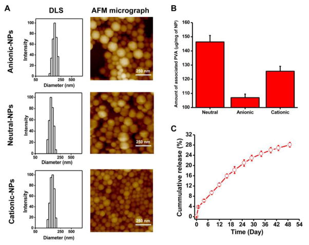
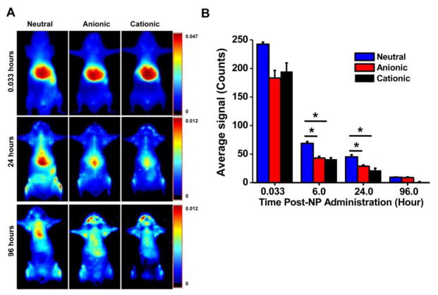
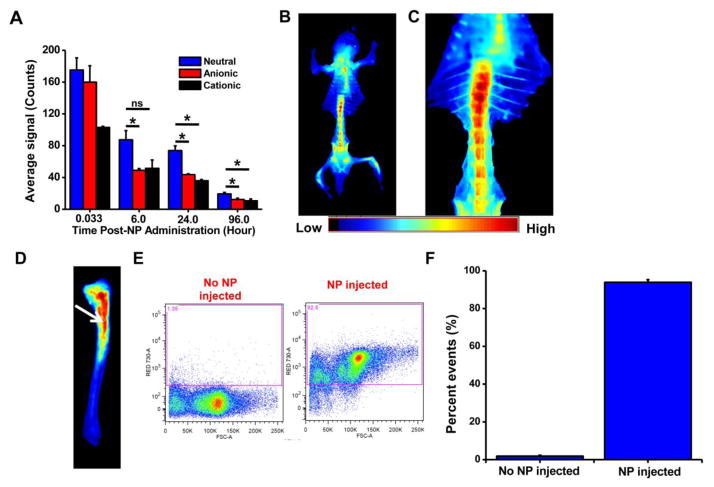
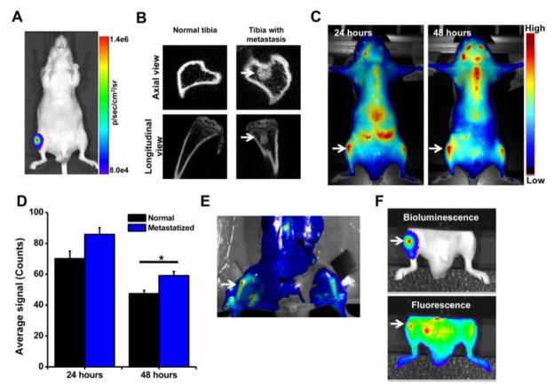
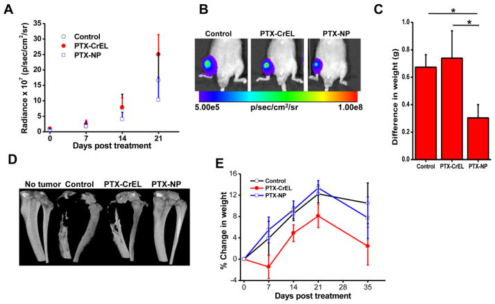
Advanced-stage prostate cancer usually metastasizes to bone and is untreatable due to poor biodistribution of intravenously administered anticancer drugs to bone. In this study, we modulated the surface charge/composition of biodegradable nanoparticles (NPs) to sustain their blood circulation time and made them small enough to extravasate through the openings of the bone's sinusoidal capillaries and thus localize into marrow. NPs with a neutral surface charge, achieved by modulating the NP surface-associated emulsifier composition, were more effective at localizing to bone marrow than NPs with a cationic or anionic surface charge. These small neutral NPs (~150nm vs. the more usual ~320nm) were also ~7-fold more effective in localizing in bone marrow than large NPs. We hypothesized that NPs that effectively localize to marrow could improve NP-mediated anticancer drug delivery to sites of bone metastasis, thereby inhibiting cancer progression and preventing bone loss. In a PC-3M-luc cell-induced osteolytic intraosseous model of prostate cancer, these small neutral NPs demonstrated greater accumulation in bone within metastatic sites than in normal contralateral bone as well as co-localization with the tumor mass in marrow. Significantly, a single-dose intravenous administration of these small neutral NPs loaded with paclitaxel (PTX-NPs), but not anionic PTX-NPs, slowed the progression of bone metastasis. In addition, neutral PTX-NPs prevented bone loss, whereas animals treated with the rapid-release drug formulation Cremophor EL (PTX-CrEL) or saline (control) showed >50% bone loss. Neutral PTX-NPs did not cause acute toxicity, whereas animals treated with PTX-CrEL experienced weight loss. These results indicate that NPs with appropriate physical and sustained drug-release characteristics could be explored to treat bone metastasis, a significant clinical issue in prostate and other cancers.

Keywords: Biodistribution; Bone marrow; Cancer therapy; Drug delivery; Nanomedicine.


Conflict of interest statement

Competing Interest: Authors declare that they have no competing interests.

Figures

Fig. 1
Fig. 1. Physical characterization of different formulations of NPs
A) Characterization of anionic, neutral, and cationic NPs for particle size and size distribution by dynamic light scattering (DLS). NP size and shape characterization was determined by atomic force microscopy (AFM). B) Amount of surface-associated PVA with anionic, neutral, and cationic NPs. Data are shown as mean ± s.e.m. n = 3. C) Release of PTX in vitro from drug-loaded neutral NPs under sink conditions. Data are shown as mean ± s.e.m., n = 3.
Fig. 2
Fig. 2. Biodistribution of NPs with different surface charges
A) Whole-body images taken over time indicated prolonged retention of neutral NPs in the body. B) Quantification of the ROIs of the skin over time as measured using Maestro, demonstrating more prolonged body retention of neutral NPs than of anionic or cationic NPs. Data are shown as mean ± s.e.m., *P < 0.05; n = 3.
Fig. 3
Fig. 3. Localization with NPs of different surface charge in bones
A) Quantification of fluorescence signals due to NP localization in tibia over time as measured using Maestro, demonstrating greater uptake and sustained retention of neutral NPs than of anionic or cationic NPs. B) Ventral view of skeleton of mouse injected with neutral NPs, showing NP localization in all bones, particularly pelvis, long bones, sternum, and vertebrae. C) Close-up view of the vertebral column shows greater NP localization in cervical than lumbar vertebrae. D) Image of the surgically resected tibia, showing localization of neutral NPs in marrow (arrow) but not in calcified bone. E) Flow cytometry analysis showing uptake of NPs by bone marrow cells. F) Quantification of the flow cytometry data shows that >90% of bone marrow cells internalize neutral NPs. Data are shown as mean ± s.e.m., *P < 0.05, ns = not significant; data shown in B to D at 24 hours post NP administration.
Fig. 4
Fig. 4. Localization of neutral NPs at site of tumor metastasis in bone
A) Bioluminescence signal due to PC-3M-luc prostate cancer cells at 7 days post inoculation as measured using IVIS® and B) micro-CT images of the bone at 7 days post cancer cell inoculation. C) Fluorescence imaging and D) quantification of signal intensity measured using Maestro, demonstrating greater localization of NPs in tibia with tumor than in normal contralateral tibia. E) Ex vivo image (by Maestro) of the bone with intraosseous tumor at 24 hrs following NP administration. Arrows indicate bone with metastatic tumors. F) Bioluminescence signals due to cancer cells and fluorescence of NPs, demonstrating localization of NPs into metastasized tumor mass. For the above colocalization study, both bioluminescence signals due to cancer cells and fluorescence signals due to NPs were captured using IVIS®. Bright spots seen next to the tumor in tibia (Fig. 4F, fluorescence signal) are due to localization of the injected NPs in lymph nodes and other tissues. Data are shown as mean ± s.e.m., *P < 0.05.
Fig. 5
Fig. 5. Efficacy of paclitaxel-loaded neutral NPs in a bone metastasis model
A) Change in bioluminescence signals due to cancer cells in bone as measured using IVIS® during 3 weeks post treatment. *P < 0.05 at 2 weeks between PTX-NPs and other groups; *P < 0.05 between PTX-NPs and PTX-CrEL at 3 weeks; **no statistical significance between PTX-NPs and saline at 3 weeks days. B) Representative bioluminescence images of the bone with tumor captured using IVIS® at 3 weeks post treatment. C) Tumor burden at 5 weeks post treatment, determined by subtracting the weight of the normal contralateral leg from that of the leg with tumor. D) Representative micro-CT of tibias of mice from different groups at 5 weeks post treatment. Animals treated with PTX-NPs showed no bone loss. E) Changes in body weight of animals post treatment. Data are shown as mean ± s.e.m., *P < 0.05 PTX-NPs and control vs. PTX-CrEL; Not significant, PTX-NPs vs. control, n = 5–6.

References

    1. Coleman RE. Metastatic bone disease: clinical features, pathophysiology and treatment strategies. Cancer Treat Rev. 2001;27:165–176. doi: 10.1053/ctrv.2000.0210. - DOI - PubMed
    1. Morrissey C, Vessella RL. The role of tumor microenvironment in prostate cancer bone metastasis. J Cell Biochem. 2007;101:873–886. doi: 10.1002/jcb.21214. Review. - DOI - PubMed
    1. Kuru B, Camlibel M, Dinc S, Gulcelik MA, Gonullu D, Alagol H. Prognostic factors for survival in breast cancer patients who developed distant metastasis subsequent to definitive surgery. Singapore medical journal. 2008;49:904–911. - PubMed
    1. Berruti A, Dogliotti L, Bitossi R, Fasolis G, Gorzegno G, Bellina M, Torta M, Porpiglia F, Fontana D, Angeli A. Incidence of skeletal complications in patients with bone metastatic prostate cancer and hormone refractory disease: Predictive role of bone resorption and formation markers evaluated at baseline. J Urol. 2000;164:1248–1253. doi: 10.1016/S0022-5347(05)67149-2. - DOI - PubMed
    1. Ramanlal Chaudhari K, Kumar A, Megraj Khandelwal VK, Ukawala M, Manjappa AS, Mishra AK, Monkkonen J, Ramachandra Murthy RS. Bone metastasis targeting: a novel approach to reach bone using Zoledronate anchored PLGA nanoparticle as carrier system loaded with Docetaxel. J Control Release. 2012;158:470–478. doi: 10.1016/j.jconrel.2011.11.020. - DOI - PubMed

Publication types

MeSH terms

Substances