Review
. 2016 Jul;30(4):317-31.
doi: 10.1016/j.blre.2016.02.003. Epub 2016 Apr 16.

FDG-PET imaging in hematological malignancies

Affiliations
Review

FDG-PET imaging in hematological malignancies

L Valls et al. Blood Rev. 2016 Jul.

Abstract

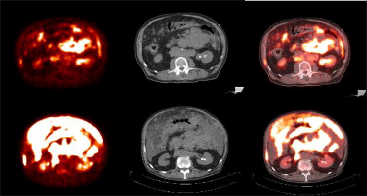
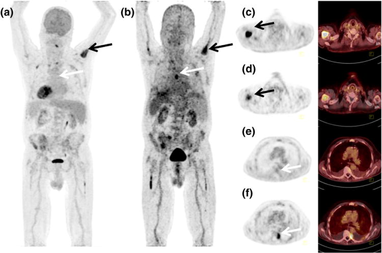
The majority of aggressive lymphomas is characterized by an up regulated glycolytic activity, which enables the visualization by F-18 FDG-PET/CT. One-stop hybrid FDG-PET/CT combines the functional and morphologic information, outperforming both, CT and FDG-PET as separate imaging modalities. This has resulted in several recommendations using FDG-PET/CT for staging, restaging, monitoring during therapy, and assessment of treatment response as well as identification of malignant transformation. FDG-PET/CT may obviate the need for a bone marrow biopsy in patients with Hodgkin's lymphoma and diffuse large B cell lymphoma. FDG-PET/CT response assessment is recommended for FDG-avid lymphomas, whereas CT-based response evaluation remains important in lymphomas with low or variable FDG avidity. The treatment induced change in metabolic activity allows for assessment of response after completion of therapy as well as prediction of outcome early during therapy. The five-point scale Deauville Criteria allows the assessment of treatment response based on visual FDG-PET analysis. Although the use of FDG-PET/CT for prediction of therapeutic response is promising it should only be conducted in the context of clinical trials. Surveillance FDG-PET/CT after complete remission is discouraged due to the relative high number of false-positive findings, which in turn may result in further unnecessary investigations. Future directions include the use of new PET tracers such as F-18 fluorothymidine (FLT), a surrogate biomarker of cellular proliferation and Ga-68 CXCR4, a chemokine receptor imaging biomarker as well as innovative digital PET/CT and PET/MRI techniques.

Keywords: FDG; FLT; Ga-68 CXCR4; Hematologic malignancies; Leukemia; Lymphoma; PET; PET/CT; PET/MRI.


Conflict of interest statement

None

Figures

Figure 1
Figure 1
69 year old male with new right tonsillar diffuse large B cell lymphoma. Baseline PET/CT study shows hypermetabolic right tonsillar mass. Mild activity in the left tonsillar fossa is reactive. No additional site of lymphoma was identified on remainder of images.
Figure 2
Figure 2
55 year old female with gastric MALT lymphoma with accompanying splenic involvement. Baseline PET/CT images (a-d) show diffuse gastric wall thickening and hypermetabolic acitivity and hypermetabolic splenomegaly.
Figure 3
Figure 3
26 year old female with nodular sclerosis type HL. Baseline PET/CT images (a, b) show large hypermetabolic anterior mediastinal masses, hypermetabolic splenic foci, and periportal adenopathy. Mild marrow hyperplasia was also noted at baseline, bone marrow biopsy was negative. Interim PET/CT (c, d) images show resolution of hypermetabolic activity in the mediastinal mass, spleen and abdominal adenopathy indicating treatment response (Deauville scale 2)
Figure 4
Figure 4
54 year old male with acute onset headache and confusion. Axial and sagittal PET images of brain fused with contrast enhanced T1 weighted MRI images show an intensely hypermetabolic mass in the suprasellar region. Additional hypermetabolic enhancing nodule is identified in the left frontal horn of lateral ventricle. Histopathology analysis confirmed the diagnosis of large B cell lymphoma.
Figure 5
Figure 5
71 year old with B cell lymphoma with bone marrow and splenic involvement at presentation PET (a) and PET/CT fused (b) images show heterogeneously hypermetabolic splenomegaly, the whole body MIP (c) shows diffuse marrow involvement. No other site of disease was identified.
Figure 6
Figure 6
47 year old female with stage IV-B diffuse large B cell lymphoma B cell lymphoma diffuse nodal involvement. PET and fused PET/CT images show hypermetabolic left iliac adenopathy. The MIP PET image (c) shows additional retroperitoneal, inguinal, mediastinal and left supraclavicular nodal involvement. Interim PET/CT study after 2 cycles of chemotherapy (d-f) shows near complete resolution of adenopathy (Deauville scale 2) with post chemotherapy marrow hyperplasia
Figure 7
Figure 7
65 year old woman with past history of thyroiditis with a rapidly growing cervical mass. Baseline PET/CT (a) PET, (b) CT and (c) fused PET/CT images show diffusely hypermetabolic thyroid mass, biopsy revealed diffuse large B cell lymphoma. Post chemotherapy PET/CT (d-f) after R-CHOP regimen shows complete response (Deauville scale 1).
Figure 8
Figure 8
76 year old female with diffuse large B cell lymphoma of right leg. Baseline PET/CT (a-c) shows hypermetabolic soft tissue mass along the lateral aspect of right knee joint. Post treatment PET/CT study (d-f) after chemotherapy and local radiation revealed near complete resolution of the mass indicating therapeutic response (Deauville scale 1)
Figure 9
Figure 9
40 year old with Burkitt's lymphoma. Baseline PET/CT (a-c) images show diffuse hypermetabolic abdominal adenopathy with a large perinephric and pelvic hypermetabolic soft tissue mass. Interim PET/CT imaging after 1 cycle of R-CHOP shows significant improvement in metabolic activity of the mass with considerable amount of mildly metabolic residual soft tissue. (Deauville scale 4)
Figure 10
Figure 10
67 year old female with diffuse large B cell lymphoma of germinal center origin. Baseline PET/CT (a-c) shows diffuse omental involvement and abdominal adenopathy. Interim PET/CT study (d-f) after 2 cycles of chemotherapy revealed significant increase in hypermetabolic disease burden indicating treatment failure (Deauville scale 5)
Figure 11
Figure 11
Maximum intensity projections from 18F-FDG (a) and Ga-68 CXCR4 (Pentixafor) PET/CT (b) of a patient with multiple myeloma. Corresponding transversal PET and fused PET/CT images are shown for FDG (c, e) and Ga-68 CXCR4 (d, f) indicating a FDG- and CXCR4 avid lesion in the right humerus (black arrows) and FDG negative but CXCR4 positive lesion left paravertrebral (white arrows)

References

    1. Seam P, Juweid ME, Cheson BD. The role of FDG-PET scans in patients with lymphoma. Blood. 2007;110:3507–16. - PubMed
    1. Kostakoglu L, Leonard JP, Kuji I, Coleman M, Vallabhajosula S, Goldsmith SJ. Comparison of fluorine-18 fluorodeoxyglucose positron emission tomography and Ga-67 scintigraphy in evaluation of lymphoma. Cancer. 2002;94:879–88. - PubMed
    1. Barrington SF, Mikhaeel NG, Kostakoglu L, Meignan M, Hutchings M, Mueller SP, et al. Role of imaging in the staging and response assessment of lymphoma: consensus of the International Conference on Malignant Lymphomas Imaging Working Group. J Clin Oncol. 2014;32:3048–58. - PMC - PubMed
    1. Cheson BD, Fisher RI, Barrington SF, Cavalli F, Schwartz LH, Zucca E, et al. Recommendations for initial evaluation, staging, and response assessment of Hodgkin and non-Hodgkin lymphoma: the Lugano classification. J Clin Oncol. 2014;32:3059–68. - PMC - PubMed
    1. Dreyling M, Thieblemont C, Gallamini A, Arcaini L, Campo E, Hermine O, et al. ESMO Consensus conferences: guidelines on malignant lymphoma. part 2: marginal zone lymphoma, mantle cell lymphoma, peripheral T-cell lymphoma. Ann Oncol. 2013;24:857–77. - PubMed

Publication types

MeSH terms

Substances

LinkOut - more resources