. 2016 Apr 19:7:11310.
doi: 10.1038/ncomms11310.

Dynamic and flexible H3K9me3 bridging via HP1β dimerization establishes a plastic state of condensed chromatin

Affiliations

Dynamic and flexible H3K9me3 bridging via HP1β dimerization establishes a plastic state of condensed chromatin

Kyoko Hiragami-Hamada et al. Nat Commun. .

Abstract

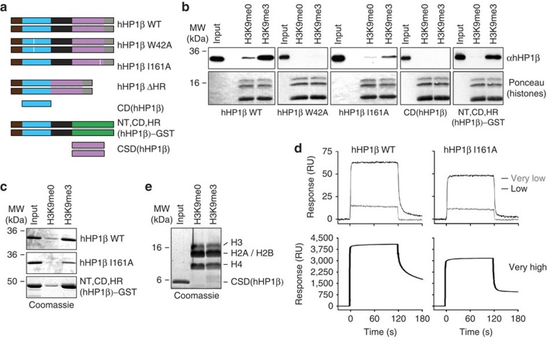
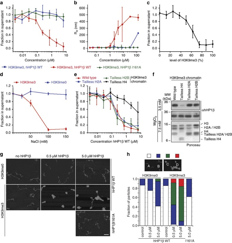
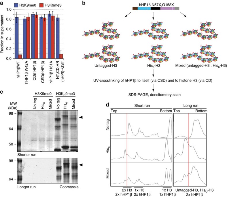
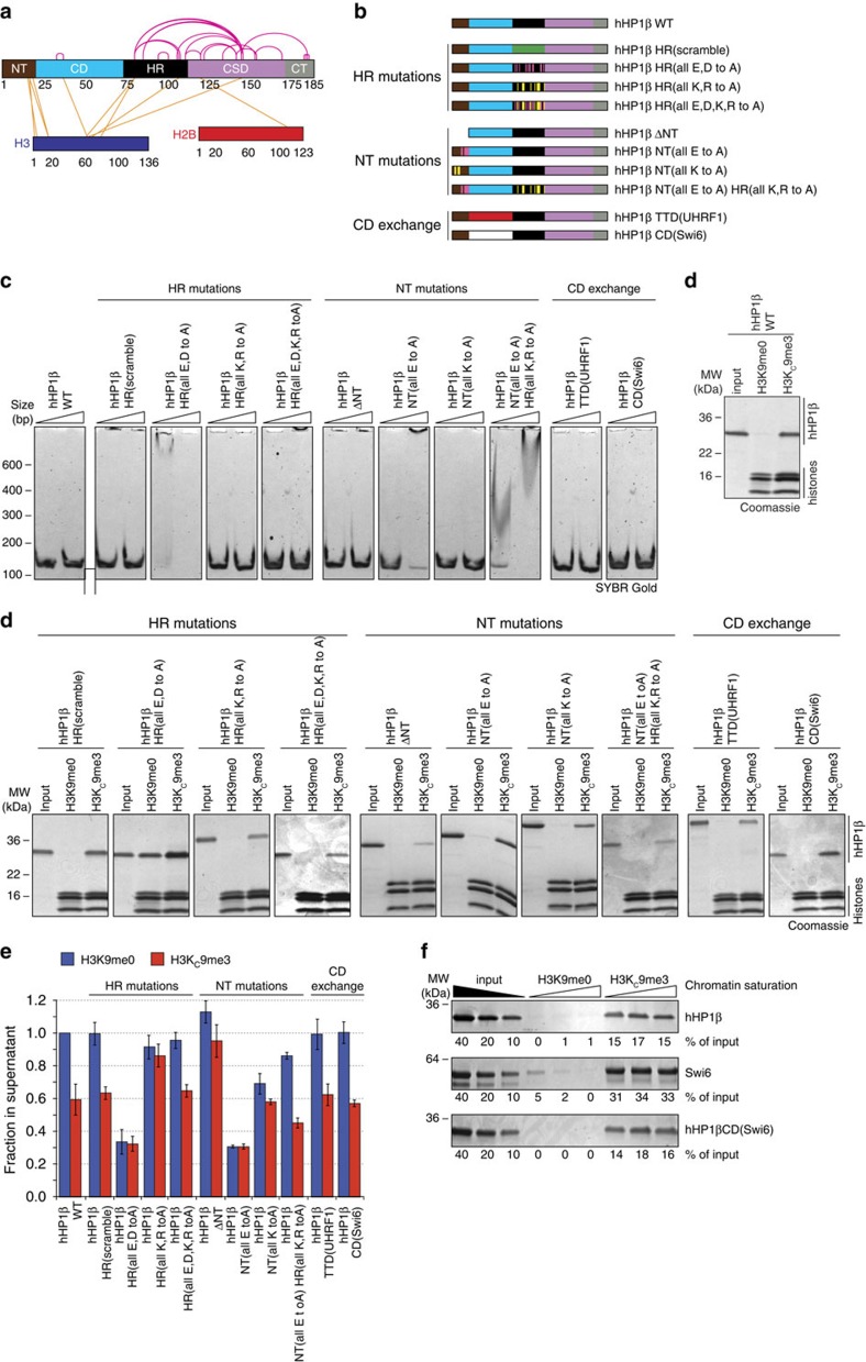
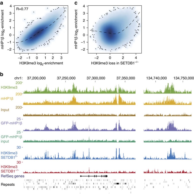
Histone H3 trimethylation of lysine 9 (H3K9me3) and proteins of the heterochromatin protein 1 (HP1) family are hallmarks of heterochromatin, a state of compacted DNA essential for genome stability and long-term transcriptional silencing. The mechanisms by which H3K9me3 and HP1 contribute to chromatin condensation have been speculative and controversial. Here we demonstrate that human HP1β is a prototypic HP1 protein exemplifying most basal chromatin binding and effects. These are caused by dimeric and dynamic interaction with highly enriched H3K9me3 and are modulated by various electrostatic interfaces. HP1β bridges condensed chromatin, which we postulate stabilizes the compacted state. In agreement, HP1β genome-wide localization follows H3K9me3-enrichment and artificial bridging of chromatin fibres is sufficient for maintaining cellular heterochromatic conformation. Overall, our findings define a fundamental mechanism for chromatin higher order structural changes caused by HP1 proteins, which might contribute to the plastic nature of condensed chromatin.


Figures

Figure 1
Figure 1. hHP1β–chromatin interaction is dependent on H3K9me3.
(a) Domain structure of hHP1β; boundaries of domains are indicated by respective amino acid positions. For more details see Supplementary Fig. 2. (b) Scheme of pull-down experiments in c using different H3K9me templates immobilized on streptavidin-coated magnetic beads via C-terminal incorporation of a biotinylated lysine (peptide) or ligation of 5′-biotinylated oligonucleotides to DNA templates used in chromatin reconstitution (mono- and oligonucleosomes). (c) Immobilized H3K9me templates according to the pull-down experimental schemes in b were incubated with recombinant hHP1β WT. Material recovered after washing was analysed by western blotting. The indicated salt concentrations were used throughout the experiment. (d) Scheme of chromatin coprecipitation assay; factors bound to oligonucleosomes are precipitated with the template when clustering is induced by addition of Mg2+ ions. (e,f) Chromatin coprecipitation of hHP1β (e) and hHP1α (f) proteins with oligonucleosomes. Precipitated material was run on SDS-PAGE and stained with Coomassie blue. Input, 10%. (g) The indicated recombinant proteins were incubated with a DNA fragment of 150 bp at 500: 1 and 1,000: 1 molar ratio. Complexes were separated by PAGE. DNA was stained with SYBR Gold.
Figure 2
Figure 2. Dimerization is necessary and sufficient for hHP1β binding of H3K9me3 chromatin.
(a) Schematic representation of hHP1β mutant proteins. White bars indicate positions of W42 and I161, respectively. (b) Chromatin coprecipitation of WT and mutant hHP1β proteins with oligonucleosomes. Precipitated material was analysed by western blotting using antibodies that recognizes the CD of hHP1β. Ponceau staining of the region of the western blot membrane containing histones is shown as loading control. Input, 10%. (c) The indicated proteins were incubated with a biotinylated H3K9me3 peptide immobilized on magnetic streptavidin beads. Material recovered after washing was run on SDS–PAGE and stained with Coomassie blue. Input, 10%. (d) SPR analysis of hHP1β WT interaction with a biotinylated H3K9me3 peptide immobilized at different density on the chip surface (very low, 5 RU; low, 24 RU; very high, 950 RU). (e) Experiment as in b using hHP1β CSD, but analyzed by SDS–PAGE and staining with Coomassie Blue.
Figure 3
Figure 3. hHP1β clusters oligonuclesomal arrays in dependence of H3K9me3.
(a) Oligonucleosomes (2.5 nM) were incubated with increasing concentrations of hHP1β WT or I161A at 150 mM NaCl. DNA remaining in the supernatant after centrifugation was incubated with EtBr and measured by fluorescence reading. Data were normalized to amounts in absence of added protein. Averages of three independent experiments are plotted; error bars represent s.d.; n=3. (b) Analysis of the hydrodynamic radius of oligonucleosomes (10 nM; DNA labelled with ATTO 610) in the presence of increasing concentrations of hHP1β WT or I161A by FCS at 150 mM NaCl. For details on experimental setup see Supplementary Fig. 3. Error bars represent s.d.; n=3. (c) Oligonucleosomes were reconstituted after mixing H3K9me0 and H3K9me3 octamers at different ratio. Chromatin precipitation analysis at 150 mM NaCl was carried out with hHP1β WT at saturating concentration. Data are presented as in a. (d) Precipitation behaviour of oligonucleosomes (2.5 nM) was analysed at different concentrations of NaCl with hHP1β WT at saturating concentration. Data are presented as in a. (e) H3K9me3-containing chromatin was reconstituted using wild-type core histones or tailless H2A, H2B or H4. Oligonucleosomes (2.5 nM) were incubated with increasing concentrations of hHP1β WT at 150 mM NaCl. Data are presented as in a. (f) Chromatin coprecipitation of hHP1β with H3K9me3-containing oligonucleosomes reconstituted with wild-type or tailless core histones at the indicated concentrations of MgCl2. Precipitated material was analysed by western blotting. The region of the western blot membrane of the experiment at 7.5 mM MgCl2 containing histones was stained with Ponceau. (g) Oligonucleosomes at 50 mM NaCl were incubated with hHP1β WT or I161A. Complexes were fixed with 0.05% (v/v) glutaraldehyde, spotted on mica surfaces and analysed by scanning force microscopy. Control, no protein added; scale bar, 100 nm. (h) Quantification of results representatively shown in g according to the classification on the top: A, extended; B, partially condensed; C, fully condensed; D; aggregates; n>50 for each condition.
Figure 4
Figure 4. hHP1β bridges nucleosomes on the same and different chromatin fibres.
(a) Chromatin precipitation analysis of oligonucleosomes with hHP1β wild-type (WT) or mutant proteins under saturating conditions. DNA remaining in the supernatant after centrifugation was incubated with EtBr and measured by fluorescence reading. Data were normalized to amounts present in the input. Error bars represent s.d.; n=3. (b) Scheme of UV-mediated hHP1β-histone H3 cross-linking experiments. (c) Photo cross-linking according to the scheme in b was done with H3K9me0 or H3KC9me3 oligonucleosomal arrays and hHP1β N57X Q158X. Samples were run on SDS–PAGE (top, shorter run; bottom, longer run) and stained with Coomassie blue. Black arrows indicate crosslinked hHP1β dimer crosslinked to one untagged histone H3 and one His6-tagged histone H3. (d) Histograms of intensity scans of Coomassie blue stained SDS–PAGE gels shown in c.
Figure 5
Figure 5. Modulation of CD and CSD functions by the HR and NT of hHP1β.
(a) Scheme representing EDC cross-links identified within the hHP1β/H3KC9me3 oligonucleosome complex under conditions of chromatin clustering (13.4 nM H3KC9me3 oligonucleosomal arrays, 15 μM hHP1β, 100 mM NaCl) using mass spectrometry. For detailed listing of the crosslinks see Supplementary Data 1. (b) Schematic representation of hHP1β mutant proteins. (c) The indicated recombinant proteins were incubated with a DNA fragment of 150 bp at 500: 1 and 1,500: 1 molar ratio. Complexes were separated by PAGE. DNA was stained with SYBR Gold. (d) Chromatin coprecipitation of the indicated wild-type (WT) and mutant hHP1β proteins with oligonucleosomes. Precipitated material was run on SDS–PAGE and stained with Coomassie blue. Input, 10%. (e) Chromatin precipitation analysis of oligonucleosomes with hHP1β WT or mutant proteins under non-saturating conditions. DNA remaining in the supernatant after centrifugation was incubated with EtBr and measured by fluorescence reading. Data were normalized to DNA levels present in the H3K9me0 chromatin/hHP1β WT sample. Averages of three independent experiments are plotted; error bars represent s.d.; n=3. (f) Chromatin coprecipitation of the indicated proteins with oligonucleosomes reconstituted to different saturation (0.8: 1.0, 1.0: 1.0 and 1.0: 1.2 ratio of octamers to positioning sequences). Precipitated material was run on SDS–PAGE and stained with Coomassie blue. Intensity of bands was quantified in relation to the input. Representative experiment is shown.
Figure 6
Figure 6. Genome-wide enrichment of HP1β correlates with SETDB1-dependent H3K9me3 deposition.
(a) Plot of genome-wide correlation of mHP1β and H3K9me3 (ref. 35) as deduced by ChIP-seq in mouse ESC and using 1 kb-sized windows. Dashed line indicates the data trend computed by loss regression. (b) Tracks displaying number of library-normalized reads per 100 bp from the indicated ChIP-seq experiments. Data obtained from antibody-based enrichments of endogenous mHP1β (top) and GFP-tagged mHP1β (middle) and the respective input tracks are shown. H3K9me3 data in SETDB1f/− and SETDB1−/− experiments are from ref. . Gene models and repetitive elements are indicated at the bottom. (c) Analysis as in a but comparing mHP1β binding to H3K9me3 loss in SETDB1−/− mESC. H3K9me3 loss is shown as the log2-difference between SETDB1f/− and SETDB1−/−.
Figure 7
Figure 7. hHP1β dimerization is required for maintaining condensed cellular chromatin states.
(a) Schematic diagram of the transgene system used to test chromatin compaction effects of hHP1β. A tandem array of the construct has been integrated into the genome of U2OS mammalian cells. Dox, doxycycline; rtTA, reverse tetracycline-controlled TET-VP16 transactivator, whose binding to tetracycline responsive elements (RE) is activated by doxycycline. (b) Together with mCherry-LacI, the indicated fusion proteins were transiently expressed in U2OS cells containing the chromatin reporter array as described in a. Representative confocal images in the absence or after induction of transcription from the array with doxycycline (Dox) are shown. DNA was stained with DAPI. Inlets show enlarged chromatin reporter array; scale bar, 10 μm. (c) Quantification of the results shown in b together with results of similar experiments using additional hHP1β mutant proteins. Averages of three independent experiments are plotted; error bars represent s.d.; n>300 for each condition; asterisks represent P<0.05 according to Student's t-test.
Figure 8
Figure 8. hHP1β is a paradigm HP1 protein.
(a) Model summarizing the functional properties (right) and interactions of different domains of hHP1β. The balance of negatively and positively charged residues within the NT and HR is essential for specific H3K9me3–chromatin interaction. (b) Due to its flexible HR the CD of an hHP1β dimer can interact with the H3K9me3 marks of the same or different nucleosomes within the same or another chromatin fibre (top). The binding is stabilized by flexible and dynamic electrostatic interaction of the NT with the H3 tail. The high concentration of H3K9me3 in condensed and clustered oligonucleosomes (bottom) establishes multiple hHP1β binding possibilities. This steadies the interaction and stabilizes the compacted chromatin state.

References

    1. Grewal S. I. & Jia S. Heterochromatin revisited. Nat. Rev. Genet. 8, 35–46 (2007). - PubMed
    1. Maison C. & Almouzni G. HP1 and the dynamics of heterochromatin maintenance. Nat. Rev. Mol. Cell Biol. 5, 296–304 (2004). - PubMed
    1. Hiragami K. & Festenstein R. Heterochromatin protein 1: a pervasive controlling influence. Cell Mol. Life Sci. 62, 2711–2726 (2005). - PMC - PubMed
    1. Fodor B. D., Shukeir N., Reuter G. & Jenuwein T. Mammalian Su(var) genes in chromatin control. Annu. Rev. Cell Dev. Biol. 26, 471–501 (2010). - PubMed
    1. Ayyanathan K. et al. Regulated recruitment of HP1 to a euchromatic gene induces mitotically heritable, epigenetic gene silencing: a mammalian cell culture model of gene variegation. Genes Dev. 17, 1855–1869 (2003). - PMC - PubMed

Publication types

MeSH terms

Associated data