. 2016 Apr 18:16:41.
doi: 10.1186/s12886-016-0218-0.

Nd:YAG laser hyaloidotomy in the management of Premacular Subhyaloid Hemorrhage

Affiliations

Nd:YAG laser hyaloidotomy in the management of Premacular Subhyaloid Hemorrhage

Deepak Khadka et al. BMC Ophthalmol. .

Abstract

Background: Premacular subhyaloid hemorrhage results in a sudden profound loss of vision. Among the modalities for its treatment, Nd:YAG laser hyaloidotomy is a non invasive method enabling rapid drainage of the obstructed macular area and improved vision within days. This study was aimed to evaluate the efficacy, visual outcome and complications following Nd:YAG laser hyaloidotomy for premacular subhyaloid hemorrhage.

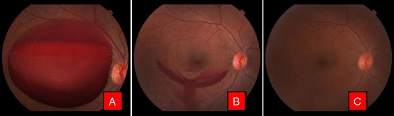
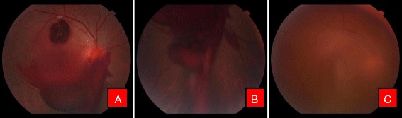
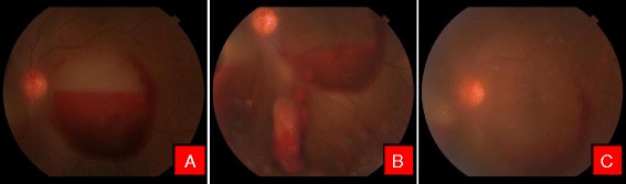
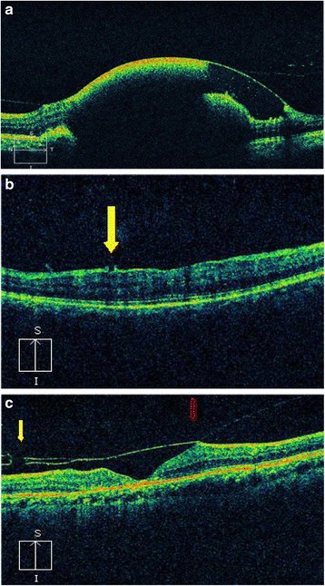
Methods: Patients with premacular subhyaloid hemorrhage of more than 3 disc diameters (DD) of various etiologies, attending Tilganga Institute of Ophthalmology, Nepal from August, 2014 to February, 2015, were included. A comprehensive ocular evaluation was conducted and fundus photographs were taken to measure the size of the subhyaloid hemorrhage. Optical coherence tomography (OCT) were performed before and after treatment and on subsequent follow up visits. Fundus fluorescence angiography was done whenever necessary. Q switched Nd:YAG laser was applied to create an opening in the posterior hyaloids membrane for draining subhyaloid hemorrhage. The main outcome measures were success rate in performing hyaloidotomy, drainage of subhyaloid blood into vitreous cavity and its resorption, improvement in visual acuity, need for further intervention and postoperative complications.

Results: There were 21 eyes of 19 patients, 17(89.48%) male and 2(10.52%) female. In 3, premacular subhyaloid hemorrhage was bilateral. Mean age was 41.68 ± 17.08 years and a mean duration of symptoms 15.04 days. Mean pretreatment hemorrhage was 6.27DD. Nd:YAG laser hyaloidotomy was successful in 19 eyes(86.4%). In 2 patients, one each with Eales' disease and retinal vein occlusion the procedure was unsuccessful, necessitating pars plana vitrectomy, while in a case with proliferative diabetic retinopathy (PDR), vitrectomy was resorted for non clearing vitreous hemorrhage. Vision improved from a median of 3/60 pre-operatively to 6/6, at 6 months follow up. At 3 months, 2 patients with Eales' disease, one developed tractional detachment at macula while the other, an epiretinal membrane. No other complications were noted at 6 months.

Conclusion: Nd:YAG laser hyaloidotomy is an inexpensive, effective and a safe outpatient procedure for premacular subhyaloid hemorrhage, producing rapid drainage with restoration of visual function avoiding more invasive procedures and enabling early assessment of the underlying retina. The final visual prognosis however, rests on the underlying cause of the subhyaloid hemorrhage and any accompanying retinal changes.

Keywords: Hyaloidotomy; Nd:YAG laser; Premacular hemorrhage; Subhyaloid hemorrhage.


Figures

Fig. 1
Fig. 1
Color fundus photographs of a patient with Valsalva retinopathy. a Demonstrating boat shaped hemorrhage. b Same patient immediately after Nd: YAG laser hyaloidotomy. Note the draining premacular hemorrhage. c Fundus picture after 6 weeks
Fig. 2
Fig. 2
Color fundus photographs of a patient with ruptured macroaneurysm. a Before treatment. Note the macroaneurysm in superotemporal quadrant. b Immediately after Nd:YAG photodisruption with blood draining into vitreous cavity inferiorly (c). Resolving hemorrhage after 1 week
Fig. 3
Fig. 3
Color fundus photographs of a patient with Eales’ disease. a At the time of presentation. b Note the liquefied blood with neovascularisation of the disc. c Patient after Nd:YAG laser hyaloidotomy. Note the draining blood. d 6 weeks after hyaloidotomy. Note the tractional detachment at macula due to regressing neovascular frond (Blue arrow) and hyaloidotomy opening (Red arrow). e Fundus flurescein angiography (FFA) showing hyperflurescence due to neovascularisation of the disc. f Late phase FFA of the same patient with characteristic leakage of dye
Fig. 4
Fig. 4
Color fundus photographs of a patient with leukemic retinopathy. a With multiple nerve fiber layer hemorrhage. b Immediately after Nd:YAG hyaloidotomy with two openings. c Day 1 after laser, with clearing premacular bleed
Fig. 5
Fig. 5
Color fundus photographs of a patient with high altitude retinopathy. a Characteristic boat shaped hemorrhage with multiple retinal hemorrhages. b Immediately after Nd:YAG laser with blood draining into vitreous cavity. c Clearing premacular hemorrhage at the end of 1 week
Fig. 6
Fig. 6
Color fundus photographs of a patient with CNVM. a Characteristic boat shaped hemorrhage. b Immediately after Nd Yag laser. c 3 months later after couple of intravitreal Bevacizumab injection
Fig. 7
Fig. 7
a Optical coherence tomogram (OCT) of a patient with Valsalva retinopathy showing hemorrhage under internal limiting membrane (ILM). b OCT of a patient with Valsalva retinopathy 6 weeks after laser photodisruption. Note the opening in ILM (yellow arrow). c OCT of a patient after Nd:YAG laser hyaloidotomy. Note the defect in posterior hyaloid membrane (yellow arrow) and the Vitreomacular traction (red arrow)

Similar articles

Cited by

References

    1. Gass JDM. Stereoscopic Atlas of Macular Diseases: diagnosis and treatment. 3. St Louis, MO: CV Mosby; 1987. pp. 560–564.
    1. Kwok AK, Lai TY, Chan NR. Epiretinal membrane formation with internal limiting membrane wrinkling after Nd:YAG laser membranotomy in valsalva retinopathy. Am J Ophthalmol. 2003;136:763–766. doi: 10.1016/S0002-9394(03)00442-2. - DOI - PubMed
    1. Meyer CH, Mennel S, Rodrigues EB, Schmidt JC. Persistent premacular cavity after membranotomy in valsalva retinopathy evident by optical coherence tomography. Retina. 2006;26:116–118. doi: 10.1097/00006982-200601000-00024. - DOI - PubMed
    1. Raymond LA. Neodymium:YAG laser treatment for hemorrhages under the internal limiting membrane and posterior hyaloid face in the macula. Ophthalmology. 1995;102:406–11. doi: 10.1016/S0161-6420(95)31008-1. - DOI - PubMed
    1. Mennel S. Subhyaloid and macular haemorrhage: localization and treatment strategies. Br J Ophthalmol. 2007;91:850–852. doi: 10.1136/bjo.2007.114025. - DOI - PMC - PubMed

LinkOut - more resources