

Coexistence of intervertebral disc herniation with intradural schwannoma in a lumbar segment: a case report
- PMID: 27091024
- PMCID: PMC4836079
- DOI: 10.1186/s12957-016-0864-y
Coexistence of intervertebral disc herniation with intradural schwannoma in a lumbar segment: a case report
Abstract
Background: Lumbar intervertebral disc herniation and spinal tumor are major pathologies that may cause back pain and radiculopathy. Neurological symptoms resulting from disc herniation and intradural spinal tumor together, however, are very rare.
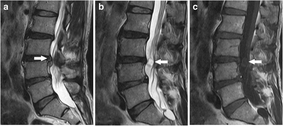
Case presentation: We report a case of lumbar disc herniation which coexists with intradural schwannoma at the same spinal level in a 67-year-old man. The patient presented with persistent low back pain, sciatica, and weakness of the lower limbs. Contrast lumbar spine magnetic resonance (MR) imaging clearly delineated an intradural lesion and an extradural herniated disc at L3/4 level. Using a single posterior approach, both pathologies were addressed. Pathological studies confirmed the intradural lesion was schwannoma.
Conclusion: The case report highlights a rare concomitance of two symptomatic pathologies in a lumbar spine, which deserves clinical attention. Complete history, careful physical examination, and investigative measures, such as contrast MR imaging, are helpful to establish throughout diagnoses.
Keywords: Intraspinal tumor; Lumbar disc herniation; Schwannoma.
Figures

References
-
- Albert FK, Oldenkott P, Bieker G, et al. Lumbar intervertebral disk herniation with a concomitant nerve root neurinoma at the same site. Case report and review of the literature. Neurochirurgia. 1988;31(6):222–5. - PubMed
Publication types
MeSH terms
LinkOut - more resources
Full Text Sources
Other Literature Sources
Medical
