

Ion channels in regulated cell death
- PMID: 27091155
- PMCID: PMC11108559
- DOI: 10.1007/s00018-016-2208-z
Ion channels in regulated cell death
Abstract
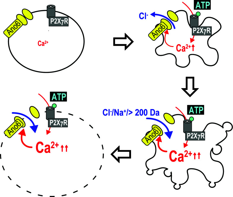
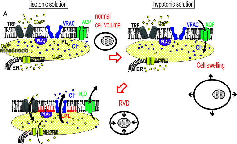
Activation of ion channels and pores are essential steps during regulated cell death. Channels and pores participate in execution of apoptosis, necroptosis and other forms of caspase-independent cell death. Within the program of regulated cell death, these channels are strategically located. Ion channels can shrink cells and drive them towards apoptosis, resulting in silent, i.e. immunologically unrecognized cell death. Alternatively, activation of channels can induce cell swelling, disintegration of the cell membrane, and highly immunogenic necrotic cell death. The underlying cell death pathways are not strictly separated as identical stimuli may induce cell shrinkage and apoptosis when applied at low strength, but may also cause cell swelling at pronounced stimulation, resulting in regulated necrosis. Nevertheless, the precise role of ion channels during regulated cell death is far from being understood, as identical channels may support regulated death in some cell types, but may cause cell proliferation, cancer development, and metastasis in others. Along this line, the phospholipid scramblase and Cl(-)/nonselective channel anoctamin 6 (ANO6) shows interesting features, as it participates in apoptotic cell death during lower levels of activation, thereby inducing cell shrinkage. At strong activation, e.g. by stimulation of purinergic P2Y7 receptors, it participates in pore formation, causes massive membrane blebbing, cell swelling, and membrane disintegration. The LRRC8 proteins deserve much attention as they were found to have a major role in volume regulation, apoptotic cell shrinkage and resistance towards anticancer drugs.
Keywords: Anoctamin 6; Apoptosis; LRRC8A; Necroptosis; TMEM16F.
Figures
Similar articles
-
Ca2+ signals, cell membrane disintegration, and activation of TMEM16F during necroptosis.Cell Mol Life Sci. 2017 Jan;74(1):173-181. doi: 10.1007/s00018-016-2338-3. Epub 2016 Aug 17. Cell Mol Life Sci. 2017. PMID: 27535660 Free PMC article.
-
Cl- channels in apoptosis.Eur Biophys J. 2016 Oct;45(7):599-610. doi: 10.1007/s00249-016-1140-3. Epub 2016 Jun 7. Eur Biophys J. 2016. PMID: 27270446
-
Relationship between TMEM16A/anoctamin 1 and LRRC8A.Pflugers Arch. 2016 Oct;468(10):1751-63. doi: 10.1007/s00424-016-1862-1. Epub 2016 Aug 12. Pflugers Arch. 2016. PMID: 27514381
-
Ion channels and cell volume in regulation of cell proliferation and apoptotic cell death.Contrib Nephrol. 2006;152:142-160. doi: 10.1159/000096321. Contrib Nephrol. 2006. PMID: 17065810 Review.
-
Role of anoctamins in cancer and apoptosis.Philos Trans R Soc Lond B Biol Sci. 2014 Feb 3;369(1638):20130096. doi: 10.1098/rstb.2013.0096. Print 2014 Mar 19. Philos Trans R Soc Lond B Biol Sci. 2014. PMID: 24493744 Free PMC article. Review.
Cited by
-
Ca2+ signals, cell membrane disintegration, and activation of TMEM16F during necroptosis.Cell Mol Life Sci. 2017 Jan;74(1):173-181. doi: 10.1007/s00018-016-2338-3. Epub 2016 Aug 17. Cell Mol Life Sci. 2017. PMID: 27535660 Free PMC article.
-
Cell death in the gut epithelium and implications for chronic inflammation.Nat Rev Gastroenterol Hepatol. 2020 Sep;17(9):543-556. doi: 10.1038/s41575-020-0326-4. Epub 2020 Jul 10. Nat Rev Gastroenterol Hepatol. 2020. PMID: 32651553 Review.
-
A 30-year journey from volume-regulated anion currents to molecular structure of the LRRC8 channel.J Gen Physiol. 2019 Feb 4;151(2):100-117. doi: 10.1085/jgp.201812138. Epub 2019 Jan 16. J Gen Physiol. 2019. PMID: 30651298 Free PMC article. Review.
-
Necroptosis Execution Is Mediated by Plasma Membrane Nanopores Independent of Calcium.Cell Rep. 2017 Apr 4;19(1):175-187. doi: 10.1016/j.celrep.2017.03.024. Cell Rep. 2017. PMID: 28380356 Free PMC article.
-
Metabolic barriers to cancer immunotherapy.Nat Rev Immunol. 2021 Dec;21(12):785-797. doi: 10.1038/s41577-021-00541-y. Epub 2021 Apr 29. Nat Rev Immunol. 2021. PMID: 33927375 Free PMC article. Review.
References
Publication types
MeSH terms
Substances
LinkOut - more resources
Full Text Sources
Other Literature Sources
