. 2016 Apr 19:6:24474.
doi: 10.1038/srep24474.

Bioprinting three-dimensional cell-laden tissue constructs with controllable degradation

Affiliations

Bioprinting three-dimensional cell-laden tissue constructs with controllable degradation

Zhengjie Wu et al. Sci Rep. .

Abstract

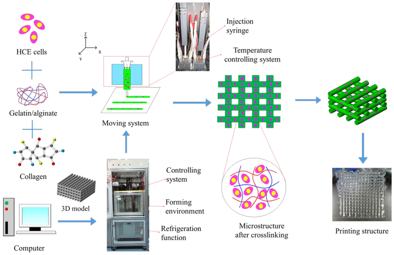
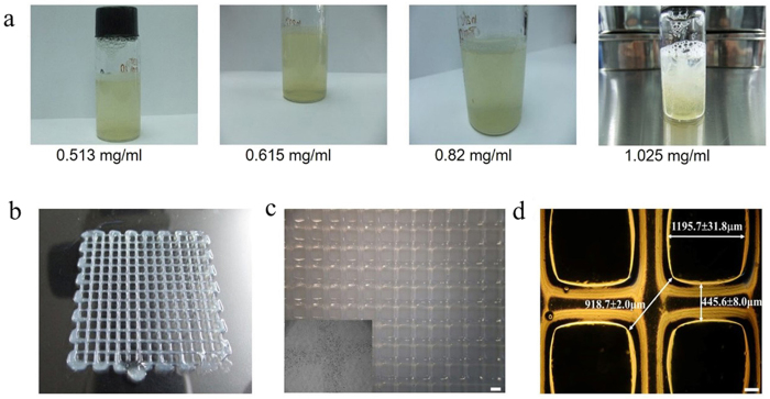
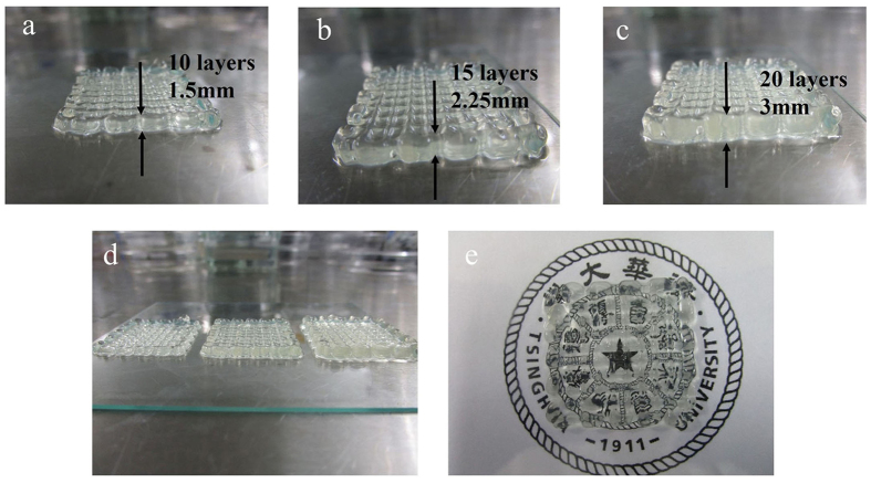
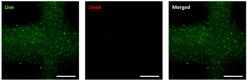
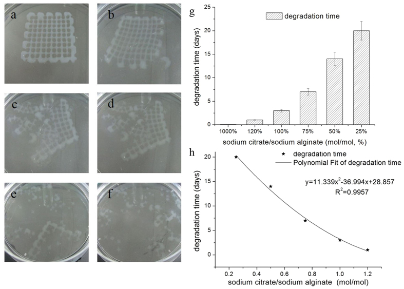
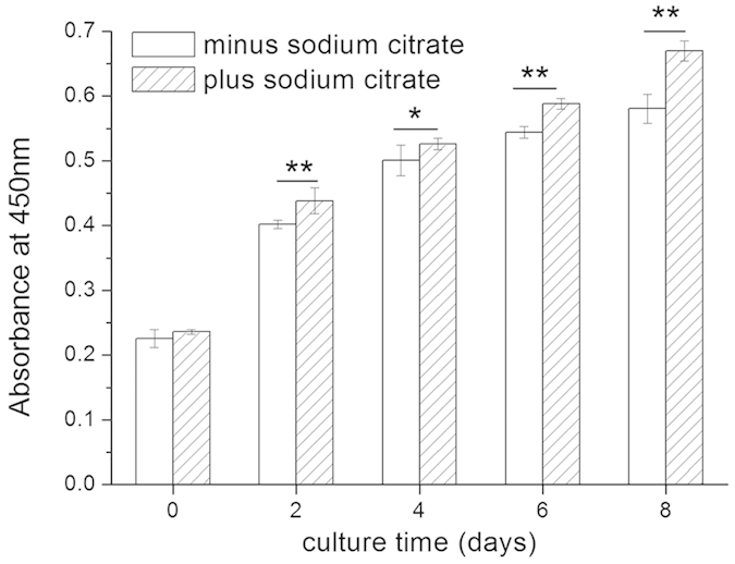
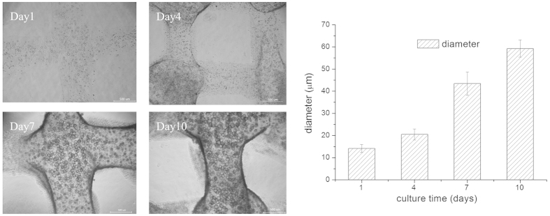
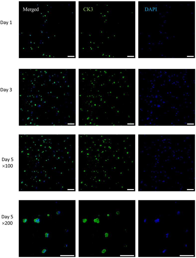
Alginate hydrogel is a popular biologically inert material that is widely used in 3D bioprinting, especially in extrusion-based printing. However, the printed cells in this hydrogel could not degrade the surrounding alginate gel matrix, causing them to remain in a poorly proliferating and non-differentiating state. Here, we report a novel study of the 3D printing of human corneal epithelial cells (HCECs)/collagen/gelatin/alginate hydrogel incubated with a medium containing sodium citrate to obtain degradation-controllable cell-laden tissue constructs. The 3D-printed hydrogel network with interconnected channels and a macroporous structure was stable and achieved high cell viability (over 90%). By altering the mole ratio of sodium citrate/sodium alginate, the degradation time of the bioprinting constructs can be controlled. Cell proliferation and specific marker protein expression results also revealed that with the help of sodium citrate degradation, the printed HCECs showed a higher proliferation rate and greater cytokeratin 3(CK3) expression, indicating that this newly developed method may help to improve the alginate bioink system for the application of 3D bioprinting in tissue engineering.


Figures

Figure 1
Figure 1. Schematic illustration of the 3D bioprinting process and optical images of the printing setup and printing constructs.
Figure 2
Figure 2. Bioprinting of HCECs in gelatin/alginate/collagen.
(a) The different concentrations of collagen added to the gelatin/alginate bioprinting materials. (b) Top view of a 3D HCECs /hydrogel construct demonstrating the porous nature of the finalised scaffold. (c,d) The overall size images of the 3D constructs, including the pore size, thread diameter and max pore distance (c: scale bar, 1 mm; d: scale bar, 200 μm).
Figure 3
Figure 3. The interconnectivity of the different layers of scaffolds in the Z-direction.
(a) Ten layers with 1.5-mm thickness, (b) 15 layers with 2.25-mm thickness, (c) 20 layers with 3-mm thickness, (d) scaffolds with different layers and thicknesses, (e) general light transmission of the scaffolds.
Figure 4
Figure 4. Cell viability after printing by live/dead staining (scale bar, 500 μm).
Figure 5
Figure 5
Images of the degradation process of the scaffolds set in the sodium citrate solution, from (a) 0 min, (b) 10 min, (c) 20 min, (d) 30 min, (e) 40 min, (f) 50 min, when the C/A is 1000% (mol/mol, %). (g) The relation of the total degradation time of the scaffolds to the mole ratio of C/A. (h) The relation curve between the degradation time of the printed constructs and amount of sodium citrate added to the culture medium.
Figure 6
Figure 6. Proliferation of HCECs within the printed constructs incubated with fresh medium containing no (minus sodium citrate) or 66.7% sodium citrate (plus sodium citrate, mole ratio of C/A), as measured by CCK-8.
Data represent means ± SD (*p < 0.01, **p < 0.005).
Figure 7
Figure 7. Morphological change in the cell aggregates within the printed constructs incubated with fresh medium containing 66.7% sodium citrate (C/A, mole ratio, %) during different culturing times (scale bar, 500 μm).
Figure 8
Figure 8. Specific marker protein expression of printed cells within constructs incubated with medium containing 66.7% sodium citrate (C/A, mole ratio, %).
Micrographs show fluorescent staining of CK3 (green) and nuclei (blue) of HCECs within the constructs after culturing for 1, 3, and 5 days (scale bar, 100 μm). (For the interpretation of the references to colour in this figure legend, the reader is referred to the web version of this article.)

References

    1. Derby B. Printing and prototyping of tissues and scaffolds. Science. 338, 921–926 (2012). - PubMed
    1. Atala A. Tissue engineering and regenerative medicine: concepts for clinical application. Rejuv. Res. 7, 15–31 (2004). - PubMed
    1. Hollister S. J. Porous scaffold design for tissue engineering. Nat. mater. 4, 518–524 (2005). - PubMed
    1. Hutmacher D. W. Scaffolds in tissue engineering bone and cartilage. Biomaterials. 21, 2529–2543 (2000). - PubMed
    1. Reiffel A. J. et al.. High-fidelity tissue engineering of patient-specific auricles for reconstruction of pediatric microtia and other auricular deformities. PloS one. 8, e56506 (2013). - PMC - PubMed

Publication types