Review
. 2016 Sep:54:49-63.
doi: 10.1016/j.preteyeres.2016.04.005. Epub 2016 Apr 16.

Goblet cells of the conjunctiva: A review of recent findings

Affiliations
Review

Goblet cells of the conjunctiva: A review of recent findings

Ilene K Gipson. Prog Retin Eye Res. 2016 Sep.

Abstract

Goblet cells within the conjunctival epithelium are specialized cells that secrete mucins onto the surface of the eye. Recent research has demonstrated new characteristics of the cells, including factors influencing their differentiation, their gene products and their functions at the ocular surface. The following review summarizes the newly discovered aspects of the role of Spdef, a member of the Ets transcription factor family in conjunctival goblet cell differentiation, the newly discovered goblet cell products including claudin2, the Wnt inhibitor Frzb, and the transmembrane mucin Muc16. The current concepts of conjunctival goblet cell function, including debris removal and immune surveillance are reviewed, as are changes in the goblet cell population in ocular surface diseases. Major remaining questions regarding conjunctival cell biology are discussed.

Keywords: Conjunctival epithelium; Dry eye; Goblet cell; Goblet cell differentiation; Ocular surface; SPDEF.


Figures

Figure 1
Figure 1
Goblet cell distribution on the human ocular surface (A, after (Kessing, 1968) and histologic appearance of goblet cells in (B) human and (C) mouse conjunctival epithelium. Note that the goblet cells in humans can appear singly in regions of sparse density and that they can also occur in clusters in the forniceal region as shown in A. Such a cluster is present in the section of human conjunctiva in B. In mice (C), clusters are present throughout the conjunctiva. Bars, B=10 µm, C=20 µm.
Figure 2
Figure 2
Confocal microscopy images of whole mounts of conjunctival epithelium labeled with phalloidin to demonstrate the actin cytoskeleton in goblet cells (after Gipson and Tisdale, 1997) in human (A), mouse (B), and rat (C, D). Note that in humans (A), goblet cells appear singly within the tissue (arrows) whereas in mice and rats, the goblet cells appear in clusters (B, C). The stacked image in C demonstrates the three dimensional structure of the cluster and demonstrates an apical band of actin around the orifice of the goblet cell cluster (arrow) suggesting an acinus like arrangement of cells. In D the apical surface of rat conjunctiva was double labeled with phalloidin in red and with fluorescein labeled UEA lectin in green to demonstrate mucin being secreted from goblet cells. Details of tissue preparation can be found in Gipson and Tisdale, 1997. Bars, A, B=10 µm, C=5µm, D=20µm.
Figure 3
Figure 3
Electron micrographs of human conjunctival goblet cells at low (A) and higher magnification (B). Note the plump appearance of the polarized goblet cell in A, with the cell content taken up by mucin granules. A thin layer of cytoplasm is present along the cell periphery with the nucleus displaced toward the base of the cell. The cytoplasm at the cell base around the nucleus is enriched in golgi and endoplasmic reticulum. A high resolution image of mucin filled mucin graules in B shows a chain-like filamentous network of the mucin. Bars, A=1.5µm, B=0.5µm.
Figure 4
Figure 4
Demonstration of the membrane –anchored mucin MUC16 protein and message in human goblet cells (A, B, C, D, E and F) and message in laser captured mouse conjunctival goblet cells (G, H). (Reprinted from Gipson et al., 2016, with permission.) Immunohistochemical localization of MUC16 in human goblet cells using two different antibodies (A) antibody OC125 and (B) antibody M11 shows the mucin to be localized to mucin granules. Bars=18um. By immunoelectron microscopy, using antibody H185, which recognizes a carbohydrate epitope on MUC16 (Argueso et al., 2003a), the mucin appears to be localized to the mucin granule membrane (C, D, inset in D shows 10 nm gold secondary antibody control). Bars=0.2um. In situ hybriization of MUC16 message is shown in human conjunctival goblet cells in E, using an S35 labeled antisense oligoprobe with F showing the control Sense probe. Bars=10um. G shows the method of dissection of goblet cell clusters from mouse conjunctiva as well as control conjunctival and corneal epithelium using laser microdissection. RNA from the goblet cells and the conjunctival and corneal keratinocytes was isolated from the dissected tissue and analyzed by qRT-PCR for levels of Muc16 ectodomain (Muc16ECD) and Muc16 cytoplasmic tail (Muc16CT). As noted in the graph, goblet cells have full length Muc16 but unlike the human, in the mouse neither the conjunctival nor corneal keratinocytes express the mucin.
Figure 5
Figure 5
Localization of claudin 2, a member of the “leaky” class of tight junction proteins to the conjunctival goblet cell in the mouse (arrows). Note that the protein appears at the apical surface but also down the lateral membrane of the cell. Inset is secondary antibody control. The primary antibody was a rabbit anti-claudin 2 polyclonal (Invitrogen, Catalog #51-6100) used at 1:100, with a secondary fluoresceinated donkey anti rabbit IgG used at 1:100. Bar=20µm.
Figure 6
Figure 6
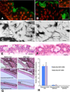
Demonstation of lack of goblet cells and phenotype of Spdef null mice. Comparison of the histology of conjunctiva of wild type (A) and Spdef−/− mice (B) show that goblet cells are lacking in the Spdef null mice. C and D demonstrate lack of overt phenotype in the null mice, but fluorescein staining revealed a significant increase in punctate staining (E, F (Marko et al., 2013). Histology demonstrated an increase in inflammatory cells within the conjunctival epithelium of the Spdef null mice (compare G and H), and enumeration of the number of CD45+ cells within the tissue by immunohistochemistry demonstrated a significant increase in their numbers within the Spdef−/− conjunctival epithelium compared to wild type control. Data and images after (Marko et al., 2013). Bar A, B=200µm, G, H =20µm.
Figure 7
Figure 7
Goblet cells express the WNT antagonist Frzb and mice null for Frzb have fewer conjunctival goblet cells. Antibodies to Frzb bind to goblet cells in the mouse conjunctiva (A). Arrows demonstrate position of goblet cells. B shows a phase contrast image of the same section as A. Frzb mRNA could not be detected in RNA isolated from conjunctival epithelium obtained by laser capture of the fornix of the conjunctiva of Spdef −/− mice contrary to wild type control (C). In wild type mice, Frzb mRNA was detected in RNA obtained from goblet cell clusters obtained by laser capture microdissection, whereas none was detected in the stratified epithelia outside the goblet cell areas (D). Goblet cell numbers were significantly decreased in conjunctivas of Frzb null mice. Counts of numbers of goblet cells were as per (Marko et al, 2014) and mice were a kind gift of Professor Frank P Luyten, Katholieke Universiteit Leuven, Leuven Belgium (Lories et al., 2007). Immunohistochemistry data and laser capture methods were as per (Marko et al., 2013). Bar=20µm. *p<0.05, **P<0.01, ***p<0.001
Figure 8
Figure 8
Diagram demonstrating factors known to date to be involved in goblet cell differentiation in the conjunctiva. Spdef appears to be a central transcription factor involved in goblet cell differentiation, being influenced by both the Notch, Wnt, and TGF beta pathways. It is not known if the IL13 induced increase in conjunctival goblet cell numbers (De Paiva et al., 2011) is through Spdef as it is in the airway epithelia (Chen et al., 2009). Similarly, a role for FoxA3 in goblet cell differentiation in airway epithelia has been demonstrated but a role for FoxA3 in conjunctival goblet cell differentiation has not been demonstrated. The FoxA3 gene is, however, highly down regulated in Spdef null mice that lack goblet cells indicating that it is Spdef regulated (Marko et al., 2013).
Figure 9
Figure 9
Accumulation of debris in conjunctival cul-de-sac of Spdef null mice compared to wild type. The conjunctival cul-de-sac of wildtype mice (A) show no debris compared to that of the Spdef null mouse (B). Debris was not present in the conjunctival cul-de-sac of wild type mice (C) even after challenge by application of Pseudomonas aeruginosa strain 6294 twice daily for three days (at 1×10⁷ colony forming units /ml in normal saline). Debris did accumulate in the Spdef mice after Pseudomonas challenge (D). See Table 2 for compilation of data. Bar=20µm.
Figure 10
Figure 10
SPDEF is localized to conjunctival goblet cells while conjunctival epithelia from patients with Sjogrens Dry Eye have decreased levels of message for both Spdef and the goblet cell mucin MUC5AC. A and B are photomicrographs of human conjunctival epithelium, with A showing fluoresceinated antibody immunolocalization of Spdef, with goblet cells indicated by arrows, and nuclei localized with Propidium iodide (PI). B is a phase contrast image of the same section as in A. Bar=20µm. C and D show significantly lower levels of SPDEF message and MUC5AC protein levels in impression cytology samples of conjunctival epithelium from normal subjects and Sjogrens dry eye subjects, respectively. (*P<0.05, **P<0.01) SPDEF immunolocalization data and SPDEF message data from Marko et al, 2013. MUC5AC data from (Argueso et al., 2002).

Similar articles

Cited by

References

    1. Argueso P, Balaram M, Spurr-Michaud S, Keutmann HT, Dana MR, Gipson IK. Decreased levels of the goblet cell mucin MUC5AC in tears of patients with Sjögren’s syndrome. Invest Ophthalmol Vis Sci. 2002;43:1004–1011. - PubMed
    1. Argueso P, Spurr-Michaud S, Russo CL, Tisdale A, Gipson IK. MUC16 mucin is expressed by the human ocular surface epithelia and carries the H185 carbohydrate epitope. Investigative ophthalmology & visual science. 2003a;44:2487–2495. http://dx.doi.org/10.1167/iovs.02-0862. - DOI - PubMed
    1. Argueso P, Tisdale A, Mandel U, Letko E, Foster CS, Gipson IK. The cell-layer- and cell-type-specific distribution of GalNAc-transferases in the ocular surface epithelia is altered during keratinization. Investigative ophthalmology & visual science. 2003b;44:86–92. http://dx.doi.org/10.1167/iovs.02-0181. - DOI - PubMed
    1. Barker N, van de Wetering M, Clevers H. The intestinal stem cell. Genes & development. 2008;22:1856–1864. http://dx.doi.org/10.1101/gad.1674008. - DOI - PMC - PubMed
    1. Bell SM, Zhang L, Xu Y, Besnard V, Wert SE, Shroyer N, Whitsett JA. Kruppel-like factor 5 controls villus formation and initiation of cytodifferentiation in the embryonic intestinal epithelium. Developmental biology. 2013;375:128–139. http://dx.doi.org/10.1016/j.ydbio.2012.12.010. - DOI - PMC - PubMed