. 2016 Jul;1863(7 Pt A):1643-52.
doi: 10.1016/j.bbamcr.2016.04.012. Epub 2016 Apr 16.

Supercomplex-associated Cox26 protein binds to cytochrome c oxidase

Affiliations

Supercomplex-associated Cox26 protein binds to cytochrome c oxidase

Valentina Strecker et al. Biochim Biophys Acta. 2016 Jul.

Abstract

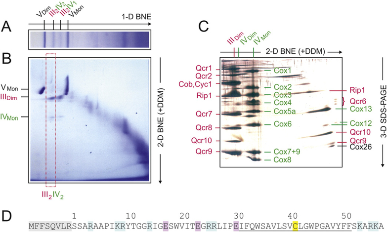
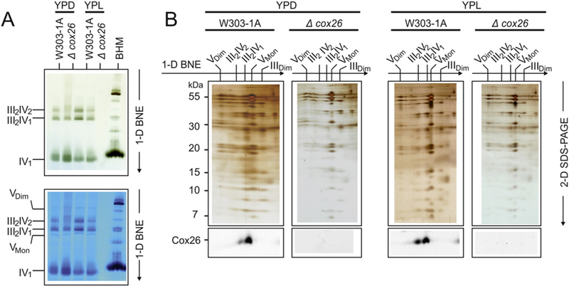
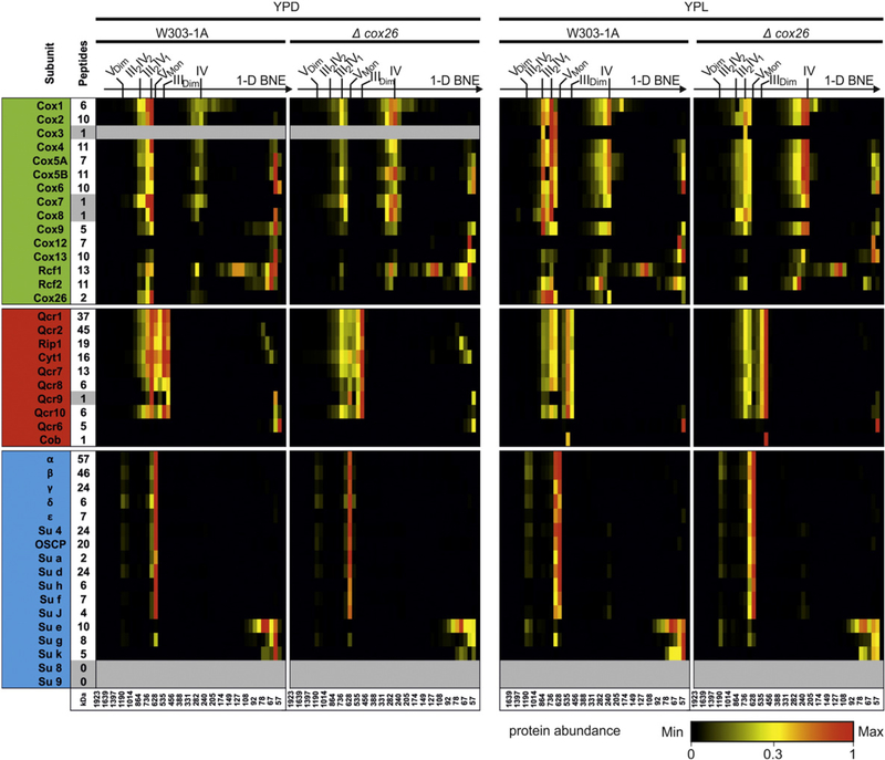
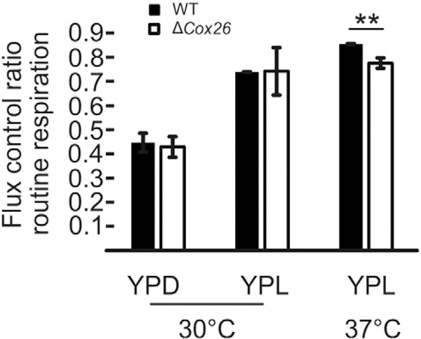
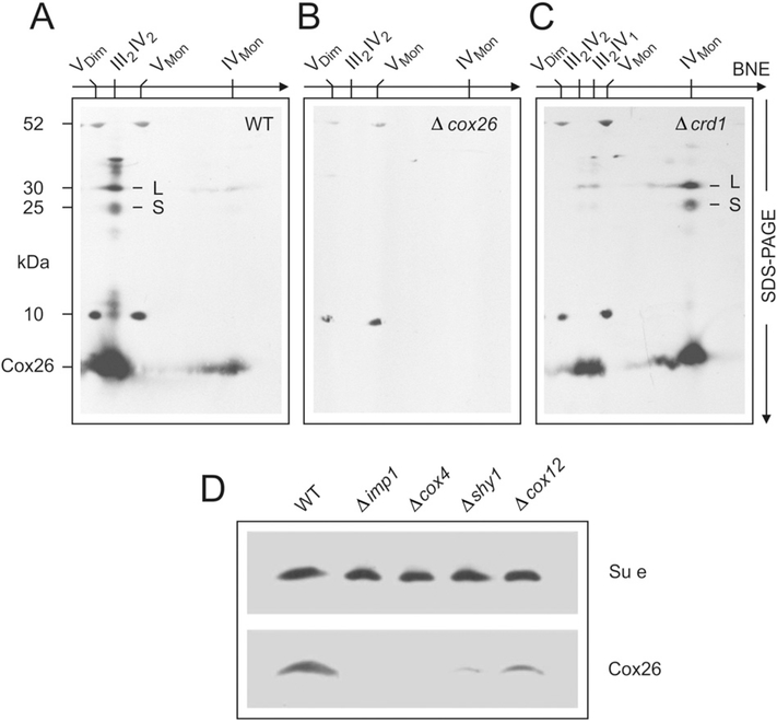
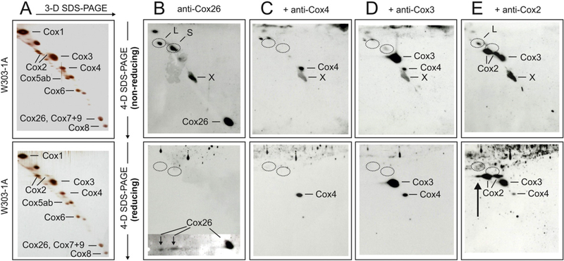
Here we identified a hydrophobic 6.4kDa protein, Cox26, as a novel component of yeast mitochondrial supercomplex comprising respiratory complexes III and IV. Multi-dimensional native and denaturing electrophoretic techniques were used to identify proteins interacting with Cox26. The majority of the Cox26 protein was found non-covalently bound to the complex IV moiety of the III-IV supercomplexes. A population of Cox26 was observed to exist in a disulfide bond partnership with the Cox2 subunit of complex IV. No pronounced growth phenotype for Cox26 deficiency was observed, indicating that Cox26 may not play a critical role in the COX enzymology, and we speculate that Cox26 may serve to regulate or support the Cox2 protein. Respiratory supercomplexes are assembled in the absence of the Cox26 protein, however their pattern slightly differs to the wild type III-IV supercomplex appearance. The catalytic activities of complexes III and IV were observed to be normal and respiration was comparable to wild type as long as cells were cultivated under normal growth conditions. Stress conditions, such as elevated temperatures resulted in mild decrease of respiration in non-fermentative media when the Cox26 protein was absent.

Keywords: Blue native electrophoresis; Cytochrome c oxidase; III–IV supercomplexes; Protein composition; Respiratory supercomplexes; Saccharomyces cerevisiae.


Conflict of interest statement

Conflict of interest

All authors have declared no conflicts of interest concerning the contents of the article.

Figures

Fig. 1.
Fig. 1.
Three-dimensional electrophoresis of yeast mitochondria to identify Cox26, a novel protein co-migrating with respiratory supercomplexes. III2IV2, III2IV1, respiratory supercomplexes containing dimeric complex III and two or one copy of complex IV, respectively. VDim. VMon, IVDim. IVMon. IIIDim, dimeric and monomeric complexes V, IV, and III. (A) Yeast mitochondria were solubilized by digitonin and the mitochondrial (super)complexes were separated by BNE. (B) A gel strip from 1-D BNE was used for 2-D resolution by modified BNE, i.e. DDM was added to the cathode buffer to release individual complexes from the supercomplexes. (C) A gel strip from 2-D BNE was used for 3-D SDS-PAGE. Subunits of dimeric complex III (IIIDim) and the detergent-labile subunits (assigned on the right side) were marked red. Subunits of dimeric and monomeric complex IV (IVDim and IVMon) and the detergent-labile subunits were marked green. The subunits were assigned according to previous Edman sequencing [29] and/or detection by specific antibodies. One single protein, Cox26, was identified as a novel protein component of supercomplexes. It was identified as the smallest protein in the group of detergent-labile subunits (assigned on the right side of Fig. 1C). (D) Sequence and structural features of Cox26 protein. The preprotein contains a presequence of eight amino acids (shaded grey). Basic and acidic residues are marked blue and red, respectively. A cysteine residue (C41 marked yellow) is located within a predicted transmembrane helix (underlined).
Fig. 2.
Fig. 2.
Cox26 stabilizes III–IV supercomplexes. (A) BNE of digitonin-solubilized mitochondrial complexes from wild type (W303–1A) and Cox26 null mutant strain (Δcox26) grown under identical conditions in YPD and YPL. Individual or supercomplex associated complex IV was detected by a specific in-gel complex IV activity stain [12]. Decreased amounts of supercomplexes were detected in the mutant strain in both conditions with even less stability in cells grown in glucose (upper panel). The same gel was stained with Coomassie to show complex V as loading control (lower panel). (B) 2-D Tricine-SDS-PAGE of complexes from wild type and Δcox26 strain using 13% acrylamide gels revealed identical subunit pattern for complexes III and IV. Decoration with an antibody against Cox26 shows presence of Cox26 in III–IV supercomplexes in wild type mitochondria. Assignment of protein complexes: according to Fig. 1. BHM, bovine heart mitochondria as native mass ladder.
Fig. 3.
Fig. 3.
Composition of III–IV supercomplexes. 1-D BNE gels were fractionated and proteins were identified and quantified by mass spectrometry. Identified proteins were quantified by intensity-based absolute quantification values (IBAQ) using only unique and razor peptides (column indicate number of peptides used for quantification). Heatmaps show protein profiles normalized to the maximum abundance of a protein within BN lanes of wild type and Δcox26 mutant for each condition, respectively. The majority of Cox12 and Cox13 was identified on the electrophoretic front indicating either higher abundance as free protein or dissociation of proteins from the complex under experimental conditions used here. Cytochrome b (Cob) was quantified only by one peptide with a modified side (methionine oxidation).Very small proteins like Cox7, Cox8, Qcr9 and the hydrophobic protein Cox3 were identified with one peptide within the complete dataset that could not give full quantitative data for the entire lane. ATP synthase subunits 8 and 9 escaped from identification as they are very hydrophobic and/or small and difficult to identify in complex samples.
Fig. 4.
Fig. 4.
Flux control ratio routine respiration ofW303–1Aand Δcox26. Cells grown in YPD or YPL and were analysed by high resolution respirometry at 30 °C and 37 °C. Error bars indicate standard deviation of three independent experiments. **, mark significant difference from studentSs t-test p < 0.01.
Fig. 5.
Fig. 5.
Cox26 protein is associated with complex IV. Assignment of complexes as in Fig. 1. Digitonin-solubilized mitochondrial complexes from (A) wild type yeast, (B) Δcox26 strain, and (C) Δcrd1 mutant with defective cardiolipin synthase were separated by BNE, followed by Tricine-SDS-PAGE in the second dimension using 13% acrylamide gels for Tricine-SDS-PAGE and electroblotted onto PVDF membranes. Anti-Cox26 antibody identified Cox26 (6.4 kDa) and two larger bands, L and S, with apparent masses around 25 kDa and 30 kDa, in the column of subunits of supercomplexes (e.g. in Figure partA). It also identified monomeric complex IV, e.g. in the Δcrd1 strain (figure partC). Detection of10 kDa and 52 kDa subunits of complex V seemed non-specific cross-reactions by the Cox26 antibody that mark the position of the monomeric and dimeric ATP synthase complexes. (D) Estimation of Cox26 protein amounts in null mutant strains not containing assembled complex IV imp1 and Δcox4) or containing reduced amounts of complex IV shy1 and Δcox12). Complex V amounts were estimated by a specific anti-subunit e antibody as loading control.
Fig. 6.
Fig. 6.
Evidence for covalent interaction of Cox26 and Cox2 proteins in isolated complex IV. Gel pieces from 2-D BNE/modified BNE containing complex IV were resolved under non-reducing conditions by 3-D Tricine-SDS-PAGE using 9% acrylamide gels. 3-D gel strips were then incubated under non-reducing (upper panels) or reducing conditions (lower panels) followed by 4-D Tricine-SDS-PAGE using 16% acrylamide gels containing 6 M urea. The 4-D gels were silver-stained (A) or blotted onto PVDF membranes (B-E). Polyclonal anti-Cox26 antibody identified individual Cox26 protein and two bands (L and S) under non-reducing conditions (B, upper panel), and two Cox26 protein spots that were dissociated from bands L and S under reducing conditions (B, lower panel). A local defect of the PVDF membrane (marked X) was also recognized by the antibody. Circles mark the actual or expected positions of bands L and S. (C and D) Anti-Cox4 and anti-Cox3 antibodies that were added consecutively to the same blot membranes (without using stripping protocols) identified individual Cox4 and Cox3 proteins but no bands L and S. Reusing the same blots, an anti-Cox2 antibody finally recognized two forms of individual Cox2 protein and also band L under non-reducing conditions (E, upper panel) in addition to the protein spots recognized before by the anti-Cox4 and anti-Cox3 antibodies. Performing 4-D SDS-PAGE under reducing conditions (E, lower panel), band L was no longer detected but a third Cox2 spot immediately below the expected position ofband L appeared (marked by an arrow).
Fig. 7.
Fig. 7.
Model for the position of Cox26 in the yeast supercomplex. (A) Model comprises cytochrome c1 (CYT1), cytochrome b (COB), RIP1,Rieske iron-sulphur protein, Core proteins 1 and 2 (COR1/2) and QCR6–9 of complex III, and all three hydrophobic subunits of complex IV, Cox1 (I),Cox2 (II), and Cox3 (III) and the subunits Cox5 (5), Cox7–9 (7–9),Cox12 (12), and Cox13 (13) of complex IV. The model was generated based on structural information of yeast complex III [49] and bovine complex IV [48] using the software PyMOL Molecular Graphics System, Version 1.2, Schrodinger, LLC. Cysteine-92 (Cys 92) which is located at the end of a transmembrane helix of Cox2 was only visible when the central complex IV subunits were inspected. After adding all other subunits Cys 92 seems to be hidden. Cysteine-8 (Cys 8) is located in the centre of a hydrophilic loop structure of Cox2. Cys 8 seems freely accessible to Cox26 protein even after complementation of the model structure with all accessory subunits of complex IV. (B, C) Cox26 was modelled using HHpred and Modellerwith the pdb files 3arc-L, 2kad_A and 2yus_A. The Cys 41 of Cox26 is located within a predicted transmembrane helix which could have direct access to Cys 8 of Cox2 at the hydrophilic boundary part of the membrane facing to the intermembrane space (IMS).

References

    1. Hatefi Y, The mitochondrial electron transport and oxidative phosphorylation system, Annu. Rev. Biochem 54 (1985) 1015–1069. - PubMed
    1. Arnold I, Pfeiffer K, Neupert W, A Stuart R, Schägger H, Yeast F1FO-ATP synthase exists as a dimer: identification of three dimer specific subunits, EMBO J. 17 (1998) 7170–7178. - PMC - PubMed
    1. Cruciat CM, Brunner S, Baumann F, Neupert W, Stuart RA, The cytochrome bc1 and cytochrome c oxidase complexes associate to form a single supracomplex in yeast mitochondria, J. Biol. Chem 275 (2000) 18093–18098. - PubMed
    1. Schägger H, Pfeiffer K, Supercomplexes in the respiratory chains of yeast and mammalian mitochondria, EMBO J. 19 (2000) 1777–1783. - PMC - PubMed
    1. Eubel H, Heinemeyer J, Braun HP, Identification and characterization of respirasomes in potato mitochondria, Plant Physiol. 134 (2004) 1450–1459. - PMC - PubMed

Publication types

MeSH terms