. 2016 Apr 25;213(2):185-200.
doi: 10.1083/jcb.201506099. Epub 2016 Apr 18.

ZRF1 mediates remodeling of E3 ligases at DNA lesion sites during nucleotide excision repair

Affiliations

ZRF1 mediates remodeling of E3 ligases at DNA lesion sites during nucleotide excision repair

Ekaterina Gracheva et al. J Cell Biol. .

Abstract

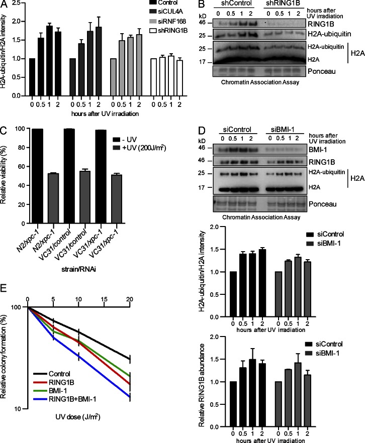
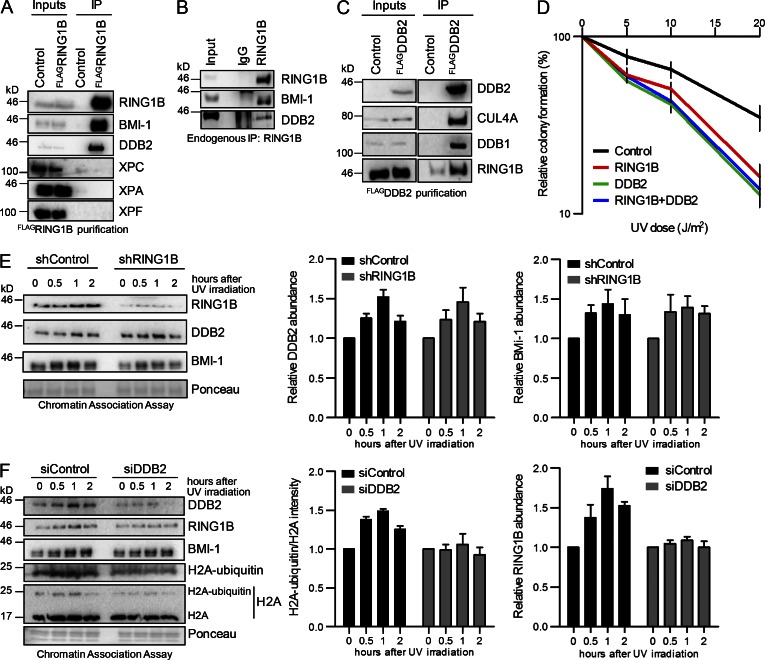
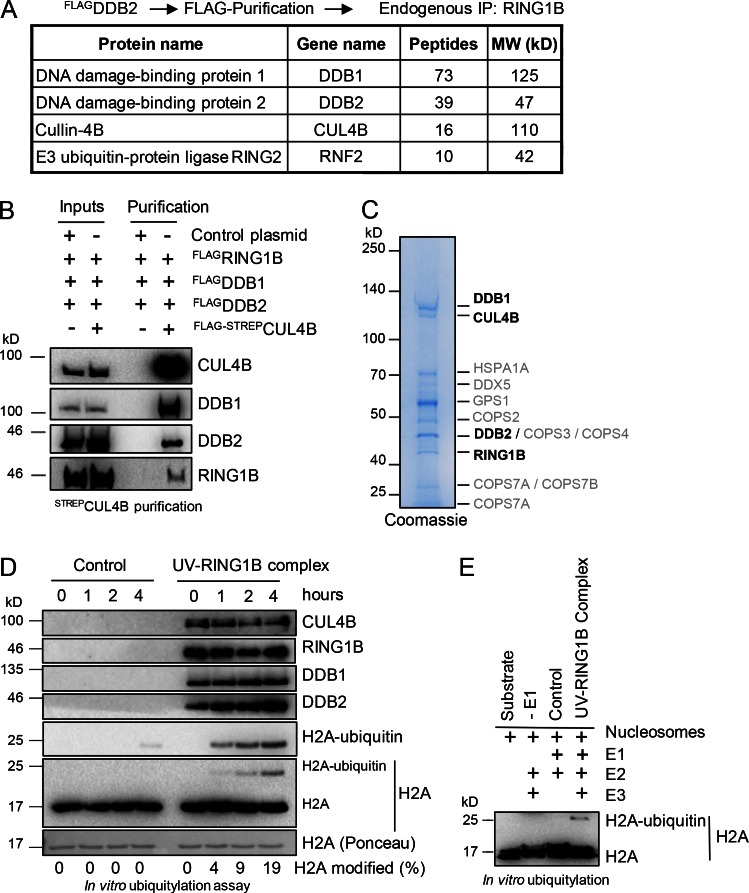
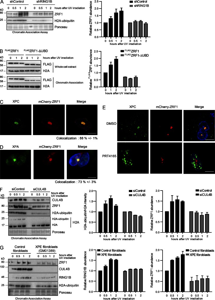
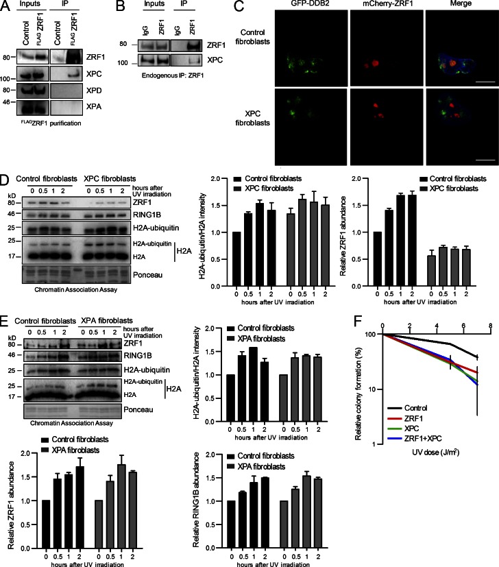
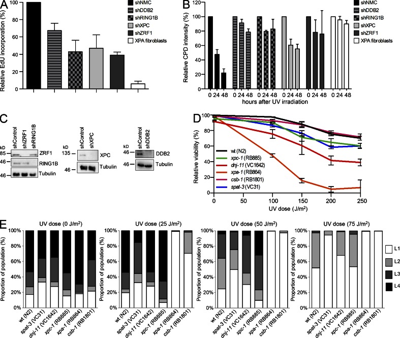
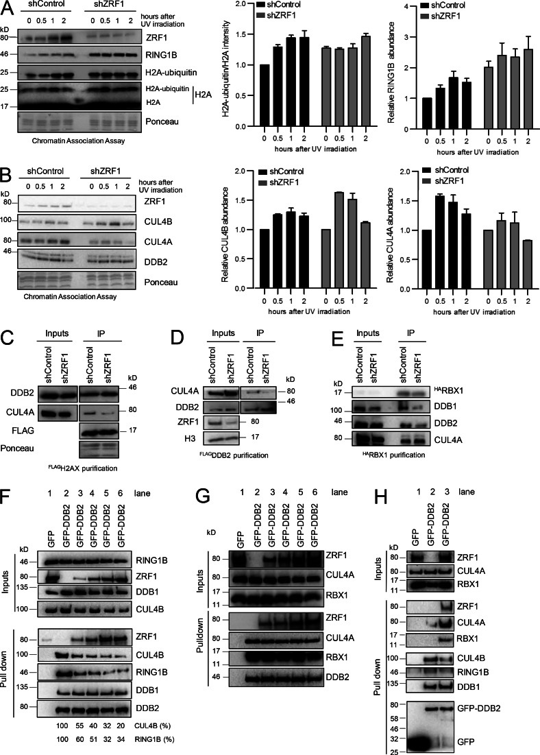
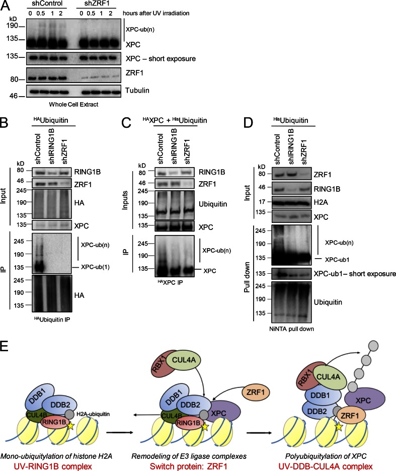
Faithful DNA repair is essential to maintain genome integrity. Ultraviolet (UV) irradiation elicits both the recruitment of DNA repair factors and the deposition of histone marks such as monoubiquitylation of histone H2A at lesion sites. Here, we report how a ubiquitin E3 ligase complex specific to DNA repair is remodeled at lesion sites in the global genome nucleotide excision repair (GG-NER) pathway. Monoubiquitylation of histone H2A (H2A-ubiquitin) is catalyzed predominantly by a novel E3 ligase complex consisting of DDB2, DDB1, CUL4B, and RING1B (UV-RING1B complex) that acts early during lesion recognition. The H2A-ubiquitin binding protein ZRF1 mediates remodeling of this E3 ligase complex directly at the DNA lesion site, causing the assembly of the UV-DDB-CUL4A E3 ligase complex (DDB1-DDB2-CUL4A-RBX1). ZRF1 is an essential factor in GG-NER, and its function at damaged chromatin sites is linked to damage recognition factor XPC. Overall, the results shed light on the interplay between epigenetic and DNA repair recognition factors at DNA lesion sites.


Figures

Figure 1.
Figure 1.
Dissection of E3 ligase functions in UV-mediated DNA damage repair. (A) Quantitative analysis of H2A-ubiquitylation levels. Immunoblots (as in B and Fig. S1, A and B) were probed with histone H2A antibody. The intensities of H2A and H2A-ubiquitin bands were quantified by the ImageJ software. The graphs illustrate the relative H2A ubiquitylation calculated as (H2A ubiquitin)/(H2A + H2A ubiquitin), normalized to Ponceau staining intensity after knockdown of the respective proteins (H2A ubiquitin/H2A). Values are normalized to the value from nonirradiated cells and are given as mean ± SEM (n = 4). (B) Monoubiquitylation of histone H2A at lysine 119 after UV irradiation is mainly catalyzed by RING1B. Chromatin association assays of control and RING1B knockdown HEK293T cells after UV irradiation. De–cross-linked material of the respective time points was subjected to Western blotting and probed with the indicated antibodies. The specificity of the H2A-ubiquitin antibody was verified (Fig. S1 C). (C) Epistatic relationship of xpc-1 and spat-3. Wild-type nematodes (N2) or spat-3 mutants (VC31) were fed with either control or xpc-1 RNAi–producing bacteria. The relative viability was analyzed after UV irradiation (200 J/m2). Values are given as mean ± SEM (n = 3). (D) Impact of BMI-1 on RING1B-mediated H2A ubiquitylation after UV irradiation. Chromatin association assays of UV-irradiated HEK293T cells treated with siRNAs (control, BMI-1). De–cross-linked material of the respective time points was subjected to Western blotting and probed with the indicated antibodies. Relative intensities of H2A ubiquitin/H2A and RING1B abundance after BMI-1 depletion were measured. Values are given as mean ± SEM (n = 4). (E) Epistatic relationship of RING1B and BMI-1 in response to UV irradiation. Relative colony formation potential of control or RING1B knockdown cell lines treated with siRNA was analyzed at different UV doses. Control cells were transfected with either control siRNA (control) or BMI-1 siRNA (BMI-1). RING1B knockdown cell lines were transfected with either control siRNA (RING1B) or BMI-1 siRNA (RING1B + BMI-1). Gene knockdown was confirmed by Western blots (not depicted). Values are given as mean ± SEM (n = 9).
Figure 2.
Figure 2.
RING1B and DDB2 cooperate in H2A ubiquitylation. (A) RING1B interacts with DDB2. Control cells and cells expressing FLAGRING1B were irradiated with UV light. After immunoprecipitation with FLAG-M2-Agarose the purified material was subjected to Western blotting and blots were incubated with the indicated antibodies. Inputs correspond to 3%. (B) Endogenous immunoprecipitations with RING1B antibodies after UV irradiation. Western blots of the precipitated material were incubated with the indicated antibodies. IgG lanes show unspecific staining of the IgG heavy chains. (C) DDB2 associates with RING1B. Control cells and cells expressing FLAGDDB2 were irradiated with UV light. After immunoprecipitation with FLAG-M2-agarose, the purified material was subjected to Western blotting and blots were incubated with the indicated antibodies. Inputs correspond to 3%. (D) Epistatic relationship of RING1B and DDB2 in response to UV irradiation. Relative colony formation potential of control or RING1B knockdown cell lines treated with siRNA was analyzed at different UV dosages. Control cells were transfected with either control siRNA (control) or DDB2 siRNA (DDB2). RING1B knockdown cell lines were transfected with either control siRNA (RING1B) or DDB2 siRNA (RING1B + DDB2). Gene knockdown was confirmed by Western blots (not depicted). Values are given as mean ± SEM (n = 6). (E) Knockdown of RING1B does not impair DDB2 recruitment. Chromatin association assays of control and RING1B knockdown HEK293T cells after UV irradiation. De–cross-linked material of the respective time points was subjected to Western blotting and probed with the indicated antibodies. The relative DDB2 and BMI-1 abundance was calculated. Values are given as mean ± SEM (n = 3). (F) Knockdown of DDB2 shows reduced H2A-ubiquitylation but unaltered BMI-1 recruitment. Chromatin association assays of UV-irradiated HEK293T cells treated with siRNAs (control, DDB2). De–cross-linked material of the respective time points was subjected to Western blotting and probed with the indicated antibodies. The relative H2A-ubiquitylation and RING1B abundance was calculated. Values are given as mean ± SEM (n = 4).
Figure 3.
Figure 3.
H2A ubiquitylation after UV irradiation is performed by the UV–RING1B complex. (A) Protein interaction partners of RING1B and DDB2. Mass spectrometry analysis after sequential immunoprecipitations with FLAG and RING1B antibodies revealed DDB1 and CUL4B as main interaction partners of DDB2 and RING1B. A comprehensive list of the identified unique peptides after RING1B and control immunoprecipitations (with or without UV irradiation) is provided in Table S5. (B) Assembly of the UV–RING1B complex. Plasmids expressing FLAGDDB1, FLAGDDB2, and FLAGRING1B were cotransfected in combination with either control plasmid or a plasmid encoding FLAG-STREPCUL4B. After immunoprecipitation with STREP-Tactin beads, the purified material was subjected to Western blotting and blots were incubated with the indicated antibodies. Inputs correspond to 5%. (C) Visualization of the UV–RING1B complex. Purified UV–RING1B complex was subjected to SDS gel electrophoresis and colloidal Coomassie staining. Mass spectrometry analysis revealed the presence of all four subunits (bold). A comprehensive list of unique peptides is provided in Table S6. (D) The UV–RING1B complex catalyzes ubiquitylation of H2A in vitro. Ubiquitylation assays were performed with recombinant H2A, E1 (UBA1), E2 (UBCH5), and either GST (control) or the UV–RING1B complex. Reactions were performed at 37°C, and samples were taken at the indicated time points. Material of the respective time points was subjected to Western blotting and probed with the indicated antibodies. (E) The UV–RING1B complex catalyzes monoubiquitylation of nucleosomal H2A. Ubiquitylation assays were performed with recombinant nucleosomes, E1 (UBA1), E2 (UBCH5), and either GST (control) or UV-RING1B complex. Reactions lacking E1 (−E1) were performed as additional controls. The ubiquitylation assays were performed at 37°C for 5 h, and samples or pure substrate (Substrate) were subjected to Western blotting and probed with H2A antibodies.
Figure 4.
Figure 4.
Function of ZRF1 in UV-mediated DNA repair. (A) ZRF1 is tethered to chromatin in a RING1B-dependent manner. Chromatin association assays of control and RING1B knockdown HEK293T cell lines after UV irradiation. De–cross-linked material of the respective time points was subjected to Western blotting and probed with the indicated antibodies. The relative ZRF1 abundance was calculated. Values are given as mean ± SEM (n = 3). (B) The ubiquitin-binding domain (UBD) is important for tethering ZRF1 to chromatin after UV irradiation. HEK293T cells expressing FLAGZRF1 and FLAGZRF1-ΔUBD were irradiated with UV light, and chromatin was isolated at the indicated time points. De-cross-linked material was subjected to Western blotting and blots were incubated with FLAG-antibody. The relative FLAGZRF1 abundance was calculated. Values are given as mean ± SEM (n = 4). (C and D) ZRF1 localizes to DNA damage sites after UV irradiation. MRC5 fibroblasts expressing mCherry-ZRF1 were UV irradiated (100 J/m2) through a micropore membrane (+ UV) 24 h after transfection. 30 min after irradiation, cells were preextracted and fixed. DNA damage sites were visualized by staining with XPC (C) or XPA (D) antibody. The colocalization of ZRF1 with XPC amounts to 88% ± 1%. The colocalization of ZRF1 with XPA amounts to 73% ± −3%. Nonirradiated control and quantification of the ZRF1 localization at the damage sites are represented in Fig. S4 A. Bar, 10 µm. (E) Inhibition of RING1B affects recruitment of ZRF1 to DNA damage sites. MRC5 fibroblasts expressing mCherry-ZRF1 were treated with PRT4165 or DMSO. Cells were UV-irradiated (100 J/m2) through a micropore membrane. 30 min after irradiation cells were preextracted and fixed. DNA damage sites were visualized by XPC antibody staining. ZRF1 localization to DNA lesions after treatment with DMSO or PRT4165 was quantified (Fig. S4 B). Bar, 10 µm. (F) Depletion of CUL4B impacts H2A ubiquitylation and ZRF1 recruitment. Chromatin association assays of UV irradiated HEK293T cells treated with siRNAs (control, CUL4B). De–cross-linked material of the respective time points was subjected to Western blotting and probed with the indicated antibodies. The relative H2A-ubiquitin and ZRF1 abundance was calculated. Values are given as mean ± SEM (n = 3). (G) Tethering of ZRF1 to chromatin depends on DDB2 during NER. Chromatin association assays in control fibroblasts (GM15876) and XPE (DDB2) fibroblasts (GM01389) after UV irradiation. De–cross-linked material of the respective time points was subjected to Western blotting and probed with the indicated antibodies. The relative RING1B and ZRF1 abundance was calculated. Values are given as mean ± SEM (n = 3).
Figure 5.
Figure 5.
ZRF1 interacts with XPC during UV-mediated DNA repair. (A) ZRF1 specifically binds to XPC. Control and FLAGZRF1-expressing cells were irradiated with UV light. After immunoprecipitation with FLAG-M2-agarose, the purified material was subjected to Western blotting and blots were incubated with the indicated antibodies. Inputs correspond to 4%. (B) Endogenous immunopreciptiations with ZRF1 antibodies. Precipitates were subjected to Western blotting, and blots were incubated with the indicated antibodies. Inputs correspond to 3%. (C) ZRF1 localization to DNA damage sites is dependent on XPC. Control fibroblasts and XPC patient fibroblasts expressing both mCherry-ZRF1 and DDB2-GFP were UV irradiated (100 J/m2) through a micropore membrane. Thirty minutes after irradiation, cells were preextracted and fixed. DNA damage sites were visualized by DDB2-GFP. (D) ZRF1 enriches at chromatin after UV irradiation in a XPC-dependent manner. Chromatin association assays with control fibroblasts (GM16248) and XPC patient fibroblasts (GM15983) after UV irradiation. De–cross-linked material of the respective time points was subjected to Western blotting and probed with the indicated antibodies. The relative H2A-ubiquitin and ZRF1 abundance was calculated. Values are given as mean ± SEM (n = 3). (E) H2A ubiquitylation is not altered in XPA patient fibroblasts. Chromatin association assays with control fibroblasts (GM15876) and XPA fibroblasts (GM04312) after UV irradiation. De–cross-linked material of the respective time points was subjected to Western blotting and probed with the indicated antibodies. Relative intensities of H2A-ubiquitin/H2A, ZRF1 and RING1B abundance were measured. Values are given as mean ± SEM (n = 3). (F) Epistasis analysis of ZRF1 and XPC. The relative colony formation potential of control or ZRF1 knockdown cell lines treated with control (Control; ZRF1) or XPC siRNA (XPC; ZRF1+XPC) was analyzed at different UV doses. Gene knockdown was confirmed by Western blots (not depicted). Values are given as mean ± SEM (n = 3).
Figure 6.
Figure 6.
ZRF1 and RING1B contribute to GG-NER. (A) RING1B and ZRF1 knockdown fibroblasts are defective in UDS after UV irradiation. UDS was measured by EdU incorporation after UV treatment in MRC5 fibroblasts with shRNA-mediated knockdown of the indicated proteins. XPA fibroblasts were used as a positive control. Values are given as mean ± SEM. Data were acquired from three independent experiments (150–300 nuclei per sample). (B) RING1B and ZRF1 knockdown fibroblasts are defective in the removal of CPDs. The CPD removal was analyzed in MRC5 fibroblasts after knockdown of the indicated proteins in MRC5 fibroblasts and in XPA fibroblasts. Cells were irradiated with 10 J/m2 and fixed immediately or 24 or 48 h after irradiation and stained with CPD antibodies. The relative fluorescence intensity was determined. Values are given as mean ± SEM. Data were acquired from three independent experiments (100–200 nuclei per sample). (C) MRC5 fibroblasts were treated with lentiviral particles containing the respective shRNA. Knockdown of the proteins levels was analyzed 48h after infection by Western blotting and incubation with the indicated antibodies. (D) C. elegans knockout mutants for ZRF1 (dnj-11) and RING1B (spat-3) show increased sensitivity toward UV irradiation. Late-L4 larval wild-type worms and the indicated mutants were irradiated with UV light at different doses, and the relative viability was determined by comparing hatched versus dead embryos (unhatched eggs). Values are given as mean ± SEM (n = 3). (E) C. elegans knockout mutants for dnj-11 and for spat-3 show only weak developmental arrest upon somatic UV irradiation. L1 larval worms were irradiated with UV light at different doses. Relative larval-stage stalling was determined after 60 h by using a large particle flow cytometer (BioSorter platform; Union Biometrica), assaying at least 1,000 worms per condition.
Figure 7.
Figure 7.
ZRF1 facilitates the assembly of the UVDDBCUL4A E3 ligase complex. (A) ZRF1 displaces RING1B from chromatin during NER. Chromatin association assays of control and ZRF1 knockdown HEK293T cell lines after UV irradiation. De–cross-linked material of the respective time points was subjected to Western blotting and probed with the indicated antibodies. The relative H2A ubiquitin and RING1B abundance was calculated. Values are given as mean ± SEM (n = 3). (B) ZRF1 regulates chromatin association of CUL4A and CUL4B. Chromatin association assays of control and ZRF1 knockdown HEK293T cell lines after UV irradiation. De–cross-linked material of the respective time points was subjected to Western blotting and probed with the indicated antibodies. The relative CUL4B and CUL4A abundance was calculated. Values are given as mean ± SEM (n = 3). (C) ZRF1 regulates CUL4A association with H2AX containing nucleosomes. Control cells and ZRF1 knockdown cells expressing FLAGH2AX were irradiated with UV. After immunoprecipitation with FLAG-M2-agarose, the purified material was subjected to Western blotting and blots were incubated with the indicated antibodies. Inputs correspond to 3%. (D) Knockdown of ZRF1 modulates CUL4A association with DDB2. Control cells and ZRF1 knockdown cells expressing FLAGDDB2 were irradiated with UV light. After immunoprecipitation with FLAG-M2-agarose, the purified material was subjected to Western blotting and blots were incubated with the indicated antibodies. Inputs correspond to 3%. (E) Assembly of the UV–DDB–CUL4A E3 ligase is facilitated by ZRF1. Control cells and ZRF1 knockdown HEK293T cells expressing HARBX1 were irradiated with UV light. After immunoprecipitation with HA-specific antibodies the precipitated material was subjected to Western blotting, and blots were incubated with the indicated antibodies. Inputs correspond to 5%. (F) ZRF1 competes with CUL4B and RING1B for DDB2 binding in vitro. GFP and GFP-DDB2 immobilized on beads were incubated with equimolar amounts of purified DDB1, CUL4B, and RING1B and increasing amounts of ZRF1. ZRF1 levels were doubled stepwise reaching an eightfold molar excess of ZRF1 over the other components (relative molarity ZRF1: DDB1–CUL4B–RING1B; lane 3, 1:1; lane 4, 2:1; lane 5, 4:1; lane 6, 8:1). Precipitated material was subjected to Western blotting and blots were incubated with the indicated antibodies. Inputs correspond to 10%. (G) ZRF1 does not compete with CUL4A and RBX1 for binding to DDB1–DDB2. GFP and GFP-DDB2 immobilized on beads were incubated with equimolar amounts of purified DDB1, CUL4A and RBX1 and increasing amounts of ZRF1. ZRF1 levels were doubled stepwise reaching an eightfold molar excess of ZRF1 (relative molarity ZRF1: DDB1–CUL4A–RBX1; lane 3, 1:1; lane 4, 2:1; lane 5, 4:1; lane 6, 8:1). Precipitated material was subjected to Western blotting and blots were incubated with the indicated antibodies. Inputs correspond to 10%. (H) ZRF1 mediates the formation of the UV-DDB-CUL4A complex in vitro. GFP and GFP-DDB2 were coupled to beads and incubated with CUL4B, DDB1 and RING1B. After washing, GFP and GFP-DDB2 (UV–RING1B complex) beads were incubated with an estimated fivefold excess of purified CUL4A and RBX1 (lanes 1–3) over the retained UV–RING1B complex. Simultaneously, ZRF1 (lanes 1 and 3) or GST (lane 2) was added to the incubations in equimolar amounts. The precipitated material was subjected to Western blotting and blots were incubated with the indicated antibodies. Inputs correspond to 5%.
Figure 8.
Figure 8.
ZRF1 regulates XPC ubiquitylation. (A) ZRF1 facilitates XPC ubiquitylation after UV irradiation. Whole-cell extracts of control and ZRF1 knockdown HEK293T cells from the stated time points were subjected to Western blotting and probed with the indicated antibodies. (B) Role of RING1B and ZRF1 in XPC ubiquitylation. Control cells and RING1B and ZRF1 knockdown HEK293T cells expressing HAUbiquitin were irradiated with UV light. After immunoprecipitation with HA-specific antibody, the precipitated material was subjected to Western blotting and blots were incubated with the indicated antibodies. Inputs correspond to 5%. (C) Control cells and RING1B and ZRF1 knockdown HEK293T cells expressing HAXPC and HISUbiquitin were irradiated with UV light. After immunoprecipitation with HA-specific antibody, the precipitated material was subjected to Western blotting and blots were incubated with the indicated antibodies. Inputs correspond to 5%. (D) Control cells and RING1B and ZRF1 knockdown HEK293T cells expressing HISUbiquitin were irradiated with UV and harvested 1 h after UV exposure. Ubiquitylated proteins were purified by NiNTA agarose under denaturing conditions, and Western blots of the purified material were incubated with the indicated antibodies. (E) The UV–RING1B complex and ZRF1 cooperate during NER. DNA lesions (yellow star) are recognized by the UV-RING1B complex (DDB1–DDB2–CUL4B–RING1B), which catalyzes ubiquitylation of histone H2A (gray sphere). ZRF1 is recruited to the lesion site by XPC and tethers to the H2A-ubiquitin mark. ZRF1 causes the assembly of the UV–DDB–CUL4A complex, which subsequently catalyzes ubiquitylation of XPC.

Comment in

Similar articles

Cited by

References

    1. Alekseev S., Kool H., Rebel H., Fousteri M., Moser J., Backendorf C., de Gruijl F.R., Vrieling H., and Mullenders L.H.. 2005. Enhanced DDB2 expression protects mice from carcinogenic effects of chronic UV-B irradiation. Cancer Res. 65:10298–10306. 10.1158/0008-5472.CAN-05-2295 - DOI - PubMed
    1. Angers S., Li T., Yi X., MacCoss M.J., Moon R.T., and Zheng N.. 2006. Molecular architecture and assembly of the DDB1-CUL4A ubiquitin ligase machinery. Nature. 443:590–593. - PubMed
    1. Araki M., Masutani C., Takemura M., Uchida A., Sugasawa K., Kondoh J., Ohkuma Y., and Hanaoka F.. 2001. Centrosome protein centrin 2/caltractin 1 is part of the xeroderma pigmentosum group C complex that initiates global genome nucleotide excision repair. J. Biol. Chem. 276:18665–18672. 10.1074/jbc.M100855200 - DOI - PubMed
    1. Bergink S., Salomons F.A., Hoogstraten D., Groothuis T.A., de Waard H., Wu J., Yuan L., Citterio E., Houtsmuller A.B., Neefjes J., et al. . 2006. DNA damage triggers nucleotide excision repair-dependent monoubiquitylation of histone H2A. Genes Dev. 20:1343–1352. 10.1101/gad.373706 - DOI - PMC - PubMed
    1. Bergink S., Toussaint W., Luijsterburg M.S., Dinant C., Alekseev S., Hoeijmakers J.H., Dantuma N.P., Houtsmuller A.B., and Vermeulen W.. 2012. Recognition of DNA damage by XPC coincides with disruption of the XPC-RAD23 complex. J. Cell Biol. 196:681–688. 10.1083/jcb.201107050 - DOI - PMC - PubMed

MeSH terms

LinkOut - more resources