. 2016 Aug;37(8):2717-35.
doi: 10.1002/hbm.23204. Epub 2016 Apr 19.

Intrinsic network activity in tinnitus investigated using functional MRI

Affiliations

Intrinsic network activity in tinnitus investigated using functional MRI

Amber M Leaver et al. Hum Brain Mapp. 2016 Aug.

Abstract

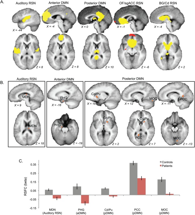
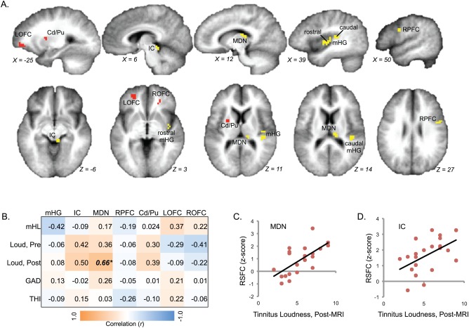
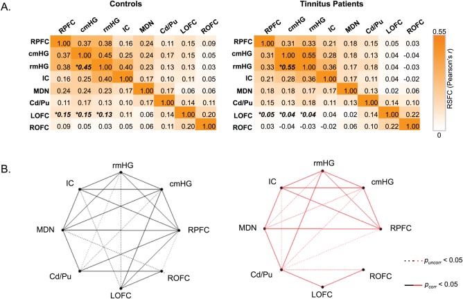
Tinnitus is an increasingly common disorder in which patients experience phantom auditory sensations, usually ringing or buzzing in the ear. Tinnitus pathophysiology has been repeatedly shown to involve both auditory and non-auditory brain structures, making network-level studies of tinnitus critical. In this magnetic resonance imaging (MRI) study, two resting-state functional connectivity (RSFC) approaches were used to better understand functional network disturbances in tinnitus. First, we demonstrated tinnitus-related reductions in RSFC between specific brain regions and resting-state networks (RSNs), defined by independent components analysis (ICA) and chosen for their overlap with structures known to be affected in tinnitus. Then, we restricted ICA to data from tinnitus patients, and identified one RSN not apparent in control data. This tinnitus RSN included auditory-sensory regions like inferior colliculus and medial Heschl's gyrus, as well as classically non-auditory regions like the mediodorsal nucleus of the thalamus, striatum, lateral prefrontal, and orbitofrontal cortex. Notably, patients' reported tinnitus loudness was positively correlated with RSFC between the mediodorsal nucleus and the tinnitus RSN, indicating that this network may underlie the auditory-sensory experience of tinnitus. These data support the idea that tinnitus involves network dysfunction, and further stress the importance of communication between auditory-sensory and fronto-striatal circuits in tinnitus pathophysiology. Hum Brain Mapp 37:2717-2735, 2016. © 2016 The Authors Human Brain Mapping Published by Wiley Periodicals, Inc.

Keywords: auditory; fMRI; functional connectivity; hearing loss; striatum; thalamus.


Figures

Figure 1
Figure 1
Comparing typical RSNs in tinnitus patients and controls. (A) Five typical RSNs were targeted for statistical analysis between groups (t‐test; P uncorr < 0.0005, k > 3), including an auditory–somatomotor RSN, anterior and posterior default‐mode networks (DMN), an RSN covering orbitofrontal and subgenual anterior cingulate cortex (OF/sgACC RSN), and a basal ganglia network emphasizing the caudate nucleus (BG/Cd RSN). Yellow and red colors indicate opposing relationships with each RSN. (B) Three RSNs demonstrated statistical differences between groups, and the resulting maps are displayed for each of these RSNs (orange). Tinnitus‐related reductions in resting‐state functional connectivity (RSFC) were noted in the mediodorsal nucleus of the thalamus (MDN), anterior parahippocampal gyrus (PHG), caudate/putamen (Cd/Pu), posterior cingulate cortex (PCC) and medal occipital cortex (MOC). (C) RSFC values (beta weights) are plotted for each cluster with respect to its RSN for controls (gray) and tinnitus patients (red). Error bars indicate standard error of mean. All maps in all figures are displayed on group‐averaged anatomical scans in neurological convention (i.e., right hemisphere is located on the right‐hand side of the image).
Figure 2
Figure 2
Spatial similarity of RSNs identified in tinnitus patients and controls. RSNs were analyzed using hierarchical cluster analysis to compare spatial similarity of networks identified using ICA separately for each group. (A) A dendrogram of map relationships is plotted, with spatial dissimilarity (1 ‐ Pearson's r) on the y axis and node‐connections reflecting average Euclidean distance. Each “leaf” node represents an RSN. (B) A heatmap of the pairwise spatial cross‐correlations between thresholded maps is displayed, with warm colors marking high spatial similarity and cool colors indicating low spatial similarity. The approximate anatomical locations of these networks is described in white text. Numbers displayed between panels A and B and black lines in B indicate the subsequent Figures in which these maps are shown. (C) The maximum or “best” pairwise spatial correlation value is plotted for each map, with respect to all pairwise comparisons made for each map (black line, left y‐axis). Mean pairwise spatial correlation values are also plotted for each map (blue dashed line, right y‐axis). Networks with particularly low values would be more likely to be atypical and unique to either tinnitus patients or controls. Letters and numbers between panels B and C mark group (T, tinnitus patients; C, controls) and RSN index used in Supporting Information Figure S5 for each column, respectively.
Figure 3
Figure 3
Atypical RSN in tinnitus patients. (A) An RSN was identified in tinnitus patients that was dissimilar to RSNs in controls. This network is displayed on group‐averaged anatomy; red and yellow depict opposing functional relationships (i.e., yellow regions are positively correlated with the RSN‐timecourse and red regions are negatively correlated). Clusters overlap with parts of the auditory system, including the inferior colliculus (IC) and two clusters on medial Heschl's gyrus (mHG). Other clusters are located in mediodorsal nucleus of the thalamus (MDN), caudate/putamen (Cd/Pu), right lateral prefrontal cortex (RPFC), and left and right orbitofrontal cortex (LOFC and ROFC). The X and Z Talairach planes are indicated for each slice. (B) Correlations between behavioral variables and network‐connectivity in these clusters are displayed in a matrix, where orange marks positive correlations and blue marks negative correlations. Significant correlations are indicated in bold and italics (*P corr < 0.05). Effects for caudal and rostral mHG clusters were similar, and were therefore analyzed together in this section (only). (C, D) Scatterplots are shown for cases where behavioral variables predict resting‐state functional connectivity (RSFC) between clusters and the tinnitus network.
Figure 4
Figure 4
Exploring the atypical tinnitus RSN. (A) Correlations in raw, denoised fMRI signal between regions of the atypical tinnitus RSN are displayed in a cross‐correlation matrix, where deeper orange colors indicate stronger positive correlations. No regions displayed strong negative correlations in raw signal with any other region of the network. Mean correlation (Pearson's r) values are displayed, derived from ROI–ROI analyses performed in single subjects using the clusters from Figure 3A and averaged for healthy controls (left) and in tinnitus patients (right). ROI–ROI relationships that exhibited differences between groups are bolded, italicized, and marked with an asterisk (P uncorr < 0.05). (B) Graphs of the atypical RSN are displayed to highlight the relationships in raw intrinsic fMRI signal between ROIs for controls and patients (at left in black and at right in red, respectively). Dotted lines mark significant correlations at P uncorr < 0.05, and solid lines mark correlations P corr < 0.05, Bonferroni‐corrected for the number of pairwise tests.
Figure 5
Figure 5
Relationships between hearing loss, tinnitus variables, and raw intrinsic connectivity in the atypical tinnitus RSN. (A) Significant correlations between mean hearing loss (mHL) and ROI–ROI correlations in raw fMRI signal across both groups were identified using a Group × mHL ANCOVA. Scatterplots of ROI pairs exhibiting significant (P uncorr < 0.05) effects of mHL are displayed. Patient data are shown in red circles, and controls in black squares. (B) Scatterplots are shown for ROI pairs demonstrating significant correlations between behavioral variables (i.e., post‐MRI tinnitus loudness ratings and anxiety rating scale scores) and ROI–ROI raw intrinsic connectivity (P uncorr < 0.05) as assessed in tinnitus patients using Pearson's r. A potential outlier is encircled in the lower right panel; statistics with this datapoint omitted are r = 0.35, P = 0.14.

Similar articles

Cited by

References

    1. Allen EA, Erhardt EB, Damaraju E, Gruner W, Segall JM, Silva RF, Havlicek M, Rachakonda S, Fries J, Kalyanam R, Michael AM, Caprihan A, Turner JA, Eichele T, Adelsheim S, Bryan AD, Bustillo J, Clark VP, Feldstein Ewing SW, Filbey F, Ford CC, Hutchison K, Jung RE, Kiehl KA, Kodituwakku P, Komesu YM, Mayer AR, Pearlson GD, Phillips JP, Sadek JR, Stevens M, Teuscher U, Thoma RJ, Calhoun VD (2011): A baseline for the multivariate comparison of resting‐state networks. Front Syst Neurosci 5:2. - PMC - PubMed
    1. Baliki MN, Geha PY, Apkarian a. V, Chialvo DR (2008): Beyond feeling: Chronic pain hurts the brain, disrupting the default‐mode network dynamics. J Neurosci 28:1398–1403. - PMC - PubMed
    1. Behrens TEJ, Johansen‐Berg H, Woolrich MW, Smith SM, Wheeler‐Kingshott CAM, Boulby PA, Barker GJ, Sillery EL, Sheehan K, Ciccarelli O, Thompson AJ, Brady JM, Matthews PM (2003): Non‐invasive mapping of connections between human thalamus and cortex using diffusion imaging. Nat Neurosci 6:750–757. - PubMed
    1. Boyen K, Langers DRM, de Kleine E, van Dijk P (2013): Gray matter in the brain: Differences associated with tinnitus and hearing loss. Hear Res 295:67–76. - PubMed
    1. Burton H, Wineland A, Bhattacharya M, Nicklaus J, Garcia KS, Piccirillo JF (2012): Altered networks in bothersome tinnitus: A functional connectivity study. BMC Neurosci 13:3. - PMC - PubMed

Publication types