Review
. 2016 Apr 20;5(8):871-88.
doi: 10.1002/adhm.201600009. Epub 2016 Mar 22.

Recent Progress of Microfluidics in Translational Applications

Affiliations
Review

Recent Progress of Microfluidics in Translational Applications

Zongbin Liu et al. Adv Healthc Mater. .

Abstract

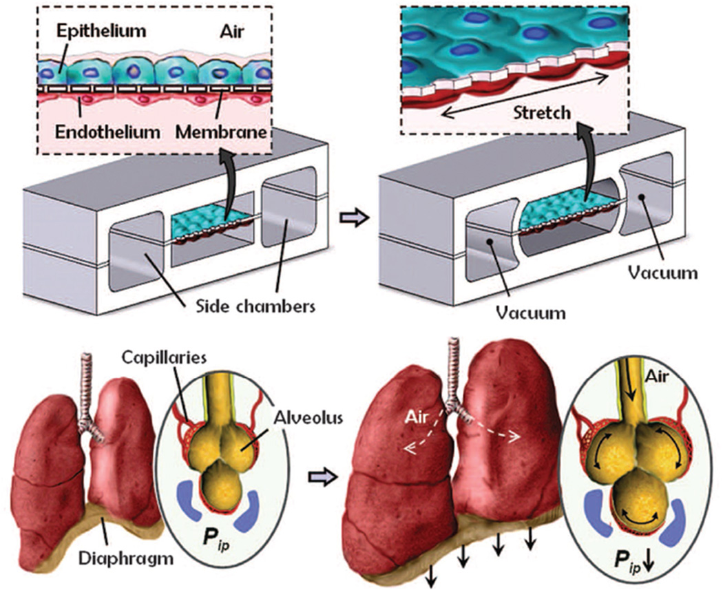
Microfluidics, featuring microfabricated structures, is a technology for manipulating fluids at the micrometer scale. The small dimension and flexibility of microfluidic systems are ideal for mimicking molecular and cellular microenvironment, and show great potential in translational research and development. Here, the recent progress of microfluidics in biological and biomedical applications, including molecular analysis, cellular analysis, and chip-based material delivery and biomimetic design is presented. The potential future developments in the translational microfluidics field are also discussed.

Keywords: cellular analysis; chip-based material delivery; microfluidics; molecular analysis; organ-on-a-chip.


Figures

Figure 1
Figure 1
Microfluidic devices for protein detection. a) Photo of microfluidic chip and schematic illustration of loading of multiple reagents to detect HIV and syphilis antigens. Reproduced with permission.[8] Copyright 2011, NPG. b) Schematic illustration of volumetric bar-chart chip. Protein concentrations are converted into travel distance of ink bars by ELISA reaction. Reproduced with permission.[11c] Copyright 2012, NPG.
Figure 2
Figure 2
Paper-based microfluidic device for ALT and AST assays. a) The device consists of multilayer papers and membranes. b) AST and ALT test zones are matched to color read guide to obtain protein concentrations. Reproduced with permission.[13b] Copyright 2012, AAAS.
Figure 3
Figure 3
Microfluidic device for the collection of DNA in as few as 100 cells. a) Schematic illustration of microfluidic oscillatory washing-based ChIP-seq to purify DNA. The five major steps are formation of a packed bed of beads, flowing of chromatin fragments, oscillatory washing, removal of the unbound chromatin, and collection of beads. b) Optimization of protocol including the amount of beads, concentration of antibody used for coating and washing duration. Reproduced with permission.[15a] Copyright 2015, NPG.
Figure 4
Figure 4
Microfluidic mechanical analysis of cells. a) Schematic illustration of deformability cytometry. A high-speed camera is used to take images of stretched cells in microchannels. Density scatter plot of size and deformability is then got. Reproduced with permission.[21] Copyright 2012, National Academy of Sciences. b) Microfluidic cytometric analysis of cancer cell transportability. The device is composed of deterministic lateral displacement and trapping barrier structures. Cancer cells are separated by their size and transportability through small gaps. Transportability and diameter of each cell is plotted. Reproduced with permission.[24] Copyright 2015, NPG.
Figure 5
Figure 5
Microfluidic devices for cell migration. a) Schematic illustration of a microfluidic chip for high throughput analysis of cancer cell migration for drug screening. Chemogradient is generated in each microchamber. Reproduced with permission.[29] Copyright 2014, Wiley-VCH. b) Schematic illustration of a microfluidic chip to mimic the biophysical environment of female reproductive tract and to study the migration behavior of sperms and pathogens. Reproduced with permission.[34] Copyright 2015, National Academy of Sciences.
Figure 6
Figure 6
Cell separation and sorting. a) Schematic illustration of a microfluidic chip utilizing hydrodynamic sorting, inertial focusing, and magnetic separation to capture CTCs. Reproduced with permission.[48] Copyright 2013, AAAS. b) A microfluidic device is coated with antibodies for capture and enumeration of HIV-related CD4+ and CD8+ leukocytes. Reproduced with permission.[52] Copyright 2013, AAAS. c) Schematic illustration of a microfluidic chip to sort flexible cells using trapping barrier structure. Cancer stem cells are enriched in flexible cell population. Reproduced with permission.[26] Copyright 2012, National Academy of Sciences.
Figure 7
Figure 7
Hand-held single-cell pipet (hSCP) for single cell isolation. a) Schematic illustration of hSCP. The hSCP tip has a hook to capture single cell. b) Demonstration of single cell isolation. c,d) Tip dimensions and work flow for single-cell isolation. Reproduced with permission.[56] Copyright 2014, American Chemical Society.
Figure 8
Figure 8
Single-cell transcriptomic analysis. a) Schematic illustration of chip structure and experimental protocol. Fluid flow is controlled by microvalves. cDNA is collected from chip, and further amplified for sequencing. Reproduced with permission.[60] Copyright 2014, National Academy of Sciences. b) A droplet microfluidic platform for sequencing and analysis of thousands of single cells. cDNA in each droplet is tagged with a barcode for sequencing. Reproduced with permission.[61] Copyright 2015, Elsevier.
Figure 9
Figure 9
Single-cell protein analysis. a) Single-cell barcode chips (SCBCs) for multiplex protein detection. Concentration of secreted proteins from individual cell is measured in microchamber. Reproduced with permission.[64c] Copyright 2011, NPG. b) A digital microfluidic platform for cell culture, stimulation, and immunocytochemistry in single droplets. This platform can achieve high time resolution for screening signaling responses of a heterogeneous cell population. Reproduced with permission.[66] Copyright 2015, NPG.
Figure 10
Figure 10
Morphological analysis of single cells. a) Block-Cell-Printing (BloC-Printing) for single cell protrusion analysis. A hook is used to capture single cell and the length of cell protrusion is measured. Reproduced with permission.[68] Copyright 2014, National Academy of Sciences. b) Trapping of single yeast cell for aging analysis. Single mother cells are trapped in microstructures and it is easy to observe the budding behavior of each mother cell because the daughter cells can be easily washed away. Reproduced with permission.[69] Copyright 2015, National Academy of Sciences.
Figure 11
Figure 11
On-chip cell–cell communication. a) Schematic illustration and images of lymphocytes pairing and activation dynamics in microfluidic channel. Reproduced with permission.[71] Copyright 2015, NPG. b) Vertical pairing of cancer cell and NK cells. The immunological synapse communication is studied. Compared to the horizontal approaches, the vertical pairing allows high-resolution imaging.[72] Copyright 2015, The American Association of Immunologists.
Figure 12
Figure 12
Chip-based material delivery. a) Schematic illustration of cell deformation in microchannels for material delivery. When cells pass through microchannels, the rapid deformation generates membrane holes enabling the delivery from outside to inside. Reproduced with permission.[77b] Copyright 2013, National Academy of Sciences. b) The same mechanism enables the delivery of CRISPR-Cas9 into cells for gene editing. Reproduced with permission.[77a] Copyright 2015, AAAS.
Figure 13
Figure 13
Lung-on-a-chip microdevice to mimic lung function. Compartmentalized PDMS microchannels enable breathing-like movements of membranes by applying vaccum. Reproduced with permission.[79d] Copyright 2010, AAAS.

Similar articles

Cited by

References

    1. Kovarik ML, Ornoff DM, Melvin AT, Dobes NC, Wang Y, Dickinson AJ, Gach PC, Shah PK, Allbritton NL. Anal. Chem. 2013;85:451. - PMC - PubMed
    2. Sackmann EK, Fulton AL, Beebe DJ. Nature. 2014;507:181. - PubMed
    1. Nge PN, Rogers CI, Woolley AT. Chem. Rev. 2013;113:2550. - PMC - PubMed
    1. Whitesides GM. Nature. 2006;442:368. - PubMed
    1. Anderson NL, Anderson NG. Mol. Cell. Proteom. 2002;1:845. - PubMed
    1. Levy RM. Science. 1988;241:234. - PubMed

Publication types