. 2016 May 3;113(18):5059-64.
doi: 10.1073/pnas.1505782113. Epub 2016 Apr 18.

Biodegradable antigen-associated PLG nanoparticles tolerize Th2-mediated allergic airway inflammation pre- and postsensitization

Affiliations

Biodegradable antigen-associated PLG nanoparticles tolerize Th2-mediated allergic airway inflammation pre- and postsensitization

Charles B Smarr et al. Proc Natl Acad Sci U S A. .

Abstract

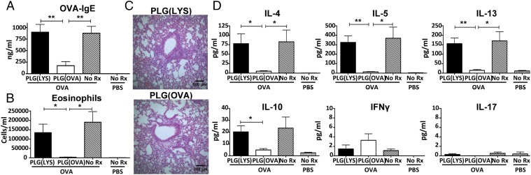
Specific immunotherapy (SIT) is the most widely used treatment for allergic diseases that directly targets the T helper 2 (Th2) bias underlying allergy. However, the most widespread clinical applications of SIT require a long period of dose escalation with soluble antigen (Ag) and carry a significant risk of adverse reactions, particularly in highly sensitized patients who stand to benefit most from a curative treatment. Thus, the development of safer, more efficient methods to induce Ag-specific immune tolerance is critical to advancing allergy treatment. We hypothesized that antigen-associated nanoparticles (Ag-NPs), which we have used to prevent and treat Th1/Th17-mediated autoimmune disease, would also be effective for the induction of tolerance in a murine model of Th2-mediated ovalbumin/alum-induced allergic airway inflammation. We demonstrate here that antigen-conjugated polystyrene (Ag-PS) NPs, although effective for the prophylactic induction of tolerance, induce anaphylaxis in presensitized mice. Antigen-conjugated NPs made of biodegradable poly(lactide-co-glycolide) (Ag-PLG) are similarly effective prophylactically, are well tolerated by sensitized animals, but only partially inhibit Th2 responses when administered therapeutically. PLG NPs containing encapsulated antigen [PLG(Ag)], however, were well tolerated and effectively inhibited Th2 responses and airway inflammation both prophylactically and therapeutically. Thus, we illustrate progression toward PLG(Ag) as a biodegradable Ag carrier platform for the safe and effective inhibition of allergic airway inflammation without the need for nonspecific immunosuppression in animals with established Th2 sensitization.

Keywords: Th2 cells; allergy; immunotherapy; nanoparticles; tolerance.


Conflict of interest statement

The authors declare no conflict of interest.

Figures

Fig. S1.
Fig. S1.
Prophylactic treatment with OVA-PS inhibits Th2-mediated airway inflammation. Naive female BALB/c mice (n = 5) were treated i.v. with 1.25 mg of OVA-PS or control lysozyme-PS (A, B, and D) or MBP-PS (C) on days −7 and +7 relative to i.p. immunization with 10 μg of OVA in 3 mg of alum or alum alone on days 0 and +14 before aerosol challenge with 10 mg/mL OVA for 20 min on days +28–30 and sample collection on day +31. (A) Concentration of serum OVA-IgE was determined by sandwich ELISA. (B) Lungs were flushed with BALF, total cell counts were determined, and samples were cytospun onto slides before DiffQuik staining for differential cell counts of bronchoalveolar lavage eosinophils. (C) Lungs were fixed in formalin and stained with H&E. (D) Cytokines from BALF supernatant were analyzed by Milliplex. (E) Mediastinal LNs were harvested and cultured with 25 μg/mL OVA. At 48 h, cells were pulsed with [3H]TdR for the final 24 h of culture before harvest. (F) Before [3H]TdR pulsing, supernatant from LN cultures was reserved and cytokines were analyzed by Milliplex. Results are mean ± SEM and are representative of two separate experiments. *P < 0.05; **P < 0.01; ***P < 0.001. Rx, treatment.
Fig. 1.
Fig. 1.
Prophylactic treatment with OVA-PLG inhibits Th2-induced airway inflammation. Naive female BALB/c mice (n = 5) were treated i.v. with 1.25 mg of OVA-PLG or control MBP-PLG on days −7 and +7 relative to i.p. immunization with 10 μg of OVA in 3 mg of alum or alum alone on days 0 and +14 before aerosol challenge with 10 mg/mL OVA for 20 min on days +28–30 and sample collection on day +31. (A) Concentration of serum OVA-IgE was determined by sandwich ELISA. (B) Lungs were flushed with BALF, total cell counts were determined, and samples were cytospun onto slides before DiffQuik staining for differential cell counts of bronchoalveolar lavage eosinophils. (C) Lungs were fixed in formalin and stained with periodic acid–Schiff. (D) Cytokines from BALF supernatant were analyzed by Milliplex. Results are mean ± SEM and are representative of four separate experiments. *P < 0.05; **P < 0.01; ***P < 0.001. Rx, treatment.
Fig. S2.
Fig. S2.
Prophylactic treatment with OVA-PLG inhibits OVA-specific recall responses. Mice were treated as outlined in Fig. 1. Mediastinal LNs were harvested and cultured with 25 μg/mL OVA. (A) At 48 h, cells were pulsed with [3H]TdR for the final 24 h of culture before harvest. (B) Before [3H]TdR pulsing, supernatant from LN cultures was reserved and cytokines were analyzed by Milliplex. Results are mean ± SEM and are representative of four separate experiments. *P < 0.05; **P < 0.01.
Fig. S3.
Fig. S3.
OVA-PS NPs, but not OVA-PLG or PLG(OVA) NPs, are anaphylactic in OVA-sensitized mice. (A and B) Female BALB/c mice (n = 5) were treated i.v. with 1.25 mg of OVA NPs or control MBP NPs on days +21 and +28 relative to i.p. immunization with 10 μg of OVA in 3 mg of alum on days 0 and +14. Mice were implanted with s.c. temperature probes, and their temperature was monitored for 1 h after the second i.v. infusion of 200 μg of soluble OVA, Ag-PS (A), or Ag-PLG (B). (C) Female BALB/c mice (n = 5) were treated i.v. with 2.5 mg of PLG(OVA) or control PLG(lysozyme) on days +28 and +42 relative to i.p. immunization with 10 μg of OVA in 3 mg of alum on days 0 and +14. Temperature was monitored with a rectal thermometer for 1 h after the second i.v. infusion of 200 μg of soluble OVA or PLG(Ag). OVA-PLG, PLG(OVA), OVA-PS or unconjugated PLG or PS was incubated with anti-OVA IgE (D) or anti-OVA IgG1 (E). Particles bound by IgE or IgG1 were tagged with biotinylated anti-isotype Abs and streptavidin-FITC before analysis by flow cytometry. Results are geometric mean fluorescence intensity (MFI) ± SEM from three individual batches of labeled particles. **P < 0.01; ***P < 0.001. n.s., nonsignificant.
Fig. 2.
Fig. 2.
Treatment of presensitized mice with OVA-PLG does not inhibit Th2-induced airway inflammation. Naive female BALB/c mice (n = 5) were treated i.v. with 1.25 mg of OVA-PLG or control lysozyme-PLG on days +21 and +28 relative to i.p. immunization with 10 μg of OVA in 3 mg of alum or alum alone on days 0 and +14 before aerosol challenge with 10 mg/mL OVA for 20 min on days +31–33 and sample collection on day +34. (A) Concentration of serum OVA-IgE was determined by sandwich ELISA. (B) Lungs were flushed with BALF, total cell counts were determined, and samples were cytospun onto slides before DiffQuik staining for differential cell counts of bronchoalveolar lavage eosinophils. (C) Cytokines from BALF supernatant were analyzed by Milliplex. (D) Mediastinal LNs were harvested and cultured with 25 μg/mL OVA. At 48 h, cells were pulsed with [3H]TdR for the final 24 h of culture before harvest. (E) Before [3H]TdR pulsing, supernatant from LN cultures was reserved and cytokines were analyzed by Milliplex. Results are mean ± SEM and are representative of two separate experiments. **P < 0.01; ***P < 0.001.
Fig. 3.
Fig. 3.
Prophylactic treatment with PLG(OVA) inhibits Th2-induced airway inflammation. Naive female BALB/c mice (n = 5) were treated i.v. with 2.5 mg PLG(OVA) or control PLG(LYS) on days −7 and +7 relative to i.p. immunization with 10 μg of OVA in 3 mg of alum or alum alone on days 0 and +14 before aerosol challenge with 10 mg/mL OVA for 20 min on days +28–30 and sample collection on day +31. LYS, lysozyme. (A) Concentration of serum OVA-IgE was determined by sandwich ELISA. (B) Lungs were flushed with BALF, total cell counts were determined, and samples were cytospun onto slides before DiffQuik staining for differential cell counts of bronchoalveolar lavage eosinophils. (C) Lungs were fixed in formalin and stained with H&E. (D) Cytokines from BALF supernatant were analyzed by Milliplex. Results are mean ± SEM and are representative of three separate experiments. *P < 0.05; **P < 0.01.
Fig. 4.
Fig. 4.
Treatment of presensitized mice with PLG(OVA) inhibits Th2-induced airway inflammation. Female BALB/c mice (n = 5) were treated i.v. with 2.5 mg PLG(OVA) or control PLG(LYS) on days +28 and +42 relative to i.p. immunization with 10 μg of OVA in 3 mg of alum or alum alone on days 0 and +14 before aerosol challenge with 10 mg/mL OVA for 20 min on days +56–58 and sample collection on day +59. (A) Concentration of serum OVA-IgE was determined by sandwich ELISA. (B) Lungs were flushed with BALF, total cell counts were determined, and samples were cytospun onto slides before DiffQuik staining for differential cell counts of BAL eosinophils. (C) Lungs were fixed in formalin and stained with periodic acid–Schiff. (D) Cytokines from BALF supernatant were analyzed by Milliplex. (E) Mediastinal LNs were harvested and cultured with 25 μg/mL OVA. At 48 h, cells were pulsed with [3H]TdR for the final 24 h of culture. (F) Before [3H]TdR pulsing, supernatant from LN cultures was reserved and cytokines were analyzed by Milliplex. Results are mean ± SEM of three separate experiments. *P < 0.05; **P < 0.005; ***P < 0.001.
Fig. S4.
Fig. S4.
Ag release from PLG(Ag). Twelve milligrams each of PLG(OVA) and PLG(LYS) were washed three times with PBS and incubated on a thermomixer in 1 mL of PBS at 37 °C for 5 d. At regular time intervals, the particle suspension was centrifuged at 7,000 × g for 5 min and the supernatant was collected. The particles were then resuspended in 1 mL of PBS. The amount of protein present in the supernatant was quantified by CBQCA. Cumulative release was calculated as the sum of protein released by that time divided by the sum of protein released and within DMSO-dissolved particles at the end of 5 d. LYS, lysozyme.
Fig. S5.
Fig. S5.
Prophylactic treatment with PLG(OVA) inhibits OVA-specific recall responses. Mice were treated as outlined in Fig. 3. Mediastinal LNs were harvested and cultured with 25 μg/mL OVA. (A) At 48 h, cells were pulsed with [3H]TdR for the final 24 h of culture before harvest. (B) Before [3H]TdR pulsing, supernatant from LN cultures was reserved and cytokines were analyzed by Milliplex. Results are mean ± SEM and are representative of three separate experiments. *P < 0.05; **P < 0.01; ***P < 0.001.

References

    1. Akdis M, et al. Interleukins, from 1 to 37, and interferon-gamma: Receptors, functions, and roles in diseases. J Allergy Clin Immunol. 2011;127:701–721.e701–e770. - PubMed
    1. Smarr CB, Bryce PJ, Miller SD. Antigen-specific tolerance in immunotherapy of Th2-associated allergic diseases. Crit Rev Immunol. 2013;33(5):389–414. - PMC - PubMed
    1. Kopp MV. Role of immunmodulators in allergen-specific immunotherapy. Allergy. 2011;66(6):792–797. - PubMed
    1. Smarr CB, Hsu CL, Byrne AJ, Miller SD, Bryce PJ. Antigen-fixed leukocytes tolerize Th2 responses in mouse models of allergy. J Immunol. 2011;187(10):5090–5098. - PMC - PubMed
    1. Lutterotti A, et al. Antigen-specific tolerance by autologous myelin peptide-coupled cells: A phase 1 trial in multiple sclerosis. Sci Transl Med. 2013;5(188):188ra75. - PMC - PubMed

Publication types

MeSH terms