

What insects can tell us about the origins of consciousness
- PMID: 27091981
- PMCID: PMC4983823
- DOI: 10.1073/pnas.1520084113
What insects can tell us about the origins of consciousness
Abstract
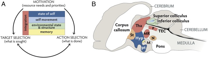
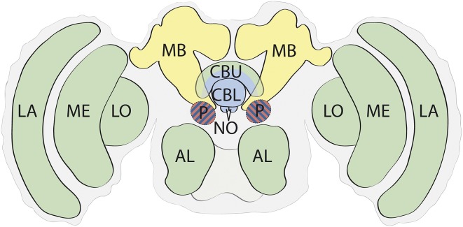
How, why, and when consciousness evolved remain hotly debated topics. Addressing these issues requires considering the distribution of consciousness across the animal phylogenetic tree. Here we propose that at least one invertebrate clade, the insects, has a capacity for the most basic aspect of consciousness: subjective experience. In vertebrates the capacity for subjective experience is supported by integrated structures in the midbrain that create a neural simulation of the state of the mobile animal in space. This integrated and egocentric representation of the world from the animal's perspective is sufficient for subjective experience. Structures in the insect brain perform analogous functions. Therefore, we argue the insect brain also supports a capacity for subjective experience. In both vertebrates and insects this form of behavioral control system evolved as an efficient solution to basic problems of sensory reafference and true navigation. The brain structures that support subjective experience in vertebrates and insects are very different from each other, but in both cases they are basal to each clade. Hence we propose the origins of subjective experience can be traced to the Cambrian.
Keywords: central complex; primary consciousness; subjective experience; vertebrate midbrain.
Conflict of interest statement
The authors declare no conflict of interest.
Figures
Comment in
-
Avoid the hard problem: Employment of mental simulation for prediction is already a crucial step.Proc Natl Acad Sci U S A. 2016 Jul 5;113(27):E3811. doi: 10.1073/pnas.1607146113. Epub 2016 Jun 28. Proc Natl Acad Sci U S A. 2016. PMID: 27357663 Free PMC article. No abstract available.
-
Insects cannot tell us anything about subjective experience or the origin of consciousness.Proc Natl Acad Sci U S A. 2016 Jul 5;113(27):E3813. doi: 10.1073/pnas.1606835113. Epub 2016 Jun 28. Proc Natl Acad Sci U S A. 2016. PMID: 27357664 Free PMC article. No abstract available.
-
Consciousness explained or consciousness redefined?Proc Natl Acad Sci U S A. 2016 Jul 5;113(27):E3812. doi: 10.1073/pnas.1606942113. Epub 2016 Jun 28. Proc Natl Acad Sci U S A. 2016. PMID: 27357665 Free PMC article. No abstract available.
References
-
- Nagel T. What is it like to be a bat? Philos Rev. 1974;83(4):435–450.
-
- Edelman DB, Seth AK. Animal consciousness: A synthetic approach. Trends Neurosci. 2009;32(9):476–484. - PubMed
-
- Pepperberg IM. The Alex Studies: Cognitive and Communicative Abilities of Grey Parrots. Harvard Univ Press; Cambridge, MA: 2002.
-
- Crick F, Koch C. Towards a neurobiological theory of consciousness. Semin Neurosci. 1990;2:263–275.
Publication types
MeSH terms
LinkOut - more resources
Full Text Sources
Other Literature Sources
