Comparative Study
. 2016 May 3;113(18):E2516-25.
doi: 10.1073/pnas.1523005113. Epub 2016 Apr 18.

Comparative systems pharmacology of HIF stabilization in the prevention of retinopathy of prematurity

Affiliations
Comparative Study

Comparative systems pharmacology of HIF stabilization in the prevention of retinopathy of prematurity

George Hoppe et al. Proc Natl Acad Sci U S A. .

Abstract

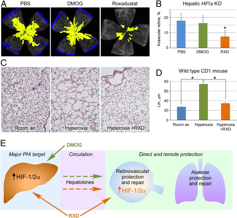
Retinopathy of prematurity (ROP) causes 100,000 new cases of childhood blindness each year. ROP is initiated by oxygen supplementation necessary to prevent neonatal death. We used organ systems pharmacology to define the transcriptomes of mice that were cured of oxygen-induced retinopathy (OIR, ROP model) by hypoxia-inducible factor (HIF) stabilization via HIF prolyl hydroxylase inhibition using the isoquinolone Roxadustat or the 2-oxoglutarate analog dimethyloxalylglycine (DMOG). Although both molecules conferred a protective phenotype, gene expression analysis by RNA sequencing found that Roxadustat can prevent OIR by two pathways: direct retinal HIF stabilization and induction of aerobic glycolysis or indirect hepatic HIF-1 stabilization and increased serum angiokines. As predicted by pathway analysis, Roxadustat rescued the hepatic HIF-1 knockout mouse from retinal oxygen toxicity, whereas DMOG could not. The simplicity of systemic treatment that targets both the liver and the eye provides a rationale for protecting the severely premature infant from oxygen toxicity.

Keywords: BPD; HIF; ROP; Roxadustat; prolyl hydroxylase inhibition.


Conflict of interest statement

The authors declare no conflict of interest.

Figures

Fig. 1.
Fig. 1.
Roxadustat targets liver and kidney. (A) Both i.p. and s.c. injections create liver- and kidney-specific luminescence in the luc-ODD mouse. (B) Quantification of luciferase activity in organ lysates demonstrates liver and kidney tropism by either i.p. or s.c. injection of Roxadustat (RXD). (C) Luminescence of organ lysates of i.p. Roxadustat indicates specificity for liver and kidney. *P < 5 × 10⁻⁴. (D) Time duration of HIF PHi by Roxadustat gives sustained stabilization of luc-ODD over 24 h. (E) Western blot analysis of HIF-1α protein after Roxadustat. (F) Integrated optical density of immunoblot analysis in E. (G) Densitometry of HIF-1α immunoblot (Insert) over time after Roxadustat i.p. injection. (H) Epo mRNA expression in organs after Roxadustat i.p. injection. (I) Serum EPO concentration versus time after i.p. Roxadustat. (J) Dose-dependent expression of Epo mRNA in the liver. (K) Dose–response of HIF-1α to Roxadustat in cultured Hep3B cells analyzed by Western blotting. (L) EPO mRNA levels in cultured Hep3B cells and EPO protein content on Hep3B culture media in response to Roxadustat.
Fig. 2.
Fig. 2.
Preservation of retinal blood vessels at P17 after Roxadustat injection. (A) Representative flat mounts comparing sham PBS injection to Roxadustat to DMOG. Each whole retinal flatmount image was obtained by taking nine overlapping microphotographs followed by merging corresponding microphotographs into one as described in SI Materials and Methods. Color coding of avascular region at P17 indicates reduction of oxygen-induced vasoobliteration protection created by Roxadustat. (B) Quantification and statistical analysis of retinal flat mounts depicting percent avascular area of total retina area. **P = 5 × 10⁻⁹, *P = 0.02. RXD, Roxadustat. (C) Isolectin and Hypoxyprobe staining indicate a decrease in ischemic, hypoxic retina at P17 after Roxadustat injection in representative retinal flat mounts. (D) Quantitative image analysis of avascular and hypoxic area expressed as percent of total retinal area (y axis). Values within the bars represent hypoxic area as percent of avascular area. (E) Rate of retinal vascular growth during two phases of the OIR model, after PBS or Roxadustat treatment. Shown are representative flat mounts with computer-assisted area calculation shown in yellow. Roxadustat decreases vascular obliteration in hyperoxia. (F) Quantitation of avascular area (*P < 1 × 10⁻⁵) demonstrates consistently less ischemic retina, which is the substrate of ROP and pathologic neovascularization. (G) Rate of retinal vascularization in control and treated animals throughout the OIR cycle. The area of ischemia is less after Roxadustat treatment, yet the slopes of regrowth are identical between control and Roxadustat-treated animals, indicating that Roxadustat protects retina during hyperoxia but does not induce abnormal or rapid regrowth of retinal blood vessels. (H) Decreased ischemia leads to decreased pathologic neovascularization. **P = 0.002, *P = 0.037.
Fig. S1.
Fig. S1.
Effect of Roxadustat on neural retina apoptosis. (A) Active caspase 3 retinal immunohistochemistry shows less apoptosis in Roxadustat-treated, hyperoxic animals. Overlapping images of entire retinal section (10 μm) were taken at 20× and photomerged in Adobe Photoshop (analogous to retinal flat mount imaging in Materials and Methods). Representative midperiphery retinal regions are shown. (B) Quantification of active caspase 3-positive cells demonstrates statistically significant reduction in apoptosis in the inner nuclear layer of animals treated with Roxadustat (RXD). **P = 8 × 10⁻⁶, *P = 0.028. At least four sections from each retina were analyzed (four pups per experimental condition), and resulting data are expressed as the mean number of positive cells per entire section ± SD for outer nuclear layer (ONL), inner nuclear layer (INL), and ganglion cell layer (GCL).
Fig. 3.
Fig. 3.
Identification and validation of gene expression similarities and differences between animals treated with DMOG versus Roxadustat (RXD). (A) Venn diagrams of differentially expressed genes in the liver and retina, PBS vs. DMOG or PBS vs. RXD; fold-change cutoff, 2.0. (B) Pie charts depicting commonly and uniquely up-regulated genes. Liver has many common genes up-regulated, whereas retina does not. The identity of the top responders (fold-change cutoff, 2.0) can be found in Tables 1 and 2 and Dataset S1, where a complete list of gene products is given, as well as online at Gene Expression Omnibus (GSE74170; NCBI tracking system no. 17567121). (C) Secondary validation of top responders—secreted liver gene products by qPCR of hepatic mRNA (*P = 9 × 10⁻⁵, **P = 0.003, ***P = 0.001) and by (D) ELISA of serum proteins (*P < 0.002). (E) Immunofluorescent microscopy (40× magnification) shows partial colocalization of MCT-4 (green) elevated by Roxadustat (RXD) with vimentin (red), specific for Muller cells. (F) Higher magnification (63×) of vimentin (red) and MCT-4 (green) demonstrates overlap within Muller cells that span the width of the retina and shows characteristic morphology of specialized retinal macroglia.
Fig. S2.
Fig. S2.
Effect of Roxadustat on Muller cell and photoreceptor SLC16A3 (MCT-4) expression. (A) Immunofluorescent microscopy (20× magnification) confirms significant elevation of MCT-4 (red) in multiple retinal layers in P10 mouse pups in response to Roxadustat. Green channel represents rhodopsin staining colocalizing with MCT-4 in the inner plexiform layer and rod inner segments. Note that rod outer segments are undeveloped at P10. Blue channel reveals retinal nuclear layers stained with Hoechst. (B) Roxadustat (10 μg/mL) was capable of >twofold increase in SLC16A3 (MCT4) expression in cultured Muller cells MIO-M1. *P = 4 × 10⁻⁷.
Fig. S3.
Fig. S3.
Pathway analysis (Metacore) of DMOG- versus Roxadustat (RXD)-treated animals highlights differences in transcriptomes. (A) Bar graph comparison of the top up-regulated pathway in retina and liver after Roxadustat or DMOG showing that mice treated with Roxadustat activate aerobic glycolysis in liver and retina, whereas mice treated with DMOG activate aerobic glycolysis in liver only. (B) Glycolysis and gluconeogenesis pathways overlaid with differentially expressed genes from all four datasets: (1) Liver_DMOGvsPBS, (2) Retinal_DMOGvsPBS, (3) Liver_ RXDvsPBS, and (4) Retinal_RXDvsPBS.
Fig. 4.
Fig. 4.
Comparison of Roxadustat to DMOG using the cre/lox hepatic Hif1a KO mouse. (A) Representative retinal flat mount showing capillary dropout after systemic treatment with DMOG or Roxadustat in the hepatic HIF-1 KO. DMOG does not rescue the KO, whereas Roxadustat does, which is shown here to prevent OIR in the KO. (B) Quantification and statistical analysis of retinal flat mounts depicting percent avascular area of total retina area. *P = 1 × 10⁻¹². (C) Lung histology after hematoxylin and eosin staining demonstrates the destructive alveolar enlargement by hyperoxia that is reduced by s.c. Roxadustat (RXD) to a size and structure comparable to age-matched controls in room air. (D) Quantification of alveolar size using Lm demonstrates statistically significant normalization in alveolar size by Roxadustat (RXD) treatment during hyperoxia in the wild-type CD1 mouse pups. *P = 1 × 10⁻⁴. (E) Schematic representation of the two pathways for retinovascular protection against OIR and bronchopulmonary dysplasia, targeting extraretinal HIF-1 in the liver in the case of DMOG, or both hepatic and retinal HIF-1 pathways in the case of Roxadustat (RXD).
Fig. S4.
Fig. S4.
Chemical structures of DMOG and Roxadustat. Our comparison of DMOG [amino dicarboxylic acid with flanking methyl groups; chemical name, N-(2-Methoxy-2-oxaoacetyl)glycine methyl ester] to Roxadustat (amino carboxylic acid with isoquinolone; chemical name, N-[(4-hydroxy-1-methyl-7-phenoxyisoquinolin-3-yl)carbonyl]glycine) demonstrates that DMOG solely targets the liver to induce retinal protection, whereas Roxadustat targets both the liver and the retina, despite the fact that Roxadustat is therapeutic at 5 times less molar concentration.

Similar articles

Cited by

References

    1. Hartnett ME, Penn JS. Mechanisms and management of retinopathy of prematurity. N Engl J Med. 2012;367(26):2515–2526. - PMC - PubMed
    1. Finer NN, et al. SUPPORT Study Group of the Eunice Kennedy Shriver NICHD Neonatal Research Network Early CPAP versus surfactant in extremely preterm infants. N Engl J Med. 2010;362(21):1970–1979. - PMC - PubMed
    1. Hartnett ME, Lane RH. Effects of oxygen on the development and severity of retinopathy of prematurity. J AAPOS. 2013;17(3):229–234. - PMC - PubMed
    1. Semenza GL, Nejfelt MK, Chi SM, Antonarakis SE. Hypoxia-inducible nuclear factors bind to an enhancer element located 3′ to the human erythropoietin gene. Proc Natl Acad Sci USA. 1991;88(13):5680–5684. - PMC - PubMed
    1. Semenza GL, Wang GL. A nuclear factor induced by hypoxia via de novo protein synthesis binds to the human erythropoietin gene enhancer at a site required for transcriptional activation. Mol Cell Biol. 1992;12(12):5447–5454. - PMC - PubMed

Publication types

MeSH terms