. 2016 Apr 7:7:86.
doi: 10.3389/fphar.2016.00086. eCollection 2016.

Adiponectin Attenuates Angiotensin II-Induced Vascular Smooth Muscle Cell Remodeling through Nitric Oxide and the RhoA/ROCK Pathway

Affiliations

Adiponectin Attenuates Angiotensin II-Induced Vascular Smooth Muscle Cell Remodeling through Nitric Oxide and the RhoA/ROCK Pathway

Wared Nour-Eldine et al. Front Pharmacol. .

Abstract

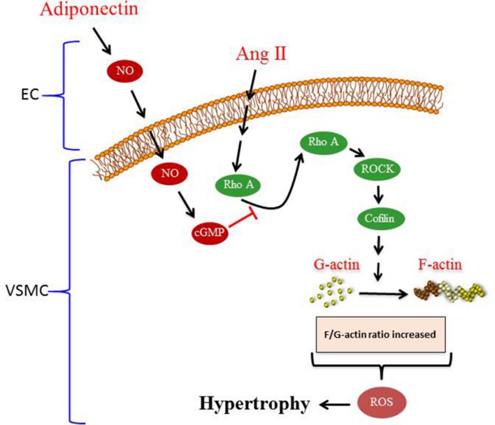
Introduction: Adiponectin (APN), an adipocytokine, exerts protective effects on cardiac remodeling, while angiotensin II (Ang II) induces hypertension and vascular remodeling. The potential protective role of APN on the vasculature during hypertension has not been fully elucidated yet. Here, we evaluate the molecular mechanisms of the protective role of APN in the physiological response of the vascular wall to Ang II.

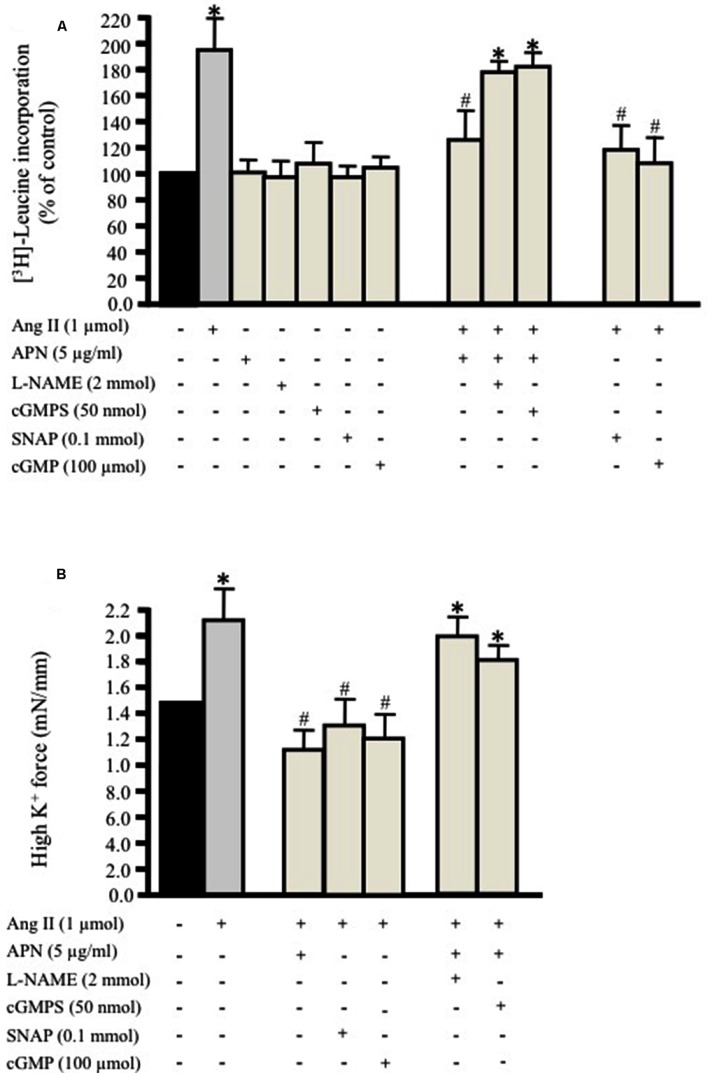
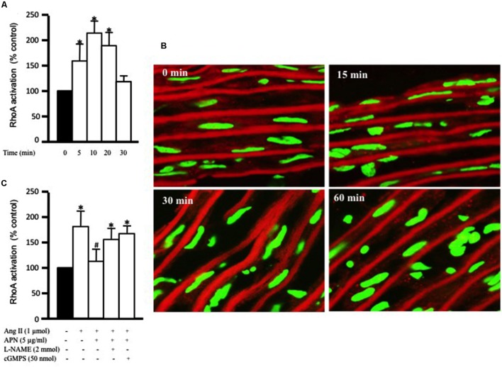
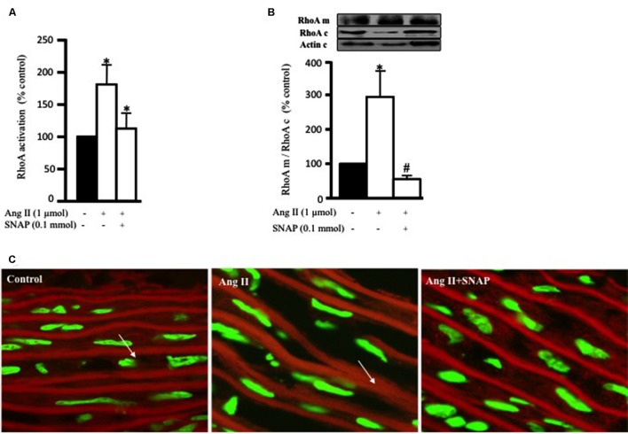
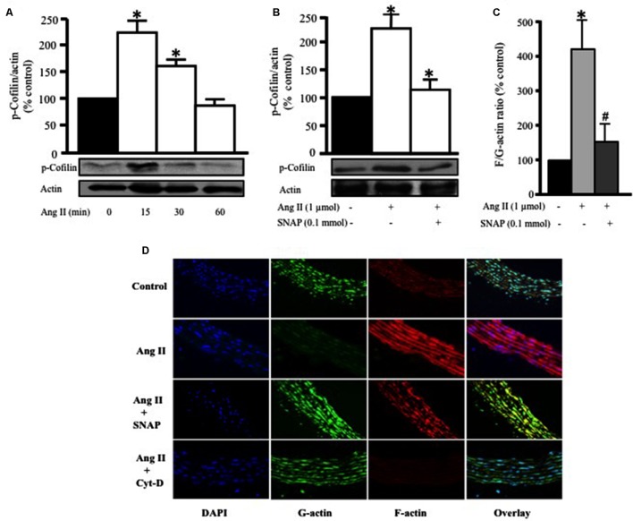
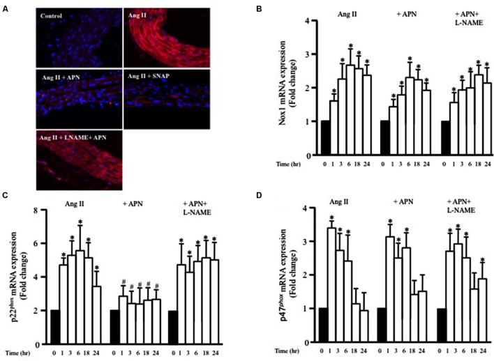
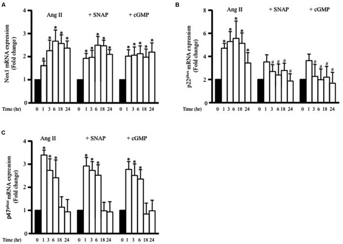
Methods and results: Rat aortic tissues were used to investigate the effect of APN on Ang II-induced vascular remodeling and hypertrophy. We investigated whether nitric oxide (NO), the RhoA/ROCK pathway, actin cytoskeleton remodeling, and reactive oxygen species (ROS) mediate the anti-hypertrophic effect of APN. Ang II-induced protein synthesis was attenuated by pre-treatment with APN, NO donor S-nitroso-N-acetylpenicillamine (SNAP), or cGMP. The hypertrophic response to Ang II was associated with a significant increase in RhoA activation and vascular force production, which were prevented by APN and SNAP. NO was also associated with inhibition of Ang II-induced phosphorylation of cofilin. In addition, immunohistochemistry revealed that 24 h Ang II treatment increased the F- to G-actin ratio, an effect that was inhibited by SNAP. Ang II-induced ROS formation and upregulation of p22(phox) mRNA expression were inhibited by APN and NO. Both compounds failed to inhibit Nox1 and p47(phox) expression.

Conclusion: Our results suggest that the anti-hypertrophic effects of APN are due, in part, to NO-dependent inhibition of the RhoA/ROCK pathway and ROS formation.

Keywords: VSMC; adiponectin; angiotensin II; nitric oxide; remodeling.


Figures

FIGURE 1
FIGURE 1
Adiponectin inhibits Ang II-induced protein synthesis and force production in rat aortic ring. Serum-starved endothelium-intact rat aortic rings were pre-treated with adiponectin (5 μg/ml), L-NAME (2 mmol/L), cGMPS (50 nmol/L), S-nitroso-N-acetylpenicillamine (SNAP; 0.1 mmol/L), 8-Br-cGMP (cGMP; 100 μmol/L) for 1 h prior to 24 h treatment with 1 μmol/L Ang II. [3H]-leucine incorporation is shown in (A). Active stress in response to high K+ is shown in (B). Data are shown as mean ± SEM with n = 5–6 for all groups. p < 0.05 vs. without Ang II (control); #p < 0.05 vs. with Ang II.
FIGURE 2
FIGURE 2
Adiponectin inhibition of Ang II-induced RhoA activation is NO-dependent. (A) Endothelium-intact rat aortic rings were treated for various times (0, 15, 30, and 60 min) with 1 μmol/L Ang II and RhoA activation was analyzed with G-LISA kit BK121. (B) Time course of Ang II-induced RhoA translocation. Confocal images for aortic frozen sections at different time points (0, 15, 30, 60 min) stained with DAPI (green) and anti-RhoA antibody (red). (C) Endothelium-intact rat aortic rings were treated for 10 min with 1 μmol/L Ang II in the presence or absence of adiponectin (5 μg/ml), L-NAME (2 mmol/L), cGMPS (50 nmol/L). RhoA activation was analyzed with G-LISA kit BK121. Data are shown as mean ± SEM and indicate fold change relative to untreated aortic ring (control). n = 6–7; p < 0.05 vs. without Ang II.
FIGURE 3
FIGURE 3
Nitric oxide (NO) inhibits Ang II-induced RhoA activation and translocation. (A) Endothelium-intact rat aortic rings were cultured for 10 min with or without 1 μmol/L Ang II pre-treated with/without SNAP and RhoA activation was analyzed with G-LISA kit BK121. (B) A representative Western blot showing RhoA membrane fraction (RhoA m), RhoA cytosolic fraction (RhoA c) and actin (actin c) after 30 min treatment with Ang II in aortic rings. Ang II caused a threefold increase in membrane bound RhoA whereas SNAP inhibited RhoA translocation. Each bar represents mean ± SEM value obtained from 3 to 7 independent experiments. p < 0.05 vs. without Ang II. #p < 0.05 vs. with Ang II. (C) Confocal images for aortic ring frozen sections stained with DAPI (green) and anti-RhoA antibody (red) treated with Ang II for 30 min with or without SNAP. Control (left panel), Ang II (middle panel), Ang II + SNAP (right panel).
FIGURE 4
FIGURE 4
Nitric oxide attenuates Ang II-induced RhoA/ROCK pathway activation and actin cytoskeleton remodeling. (A) Endothelium-intact rat aortic rings were treated for various times (0, 15, 30, and 60 min) with 1 μmol/L Ang II and cofilin-2 phosphorylation was analyzed by Western Blotting. n = 5; p < 0.05. (B) SNAP significantly inhibited Ang II-induced cofilin-2 phosphorylation after 15 min. Endothelium-intact rat aortic rings were treated with Ang II for 24 h with or without SNAP followed by quantification of F/G-actin ratio (C). Confocal images of aortic ring sections stained with DAPI (blue, nucleus), Deoxyribonuclease-I Alexa Fluor 488 (green, G-actin) and F-Phalloidin-555 actin stain (red, F-actin). Rat aortas were treated for 24 h with 1 μmol/L Ang II in the presence or absence of SNAP (0.1 mmol/L) or Cyt-D (1 μmol/L) and relative amounts of F- and G-actin were assessed (D). n = 5–7; p < 0.05 vs. without Ang II; #p < 0.05 vs. with Ang II.
FIGURE 5
FIGURE 5
Nitric oxide mediates the inhibitory effect of adiponectin on Ang II-induced ROS formation and p22phox mRNA overexpression. (A) Representative confocal microscopic images of aortic wall treated with Ang II for 1 h, with or without inhibitors and agonists, followed by staining with DHE (red). DAPI stained the nuclei blue. Fold change of Nox1 (B), p22phox (C), and p47phox (D) mRNA expression levels in the aortic rings after 0, 1, 3, 6, 18, 24 h of Ang II (1 μmol/L) treatment. n = 8–10 in each group; p < 0.05 vs. control, #p < 0.05 vs. with Ang II.
FIGURE 6
FIGURE 6
S-nitroso-N-acetylpenicillamine and cGMP attenuate Ang II-induced p22phox mRNA expression. Fold change of Nox1 (A), p22phox (B), and p47phox (C) mRNA expression levels in the endothelium-intact rat aortic rings after 0, 1, 3, 6, 18, 24 h of Ang II (1 μmol/L) treatment. n = 8–10 in each group; p < 0.05 vs. control; #p < 0.05 vs. with Ang II.
FIGURE 7
FIGURE 7
Proposed model of adiponectin-mediated inhibition of Ang II-induced VSMC remodeling. Treatment with Ang II induces hypertrophy in VSMC. Adiponectin suppresses AngII-induced VSMC hypertrophy through signaling pathways that activates NO synthesis in endothelial cells (ECs). The inhibitory effect of adiponectin on Ang II-induced VSMC hypertrophy is mediated through modulating the RhoA/ROCK signaling pathway, F-actin to G-actin ratio, and ROS formation.

Similar articles

Cited by

References

    1. Bagi Z., Feher A., Cassuto J., Akula K., Labinskyy N., Kaley G., et al. (2011). Increased availability of angiotensin AT 1 receptors leads to sustained arterial constriction to angiotensin II in diabetes – role for Rho-kinase activation. Br. J. Pharmacol. 163 1059–1068. 10.1111/j.1476-5381.2011.01307.x - DOI - PMC - PubMed
    1. Carbone M. L., Bregeon J., Devos N., Chadeuf G., Blanchard A., Azizi M., et al. (2015). Angiotensin II activates the RhoA exchange factor Arhgef1 in humans. Hypertension 65 1273–1278. 10.1161/HYPERTENSIONAHA.114.05065 - DOI - PubMed
    1. Caselli C., D’amico A., Cabiati M., Prescimone T., Del Ry S., Giannessi D. (2014). Back to the heart: the protective role of adiponectin. Pharmacol. Res. 82 9–20. 10.1016/j.phrs.2014.03.003 - DOI - PubMed
    1. Coles B., Fielding C. A., Rose-John S., Scheller J., Jones S. A., O’donnell V. B. (2007). Classic interleukin-6 receptor signaling and interleukin-6 trans-signaling differentially control angiotensin II-dependent hypertension, cardiac signal transducer and activator of transcription-3 activation, and vascular hypertrophy in vivo. Am. J. Pathol. 171 315–325. 10.2353/ajpath.2007.061078 - DOI - PMC - PubMed
    1. Dai D. F., Johnson S. C., Villarin J. J., Chin M. T., Nieves-Cintron M., Chen T., et al. (2011). Mitochondrial oxidative stress mediates angiotensin II-induced cardiac hypertrophy and Galphaq overexpression-induced heart failure. Circ. Res. 108 837–846. 10.1161/CIRCRESAHA.110.232306 - DOI - PMC - PubMed

LinkOut - more resources