. 2016 Apr 19;12(4):e1004880.
doi: 10.1371/journal.pcbi.1004880. eCollection 2016 Apr.

Hippocampal CA1 Ripples as Inhibitory Transients

Affiliations

Hippocampal CA1 Ripples as Inhibitory Transients

Paola Malerba et al. PLoS Comput Biol. .

Abstract

Memories are stored and consolidated as a result of a dialogue between the hippocampus and cortex during sleep. Neurons active during behavior reactivate in both structures during sleep, in conjunction with characteristic brain oscillations that may form the neural substrate of memory consolidation. In the hippocampus, replay occurs within sharp wave-ripples: short bouts of high-frequency activity in area CA1 caused by excitatory activation from area CA3. In this work, we develop a computational model of ripple generation, motivated by in vivo rat data showing that ripples have a broad frequency distribution, exponential inter-arrival times and yet highly non-variable durations. Our study predicts that ripples are not persistent oscillations but result from a transient network behavior, induced by input from CA3, in which the high frequency synchronous firing of perisomatic interneurons does not depend on the time scale of synaptic inhibition. We found that noise-induced loss of synchrony among CA1 interneurons dynamically constrains individual ripple duration. Our study proposes a novel mechanism of hippocampal ripple generation consistent with a broad range of experimental data, and highlights the role of noise in regulating the duration of input-driven oscillatory spiking in an inhibitory network.


Conflict of interest statement

The authors have declared that no competing interests exist.

Figures

Fig 1
Fig 1. CA1 ripples have standardized durations.
From a 25hr experiment recording, ripples are identified as large amplitude excursion of the band-passed LFP. (a) The leftmost plots show wide band (1-400Hz) LFP recordings from CA1. The rightmost plots show the same LFPs band-pass filtered between 50Hz and 350Hz. (b) Histogram of ripple frequencies (count normalized to total number of ripples). The blue line is its Gaussian distribution fit. (c) Histogram of ripple inter-arrival times, and exponential fit in red. (d) Histogram of ripple durations, showing a high kurtosis, hence highlighting that ripples are very different in frequency and have almost memory-less arrival times, but their duration has small variability.
Fig 2
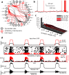
Fig 2. Computational model of CA1 ripples.
(a) Model schematic. Parameters: see Materials and Methods. (b) Example voltage traces of pyramidal cell (black) and interneuron (red). (c) Distributions of synaptic weights. (d) A simulation, described from the top. Traces of current inputs from CA3 to pyramidal cells (black) and to inhibitory interneurons (red). Rastergram where cells are stacked along the y-axis and crosses represent spikes of pyramidal cells (black) and interneurons (red). Probability to spike for pyramidal cells (black) and interneurons (red) in 1ms bins. LFP wide-band, and band-passed filtered (50-350Hz).
Fig 3
Fig 3. Properties of ripples in the model.
Histograms of ripple frequency (a), durations (b), number of spikes of each pyramidal cell during ripples (c) and percent of pyramidal cells spiking in a ripple (d). (e) Average cross-correlations between firing probability of interneurons (red) or pyramidal cells (black) and the band-passed LFP.
Fig 4
Fig 4. Predictions from ripple network model.
(a) Stereotypical ripple LFP has a duration independent from CA3 input length. On the left, the duration of CA3 input is reported. In each group, shown in gray are the actual band-passed LFPs for 40 ripples, while the black line is their average. The black dot marks where each average ripple ends. Next to the LFPs, a schematic of the full network used in this figure (as in Fig 2) (b). The percentage of pyramidal cells spiking on every ripple increases with the magnitude of CA3 input to pyramidal cells population. (c) Effect of inhibition on pyramidal cell recruitment to ripples. Histograms of the percent of pyramidal cells spiking in any given ripple in three conditions: default (black bars), inhibition on interneurons increased to 5ms (red bars), inhibition on pyramidal cells increased to 6ms (green bars). Bars heights have been normalized by total number of ripples. Note that increasing inhibition on interneurons un-inhibits pyramidal cell spiking, while increased inhibition on pyramidal cells predictably suppresses pyramidal cell firing during ripples. (d) Increasing inhibition time scale has almost no effect on ripple frequency. Histograms of ripple frequency under the same three conditions as in panel c.
Fig 5
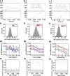
Fig 5. Transients in inhibitory network explain ripple mechanism.
(a) Schematic representation of the model considered, only composed of inhibitory neurons. (b) Histograms of spike probabilities, with step current input (blue). The time axes is binned in 1ms bins. Changing parameters: decay time scale of inhibitory synapses (τ) and scaling (α) of maximal inhibitory synapses conductance. (c) Transient duration (in ms) as a function of α and τ. (d) Transient frequency. (e) Number of peak cycles in the transient, before the distribution flattens (see Materials and Methods).
Fig 6
Fig 6. Transients in inhibitory network depend on input strength and noise size.
(a, b) Effect of the input amplitude on oscillation properties. (a) Histograms of firing probabilities for a model shown in Fig 5, only with a DC step half the size (350 pA). The time axes is binned in 1ms bins. Note that synchrony is strongly affected, and the transient is composed by a drastically reduced number of cycles (b). (c, d) Effect of noise and strength of inhibition on transient properties. (c) Histograms of firing probabilities, for changing synaptic strength (scaled by α) and noise standard deviation (scaled by σ). The time axes is binned in 1ms bins. (d) Top panel: number of cycles composing the transient event. Bottom panel: network frequency.
Fig 7
Fig 7. Effect of driving only Pyramidal cells or only Interneurons on ripple oscillations.
(a) Schematic of the model, in which input current is delivered only to inhibitory interneurons. (b) Examples of model behavior. Top plot: input current. Middle plot: firing probabilities of interneurons (red) and pyramidal cells (black)–note that the pyramidal cells are not spiking during stimulation. Lower plots: LFP, wide band above and band-passed (50–350 Hz) below. (c) The ratio of interneuron receiving input current affects the size of filtered LFP. Note small amplitude of LFP when only a fraction of interneurons in activated. (d) Schematic of the model, in which only pyramidal cells receive input current. (e) Examples of model behavior. Same as in b. Note that reducing adaptation shows an increased duration of the high frequency event, which quickly (after less than 30ms) defaults to a gamma frequency oscillation. (f) Spike-frequency adaptation in pyramidal cells regulates the duration of HFO triggered by input only on pyramidal cells.
Fig 8
Fig 8. Selective input from CA3 induces sequential activation of CA1 pyramidal cells in model.
(a i) Top panel, the CA3 input currents delivered to selected cells (sequence cells) during a ripple. Bottom panel, simultaneous rastergram of sequence cells. (a ii) Distribution of the input drives to all pyramidal cells in the model. In red, the values for sequence cells. (a iii) Average spike time differences for all sequence cells: each line is the average spike time difference between a given cell and all cells in the sequence, averaged across 40 ripples. The lines cross zero when the spike time difference with itself is reported. Note that in this case sequential spiking behavior during ripples is not present. (a iv) Plot of each pyramidal cell vs the fraction of ripples it visited. In black all cells, circled in red are the sequence cells. For every cell, the number of ripples in which it spiked (ripple visited) can be found and divided by the total number of ripples in the simulation. (b) Same plots as in a, but in this case selected cells only receive selective CA3 input (b i), without enhanced intrinsic excitability provided by constant direct current (see b ii). Note that in this case, the orderliness of spiking across ripples is overall preserved (see b iii). Note that most cells spike in less than 20% of the ripples, while selected “sequence” cells all visit about 70% of the ripples (see b iv). (c) Same as a, but in this case selected cells receive both enhanced intrinsic excitability (see c ii) and selective temporal ordering in CA3 input (see c i). The orderliness of the average spike time difference curves emphasizes that cells spike in order across multiple ripples (see c iii), and sequence cells spike in more ripples than all other cells (see c iv).

Similar articles

Cited by

References

    1. Diekelmann S, Born J. The memory function of sleep. Nature reviews Neuroscience. 2010;11(2):114–26. Epub 2010/01/05. 10.1038/nrn2762. . - DOI - PubMed
    1. Schonauer M, Pawlizki A, Kock C, Gais S. Exploring the effect of sleep and reduced interference on different forms of declarative memory. Sleep. 2014;37(12):1995–2007. doi: 10.5665/sleep.4258. 10.5665/sleep.4258 - DOI - DOI - PMC - PubMed
    1. Stickgold R, James L, Hobson JA. Visual discrimination learning requires sleep after training. Nat Neurosci. 2000;3(12):1237–8. Epub 2000/12/02. 10.1038/81756 . - DOI - PubMed
    1. Plihal W, Born J. Effects of early and late nocturnal sleep on priming and spatial memory. Psychophysiology. 1999;36(5):571–82. Epub 1999/08/12. . - PubMed
    1. Wagner U, Gais S, Born J. Emotional memory formation is enhanced across sleep intervals with high amounts of rapid eye movement sleep. Learn Mem. 2001;8(2):112–9. Epub 2001/03/29. 10.1101/lm.36801 - DOI - PMC - PubMed

Publication types

LinkOut - more resources