. 2016 Apr 18;37(2):162-73.
doi: 10.1016/j.devcel.2016.03.012.

Calmodulin Promotes N-BAR Domain-Mediated Membrane Constriction and Endocytosis

Affiliations

Calmodulin Promotes N-BAR Domain-Mediated Membrane Constriction and Endocytosis

Margaret D Myers et al. Dev Cell. .

Abstract

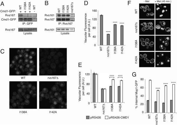
Membrane remodeling by BAR (Bin, Amphiphysin, RVS) domain-containing proteins, such as endophilins and amphiphysins, is integral to the process of endocytosis. However, little is known about the regulation of endocytic BAR domain activity. We have identified an interaction between the yeast Rvs167 N-BAR domain and calmodulin. Calmodulin-binding mutants of Rvs167 exhibited defects in endocytic vesicle release. In vitro, calmodulin enhanced membrane tubulation and constriction by wild-type Rvs167 but not calmodulin-binding-defective mutants. A subset of mammalian N-BAR domains bound calmodulin, and co-expression of calmodulin with endophilin A2 potentiated tubulation in vivo. These studies reveal a conserved role for calmodulin in regulating the intrinsic membrane-sculpting activity of endocytic N-BAR domains.


Figures

Figure 1
Figure 1. Rvs167 N-BAR domain binds calmodulin
(A) Diagram of Rvs167 fragments. The black box represents the N-terminal amphipathic helix. The grey box represents a spacer region. (B) Rvs167 binds calmodulin. The indicated Rvs167 fragments in bacterial lysates (Input = 5%) were tested for binding to calmodulin-Agarose (CMD) or protein A sepharose (PAS). Proteins separated by SDS-PAGE were visualized with Coomassie blue. (C) Rvs167 1-265 binds directly to calmodulin. The indicated purified Rvs167 fragments (Input = 3%) were tested for binding to CMD. Proteins were analyzed as in (B). (D) Rvs167 binds Cmd1 in vivo. Yeast cell extracts (Lysate = 2%) expressing calmodulin with (+) or without (−) a FLAG tag were subjected to immunoprecipitation with anti-FLAG antibodies. Immunoprecipitated proteins were separated by SDS-PAGE and visualized by immunoblotting for Rvs167 or Cmd1.
Figure 2
Figure 2. Rvs167 N-BAR domain contains two calmodulin binding regions
(A) Diagram of Rvs167 fragments. (B) Helices 1 and 2 of Rvs167 bind calmodulin. The indicated Rvs167 fragments in bacterial cell lysates were tested for binding to CMD or PAS (input = 3%) as in Figure 1B. (C) Optimal 1-265 binding to both calmodulin and liposomes requires the N-terminal amphipathic helix. The indicated Rvs167 fragments in bacterial lysates were tested for binding to CMD (input = 2%) or liposomes (input = 10%). Proteins were analyzed as in Figure 1B. (D) Potential calmodulin binding region in helix 2. Sequence predicted to bind calmodulin by the Calmodulin Target Database is highlighted in grey. Underlined region is required for calmodulin binding in vitro. Also see Figure S1. Alanine mutations used in this study are indicated. (E) Mutations of hydrophobic residues in the helix 2 binding region decrease binding to calmodulin. The indicated Rvs167 mutants (in 1-265) from bacterial lysates (input = 20%) were tested for calmodulin or liposome binding as in (C).
Figure 3
Figure 3. Calmodulin binding mutants of Rvs167 are defective in endocytosis
(A) Rvs167 mutants are defective in calmodulin binding in vivo. Lysates (1%) from wild-type (WT) or indicated Rvs167 mutant yeast cells expressing Cmd1-GFP (+) or untagged Cmd1 (−) were subjected to immunoprecipitation with anti-GFP antibodies. Immunoprecipitated proteins were separated by SDS-PAGE and visualized by immunoblotting for Rvs167 or GFP. (B) Rvs167 mutants bind Rvs161. Lysates (1%) from WT or indicated Rvs167 mutant yeast cells were subjected to immunoprecipitation with anti-Rvs167 antibodies. Immunoprecipitated proteins were separated by SDS-PAGE and visualized by immunoblotting for Rvs161 or Rvs167. As previously reported (Lombardi and Riezman, 2001), expression of Rvs161 is decreased in rvs167Δ cells. (C) Calmodulin binding mutants of Rvs167 are defective in LY endocytosis. The indicated strains were assessed for LY internalization by confocal microscopy. (D) Quantitation of vacuolar LY in strains presented in (C). Bars indicate the mean total vacuolar fluorescence per cell ± s.e.m. in arbitrary units (A.U.). **** p < 0.0001 compared to WT as determined by t-test. (E) Calmodulin overexpression partially rescues LY endocytosis in I138A and I142A mutant cells. The indicated strains expressing CMD1 from a multicopy vector (pRS426-CMD1) or vector alone (pRS426) were tested for LY uptake and the results quantified as in (D). **** p < 0.0001 as compared to empty vector as determined by t-test for each strain. Also see Figure S2. (F) Impaired Mup1-GFP endocytosis in Rvs167 mutants. Endocytosis of Mup1-GFP in the indicated strains was monitored by confocal microscopy 45 minutes after incubation in the presence (+Met) or absence of methionine (−Met). (G) Quantitation of Mup1-GFP internalization in strains presented in (F). Mup1-GFP internalization was quantified in the absence of methionine (closed bars) and 45 minutes after methionine addition (open bars) by determining the percentage of internalized Mup1-GFP signal as compared to total cell fluorescence. Bars show the mean ± s.e.m. of multiple independent transformants. **** p < 0.0001 compared to WT as determined by t-test.
Figure 4
Figure 4. Defective endocytic vesicle formation in Rvs167 calmodulin-binding mutants
(A) Cmd1 colocalizes with Rvs167 and Abp1 at sites of endocytosis. Yeast expressing Cmd1-GFP and either Rvs167-2XRFP or Abp1-RFP were visualized using spinning disc confocal microscopy. (B) Cmd1 localizes to sites of endocytosis with dynamics similar to Abp1. Yeast co-expressing Abp1-RFP with either Cmd1-GFP or Rvs167-GFP were imaged over time by spinning disc confocal microscopy. Images show consecutive frames from movies of representative endocytic events. Each frame represents 1 second. (C) Rvs167 mutants display increased Sla1 retractions. Sla1-GFP in WT, rvs167Δ and I138A mutant cells was imaged using spinning disc confocal microscopy. Kymographs show representative retraction events in mutant cells. (D) Quantitation of retraction events. Mean percentage retraction events ± s.e.m. was determined by analyzing endocytic events in WT, rvs167Δ and I138A mutants for three independent experiments. ** and * p < 0.01 or 0.05 compared to WT, respectively, as determined by t-test. (E) Rvs167 localizes to endocytic sites normally in I138A mutant cells. Yeast strains expressing Rvs167-GFP or I138A-GFP were imaged by confocal microscopy. (F) Rvs167-GFP lifetime is slightly longer in I138A mutant cells. Mean lifetimes of Rvs167-GFP ± s.e.m. were calculated from images acquired by spinning disc confocal microscopy. *** p< 0.001 as compared to WT as determined by t-test.
Figure 5
Figure 5. Calcium is required for calmodulin-Rvs167 binding and optimal endocytosis
(A,B) Rvs167 binding to calmodulin requires calcium. Rvs167 aa1-265 in bacterial lysate (Input = 2.5%) was tested for binding to calmodulin-agarose (A) or GST, GST-Cmd1, GST-Cmd1-3 and GST-Cmd1-6 (B) in the absence or presence of CaCl2 and/or EGTA. Bound proteins were separated and detected by Coomassie blue as in Figure 1B. Arrowhead in (B) indicates Rvs167 aa1-265. (C) Calmodulin residues required for endocytosis are also required for Rvs167 binding. Purified Rvs167 aa1-265 (Input = 4%) was tested for binding to GST, GST-Cmd1 or the indicated mutant forms of GST-Cmd1 immobilized on glutathione-sepharose. Bound proteins were analyzed as in (A). Arrowhead indicates bound Rvs167 aa1-265. (D) Calcium binding by Cmd1 is required for optimal LY endocytosis. LY uptake was assessed by confocal microscopy in cells expressing the calcium-binding calmodulin mutant cmd1-3 or the temperature-sensitive calmodulin mutant cmd1-1 at the indicated temperatures. Cell death accounts for the bright, fully-stained cmd1-1 cells at 37°C. Also see Figure S3. (E) Quantitatio n of vacuolar LY in cmd1-1 or cmd1-3 in strains presented in (D). **** p < 0.0001 as determined by t-test.
Figure 6
Figure 6. Calmodulin lengthens and constricts tubules generated by Rvs167
(A) Calmodulin does not affect binding of Rvs167/161 to liposomes. Rvs167 1-265 or I138A Rvs167 1-265 was co-expressed with Rvs161 in bacteria and tested for liposome binding in the presence of GST or GST-Cmd1. Liposome-bound proteins were analyzed as in Figure 2C. Also see Figure S4A. Closed arrowhead indicates GST-Cmd1. Open arrowheads indicate Rvs167 (1-265) and Rvs161. (B) Calmodulin enhances tubulation by Rvs167 (1-265)/161. Liposomes incubated with Rvs167 (1-265)/161 (left column) or I138A/Rvs161 (right column) in the absence (top row) or presence of calmodulin (middle row) or Cmd1-6 (bottom row) were analyzed by electron microscopy. See also Figure S4B,C. (C,D) Tubule lengths (C) and diameters (D) generated by Rvs167 (1-265)/161 or I138A/Rvs161 in the absence (−Cmd1) or in the presence of wild-type calmodulin (+Cmd1) or Cmd1-6. Bars in (C) and (D) show the mean tubule length or diameter ± s.e.m. ** and **** p < 0.01 or 0.0001 compared to Rvs167 (1-265)/161 without calmodulin as determined by t-test. Also see Figure S4D,E.
Figure 7
Figure 7. A subset of mammalian N-BAR domains are regulated by calmodulin
(A) Amphiphysin, BIN1 and endophilins A1 and A2 bind calmodulin. The indicated N-BAR domain proteins in bacterial lysates (Input = 2.5%) were tested for binding to CMD or PAS as in Figure 1B. (B) Mammalian N-BAR domains bind calmodulin. The N-BAR domain or C-terminal regions of the indicated proteins in bacterial lysates (Input = 2.5%) were tested for binding to CMD as in (A). (C) Co-expression of calmodulin with endophilin A2 promotes tubulation in vivo. Endophilin A2-GFP or endophilin B2-GFP was co-expressed with mCherry or calmodulin-mCherry in COS7 cells. Cells were imaged by confocal microscopy. Arrowheads indicate examples of tubules labeling with both endophilin A2 and calmodulin. Scale bar = 10 μm. Lower two rows display enlargements of boxed areas in upper two rows. Also see Figure S5. (D) Quantitation of tubulation in cells expressing endophilin A2-GFP or endophilin B2-GFP without (mCherry) or with calmodulin-mCherry (calmodulin). For each population, the percentage of cells exhibiting GFP-labeled tubules that were predominantly <2 μm, 2-5 μm, or >5 μm was determined for three independent experiments ± s.e.m. **** p < 0.0001 as determined by logistic regression.

References

    1. Artalejo CR, Elhamdani A, Palfrey HC. Calmodulin is the divalent cation receptor for rapid endocytosis, but not exocytosis, in adrenal chromaffin cells. Neuron. 1996;16:195–205. - PubMed
    1. Bauer F, Urdaci M, Aigle M, Crouzet M. Alteration of a yeast SH3 protein leads to conditional viability with defects in cytoskeletal and budding patterns. Mol Cell Biol. 1993;13:5070–5084. - PMC - PubMed
    1. Berman H, Henrick K, Nakamura H. Announcing the worldwide Protein Data Bank. Nat Struct Biol. 2003;10:980. - PubMed
    1. Bonilla M, Nastase KK, Cunningham KW. Essential role of calcineurin in response to endoplasmic reticulum stress. Embo J. 2002;21:2343–2353. - PMC - PubMed
    1. Carroll SY, Stirling PC, Stimpson HE, Giesselmann E, Schmitt MJ, Drubin DG. A yeast killer toxin screen provides insights into a/b toxin entry, trafficking, and killing mechanisms. Dev Cell. 2009;17:552–560. - PMC - PubMed

Publication types

MeSH terms