. 2016 Jun;30(6):630-44.
doi: 10.1210/me.2016-1026. Epub 2016 Apr 19.

Optogenetic Stimulation of Arcuate Nucleus Kiss1 Neurons Reveals a Steroid-Dependent Glutamatergic Input to POMC and AgRP Neurons in Male Mice

Affiliations

Optogenetic Stimulation of Arcuate Nucleus Kiss1 Neurons Reveals a Steroid-Dependent Glutamatergic Input to POMC and AgRP Neurons in Male Mice

Casey C Nestor et al. Mol Endocrinol. 2016 Jun.

Abstract

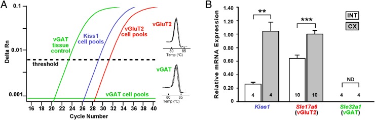
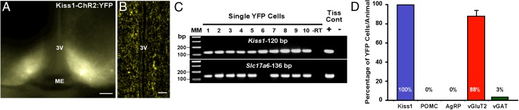
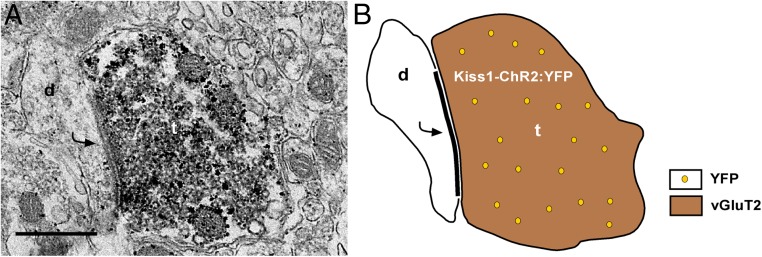
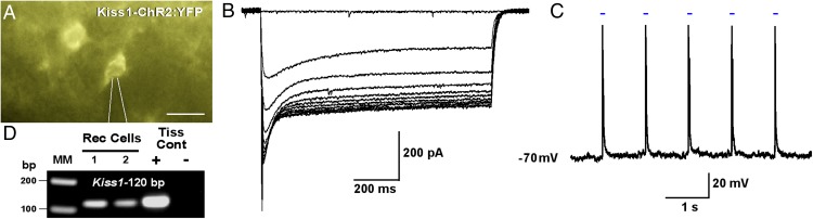
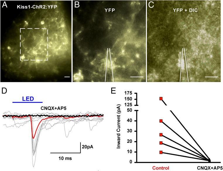
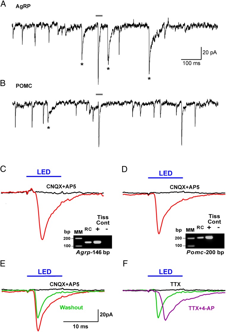
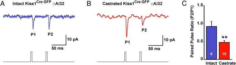
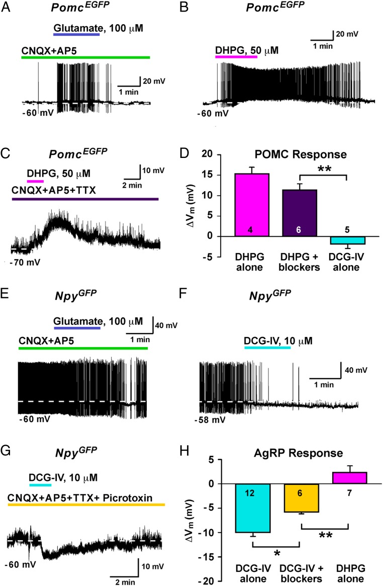
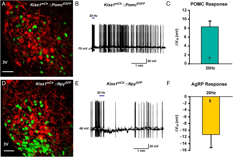
Kisspeptin (Kiss1) neurons are essential for reproduction, but their role in the control of energy balance and other homeostatic functions remains unclear. Proopiomelanocortin (POMC) and agouti-related peptide (AgRP) neurons, located in the arcuate nucleus (ARC) of the hypothalamus, integrate numerous excitatory and inhibitory inputs to ultimately regulate energy homeostasis. Given that POMC and AgRP neurons are contacted by Kiss1 neurons in the ARC (Kiss1(ARC)) and they express androgen receptors, Kiss1(ARC) neurons may mediate the orexigenic action of testosterone via POMC and/or AgRP neurons. Quantitative PCR analysis of pooled Kiss1(ARC) neurons revealed that mRNA levels for Kiss1 and vesicular glutamate transporter 2 were higher in castrated male mice compared with gonad-intact males. Single-cell RT-PCR analysis of yellow fluorescent protein (YFP) ARC neurons harvested from males injected with AAV1-EF1α-DIO-ChR2:YFP revealed that 100% and 88% expressed mRNAs for Kiss1 and vesicular glutamate transporter 2, respectively. Whole-cell, voltage-clamp recordings from nonfluorescent postsynaptic ARC neurons showed that low frequency photo-stimulation (0.5 Hz) of Kiss1-ChR2:YFP neurons elicited a fast glutamatergic inward current in POMC and AgRP neurons. Paired-pulse, photo-stimulation revealed paired-pulse depression, which is indicative of greater glutamate release, in the castrated male mice compared with gonad-intact male mice. Group I and group II metabotropic glutamate receptor agonists depolarized and hyperpolarized POMC and AgRP neurons, respectively, which was mimicked by high frequency photo-stimulation (20 Hz) of Kiss1(ARC) neurons. Therefore, POMC and AgRP neurons receive direct steroid- and frequency-dependent glutamatergic synaptic input from Kiss1(ARC) neurons in male mice, which may be a critical pathway for Kiss1 neurons to help coordinate energy homeostasis and reproduction.


Figures

Figure 1.
Figure 1.
Kiss1, vGluT2, and vGAT expression in Kiss1ARC neurons. A, qPCR assay with amplification curves for Kiss1, vGluT2, and vGAT (all analyzed in 5-cell pools). Cycle number of castrated males was plotted against the normalized fluorescence intensity (ΔRn) to visualize the PCR amplification. The cycle threshold (dashed line) is the point in the amplification at which sample values were calculated. Melting curves depict single-product melting at 82.8°C and 83.7°C for vGluT2 and vGAT, respectively, illustrating that only 1 product was formed from each transcript in the Kiss1ARC neuronal pools. B, Bar graph summarizing mRNA expression of Kiss1, Slc17a6 (vGluT2), and Slc32a1 (vGAT) from pooled Kiss1Cre:GFP neurons relative to the calibrator (see Materials and Methods) of gonad-intact (INT) and castrated (CX) male mice. Animal numbers are indicated. Using Newman-Keuls multiple comparison test; **, P < .01; ***, P < .001. Slc32a1 mRNA in pooled Kiss1ARC neurons was not detectable (ND).
Figure 2.
Figure 2.
ARC YFP cells contain mRNA for Kiss1 and vGluT2. A, Photomicrograph of the ARC 14 days after bilateral ARC injection of AAV1-EF1α-DIO-ChR2:YFP in a Kiss1Cre:GFP castrated male mouse at a low-power magnification (scale bar, 200 μm). 3V, third ventricle; ME, median eminence. B, Photomicrograph of the periventricular nucleus illustrating YFP fibers (scale bar, 20 μm). 3V, third ventricle. C, Representative gels illustrating mRNA expression of Kiss1 and Slc17a6 (vGluT2) in single YFP cells. The expected base pair (bp) sizes for Kiss1 and Slc17a6 are 120 and 136 bp, respectively. Exclusion of reverse transcriptase (−RT) in a reacted cell was used as a negative control. RNA extracted from the medial basal hypothalamic tissue was also included as positive (+, with RT) and negative (−, without RT) tissue controls. MM, molecular marker. D, Bar graph summarizing the percentage (mean ± SEM) of YFP cells that expressed mRNAs that encode for Kiss1, POMC, AgRP, vGluT2, and vGAT. Note: only 1 out of 30 cells expressed mRNA for vGAT.
Figure 3.
Figure 3.
EM revealed vGluT2-immunoreactivity in Kiss1ARC axon terminals. A, A dual-labeled ARC Kiss1-ChR2:YFP axon terminal (t) containing both vGluT2 (immunoperoxidase) and YFP (immunogold particles) and forms an asymmetrical synapse with a postsynaptic density (curved arrow) onto an unlabeled ARC dendrite (d). Scale bar, 500 nm. B, Schematic representation of A with a Kiss1ARC terminal (containing vGluT2 and YFP) forming an asymmetrical (glutamatergic) synapse with an unlabeled ARC dendrite.
Figure 4.
Figure 4.
Direct ChR2 activation of Kiss1ARC neurons. A, Photomicrograph showing a recording pipette (outlined by white lines) patched onto an ARC Kiss1-ChR2:YFP cell. Scale bar, 20 μm. B, Whole-cell voltage clamp recording (Vh = −60 mV) of light-evoked inward currents with LED intensities increasing from 33 to 330 μW. C, Whole-cell current clamp recording of depolarizations/action potentials induced by 1-Hz photo-stimulation (intensity, 660 μW). Blue bar above recording indicates LED stimulus. D, Representative gel illustrating Kiss1 mRNA expression in ChR2:YFP recorded cells (Rec Cells). RNA extracted from the medial basal hypothalamic tissue was included as positive (+, with RT) and negative (−, without RT) controls. MM, molecular marker.
Figure 5.
Figure 5.
Photo-stimulation of ARC Kiss1-ChR2:YFP neurons evoked excitatory glutamatergic PSCs. A, Low-power magnification photomicrograph of Kiss1-ChR2:YFP neurons in the ARC. Outlined area in A (scale bar, 20 μm) enlarged to show Kiss-ChR2:YFP projections (B) (YFP; scale bar, 20 μm) near a nonfluorescent postsynaptic cell (C) (YFP + DIC; scale bar, 20 μm). White lines outline recording pipette (B and C). D, Overlay of individual (gray) whole-cell recordings where photo-stimulation (intensity, 660 μW; duration, 10 ms) of ARC Kiss1-ChR2:YFP neurons induced PSCs in a nonfluorescent, ARC neuron. The averaged responses before (red trace) and after (black trace) the application of CNQX (10μM) and AP5 (50μM). Solid blue line above recordings indicates light stimulus. E, Line graph illustrates light-induced inward currents before (control) and after the application of CNQX and AP5 in 5 nonfluorescent ARC cells.
Figure 6.
Figure 6.
Photo-stimulation of glutamate, not GABA, release from Kiss1ARC neurons onto AgRP and POMC neurons. Whole-cell, voltage-clamp recording (Vh = −60 mV) using high chloride internal solution from an AgRP neuron (A) and a POMC neuron (B) near Kiss-ChR2:YFP fibers. Based on the decay time constant (13.1 ± 1.8 ms), GABA PSCs are noted with an asterisk. Averaged traces for photo-stimulation (intensity, 660 μW; duration, 10 ms) of ARC Kiss1-ChR2:YFP neurons while recording (high chloride internal solution) from an AgRP neuron (C) and a POMC neuron (D) before (red trace) and after (black trace) CNQX (10μM) and AP5 (50μM) application. E, Photo-stimulation after the washout (green trace) of CNQX and AP5 from (D). F, Photo-stimulation in the presence of TTX (black trace; 1μM) followed by photo-stimulation with addition of 4-AP (purple trace; 100μM). The average of recordings for each experiment is displayed as a trace line with color (red, black, green, or purple). Cells were identified post hoc using scRT-PCR. RC, recorded cell. Gray bar above recordings in A and B correspond to recordings in C and D. Blue bar above recordings indicate LED stimulus (C–F). Scale in E also applies to C–F.
Figure 7.
Figure 7.
Steroid-dependent change in glutamate release from Kiss1ARC neurons. Whole-cell, voltage-clamp recording (Vh = −60 mV) of nonfluorescent AgRP neurons (post hoc identified) using a photo-stimulation paired-pulse paradigm (pulse duration, 5 ms; interpulse interval, 50 ms) in male Kiss1Cre:GFP::Ai32 mice and AAV-EF1α-DIO-ChR2:YFP injected Kiss1Cre:GFP mice. A representative trace that averages 10 sequential paired-pulse recordings is shown for a gonad-intact (A) and castrated (B) male Kiss1Cre:GFP::Ai32 mouse. C, Bar graph summarizes the averaged (±SEM) PPR (pulse 2 amplitude to pulse 1 amplitude) of the induced EPSC from gonad-intact and castrated male Kiss1Cre:GFP::Ai32 and AAV injected Kiss1Cre:GFP mice. Cell numbers are indicated. Using an unpaired Student's t test; **, P < .01.
Figure 8.
Figure 8.
Glutamate differentially regulates POMC and AgRP neurons via mGluR signaling. A, Whole-cell current clamp recording of a PomcEGFP neuron in the presence of CNQX (10μM) and AP5 (50μM) with the fast perfusion of glutamate (100μM). Whole-cell current clamp recording of a PomcEGFP neuron with (B) DHPG (50μM) alone or (C) DHPG in the presence of CNQX (10μM), AP5 (50μM), and TTX (1μM). D, Bar graph summarizing the effect of DHPG alone, DHPG with blockers (CNQX, AP5, and TTX), and DCG-IV alone on POMC neurons. Cell numbers are indicated. E, Whole-cell current clamp recording of an ARC NpyGFP neuron in the presence of CNQX (10μM) and AP5 (50μM) with the fast perfusion of glutamate (100μM). Whole-cell current clamp recording of an ARC NpyGFP neuron with (F) DCG-IV (10μM) alone or (G) DCG-IV (10μM) in the presence of CNQX (10μM), AP5 (50μM), TTX (1μM), and picrotoxin (100μM). H, Bar graph summarizing the effect of DCG-IV alone, DCG-IV with blockers (CNQX, AP5, and TTX), and DHPG alone on AgRP neurons. Cell numbers are indicated. Using Newman-Keuls multiple comparison test; *, P < .05; **, P < .01.
Figure 9.
Figure 9.
High frequency photo-stimulation of ChR2 Kiss1ARC neurons stimulates POMC neurons and inhibits AgRP neurons. A, Representative confocal image of the ARC from a Kiss1Cre:GFP::PomcEGFP male mouse injected with AAV1-EF1α-DIO-ChR2:mCherry. Scale bar, 50 μm. 3V, third ventricle. B, High frequency photo-stimulation of ChR2 (20 Hz, solid blue bar) evoked a slow depolarization (EPSP) in a PomcEGFP neuron. C, Bar graph summarizing the effect of 20-Hz photo-stimulation in POMC neurons (n = 7). D, Representative confocal image of the ARC from a Kiss1Cre:GFP::NpyGFP male mouse injected with AAV1-EF1α-DIO-ChR2:mCherry. Scale bar, 50 μm. 3V, third ventricle. E, High frequency photo-stimulation of ChR2 (20 Hz, solid blue bar) evoked a slow hyperpolarization (IPSP) in an ARC NpyGFP neuron. F, Bar graph summarizing the effect of 20 Hz LED stimulation in AgRP neurons (n = 5). Note the hyperpolarization typical for activation of Gαi.o-coupled receptors.

References

    1. Seminara SB, Messager S, Chatzidaki EE, et al. The GPR54 gene as a regulator of puberty. N Engl J Med. 2003;349:1614–1627. - PubMed
    1. de Roux N, Genin E, Carel JC, Matsuda F, Chaussain JL, Milgrom E. Hypogonadotropic hypogonadism due to loss of function of the KiSS1-derived peptide receptor GPR54. Proc Natl Acad Sci USA. 2003;100:10972–10976. - PMC - PubMed
    1. Mayer C, Boehm U. Female reproductive maturation in the absence of kisspeptin/GPR54 signaling. Nat Neurosci. 2011;14:704–710. - PubMed
    1. de Bond JA, Smith JT. Kisspeptin and energy balance in reproduction. Reproduction. 2014;147:R53–R63. - PubMed
    1. Roa J, Tena-Sempere M. Connecting metabolism and reproduction: roles of central energy sensors and key molecular mediators. Mol Cell Endocrinol. 2014;397:4–14. - PubMed

MeSH terms