. 2016 Apr 19;12(4):e1005581.
doi: 10.1371/journal.ppat.1005581. eCollection 2016 Apr.

Sulforaphane Inhibits HIV Infection of Macrophages through Nrf2

Affiliations

Sulforaphane Inhibits HIV Infection of Macrophages through Nrf2

Andrea Kinga Marias Furuya et al. PLoS Pathog. .

Abstract

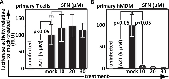
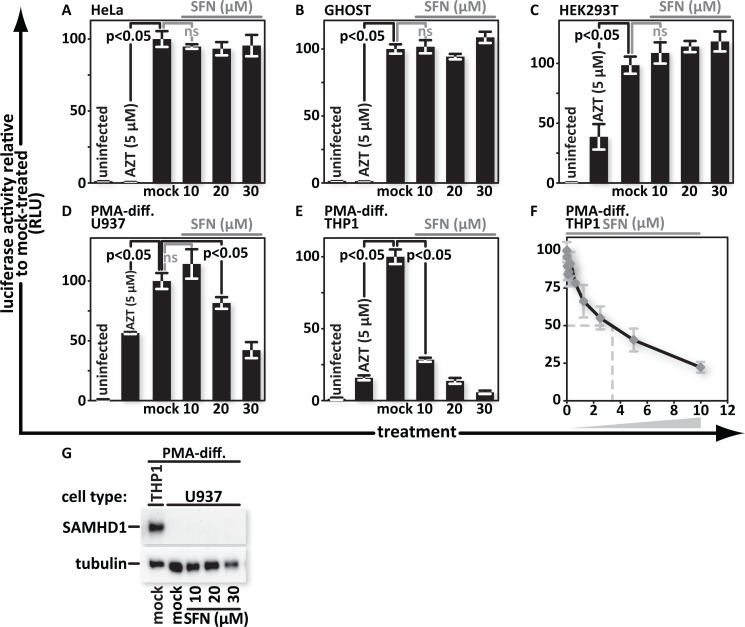
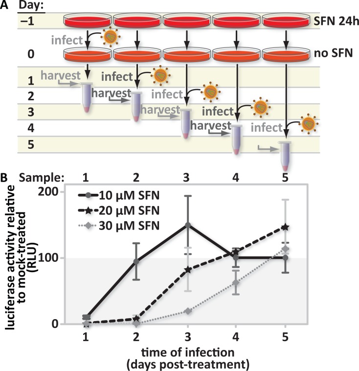
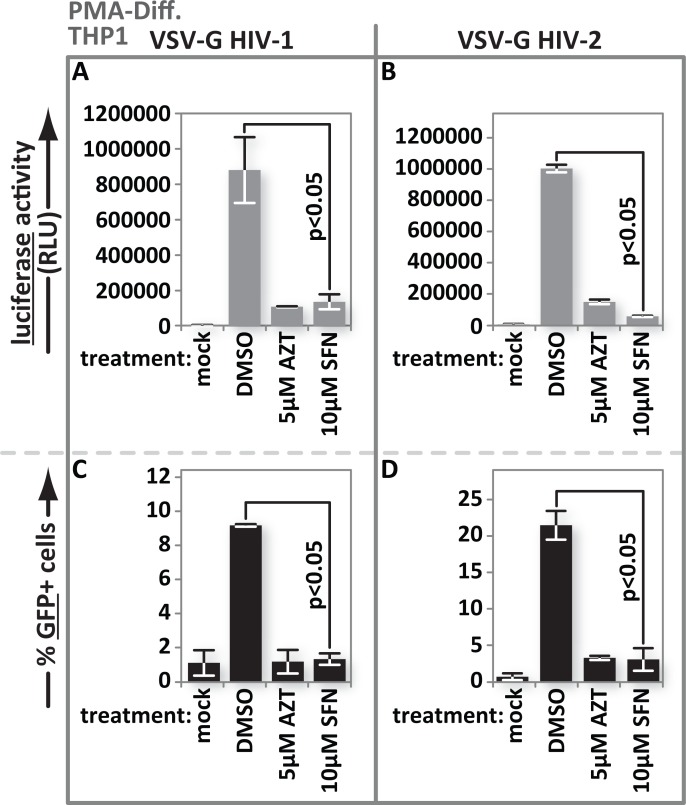
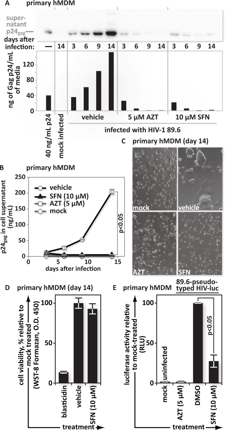
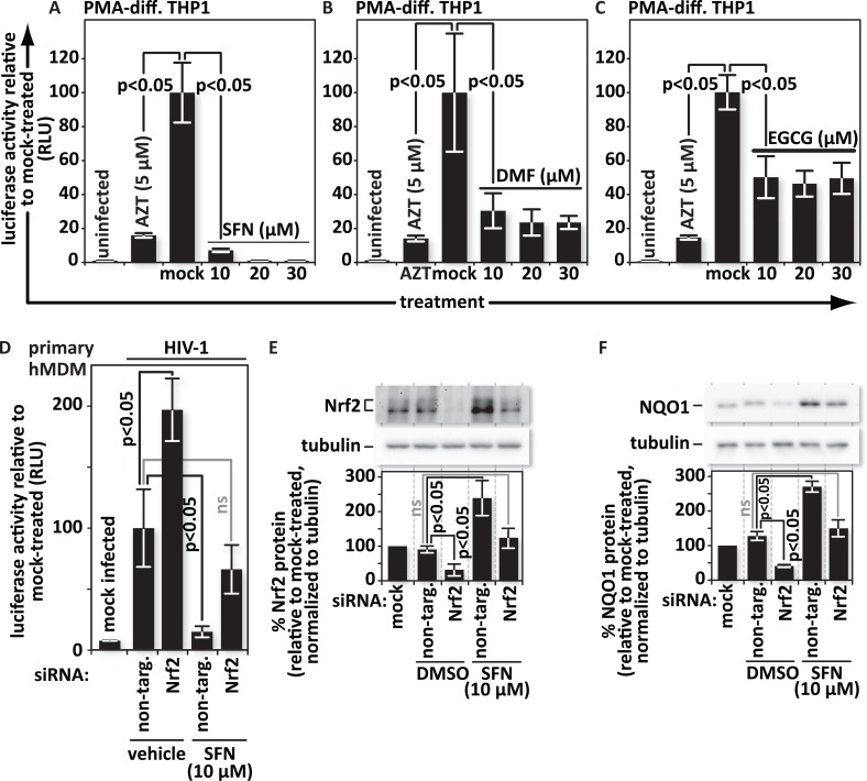
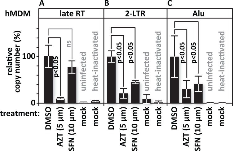
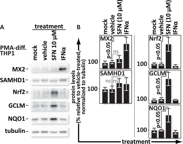
Marburg virus, the Kaposi's sarcoma-associated herpesvirus (KSHV) and Dengue virus all activate, and benefit from, expression of the transcription regulator nuclear erythroid 2-related factor 2 (Nrf2). The impact of Nrf2 activation on human immunodeficiency virus (HIV) infection has not been tested. Sulforaphane (SFN), produced in cruciferous vegetables after mechanical damage, mobilizes Nrf2 to potently reprogram cellular gene expression. Here we show for the first time that SFN blocks HIV infection in primary macrophages but not in primary T cells. Similarly SFN blocks infection in PMA-differentiated promonocytic cell lines, but not in other cell lines tested. siRNA-mediated depletion of Nrf2 boosted HIV infectivity in primary macrophages and reduced the anti-viral effects of SFN treatment. This supports a model in which anti-viral activity is mediated through Nrf2 after it is mobilized by SFN. We further found that, like the type I interferon-induced cellular anti-viral proteins SAMHD1 and MX2, SFN treatment blocks infection after entry, but before formation of 2-LTR circles. Interestingly however, neither SAMHD1 nor MX2 were upregulated. This shows for the first time that Nrf2 action can potently block HIV infection and highlights a novel way to trigger this inhibition.


Conflict of interest statement

The authors have declared that no competing interests exist.

Figures

Fig 1
Fig 1. SFN blocks HIV in primary macrophages but not primary T cells.
(A), Primary T cells and (B), hMDMs were treated with media supplemented with vehicle only (DMSO) or with 10 μM, 20 μM or 30 μM SFN. Pretreatment of cultures with 5 μM of the reverse transcription inhibitor zidovudine (AZT) served as a positive control for viral inhibition. Twenty-four hours after treatment, cells were either mock infected or infected with VSV-G-pseudotyped HIV-1, encoding firefly luciferase in place of nef. Twenty-four hours after infection, the cells were harvested and luciferase activity was measured by photon emission. The bar graphs represent the data for replicate experiments (n = 3).
Fig 2
Fig 2. SFN blocks HIV infection in monocytoid cell lines but does not increase SAMHD1 expression.
(A), HeLa, (B), GHOST, (C), HEK293T, (D), PMA-differentiated U937, and (E), PMA-differentiated THP1, cells were treated with media supplemented with vehicle only (DMSO) or with 10 μM, 20 μM or 30 μM SFN. Pretreatment of cell cultures with 5 μM AZT served as a positive control for viral inhibition. Twenty-four hours after treatment, the samples were infected with VSV-G pseudotyped HIV-1 encoding firefly luciferase in place of nef. Twenty-four hours after infection, the cultures were harvested and luciferase activity was measured by photon emission. (F), PMA-differentiated THP1 cells were treated with SFN that underwent a twofold serial dilution with 10μM of SFN being the highest concentration. Twenty-four hours after treatment, the samples were either mock infected or infected with VSV-G pseudotyped HIV-1 encoding firefly luciferase in place of nef. Twenty-four hours after infection, luciferase activity was measured. The bar graphs represent the data for replicate experiments (n = 3). (G), SAMHD1 protein was detected by western blot analysis from lysates of mock- and SFN treated PMA-differentiated U937 cells. Lysates from mock-treated, PMA-differentiated THP1 cells served as a positive control for SAMHD1 production.
Fig 3
Fig 3. The SFN-mediated HIV infection block is reversible.
(A), Study design: PMA-differentiated THP1 cells were pretreated with media supplemented with vehicle only (DMSO) or with 10 μM, 20 μM or 30 μM SFN. After twenty-four hours of treatment, the culture media was replaced with fresh SFN-free media. Cultures were infected 1, 2, 3, 4, or 5 days after treatment with freshly thawed aliquots of VSV-G pseudotyped HIV-1 encoding firefly luciferase in place of nef. Twenty-four hours after the start of each time-point infection, the cells were harvested and (B), luciferase activity was measured by photon emission. The bar graph represents the data for replicate experiments (n = 3).
Fig 4
Fig 4. SFN action impacts HIV-2 as well as HIV-1 and is not reporter dependent.
PMA-differentiated THP1 cells were treated with media supplemented with vehicle only (DMSO), 5 μM AZT or with 10 μM SFN. Twenty-four hours after treatment, the samples were either mock infected or infected with (A), VSV-G pseudotyped HIV-1 encoding firefly luciferase in place of nef or (B), VSV-G pseudotyped HIV-2 encoding firefly luciferase in place of nef. Twenty-four hours after infection, luciferase activity was measured by photon emission. (C), In parallel, the same experiment as in (A) and (B) was performed except that THP1 cells were infected with VSV-G-pseudotyped HIV-1 with GFP in place of nef or (D), VSV-G pseudotyped HIV-2 with GFP in place of nef. The samples with GFP-reporter viruses were fixed and harvested 24 h after infection and the fraction of GFP(+) cells was enumerated by flow cytometry. Bar graphs represent the data for replicate experiments (n = 3).
Fig 5
Fig 5. SFN action blocks spreading infections that rely on the HIV envelope for viral entry.
hMDMs were pretreated with vehicle (DMSO), 5 μM AZT, or 10 μM SFN. All cultures were subsequently maintained in their respective treatments for the duration of the experiment. Twenty four hours after initial treatment, the cultures were infected with the HIV-1 clinical isolate 89.6. Culture supernatants were collected 3, 6, 9, and 14 days after infection. (A), Western blots and (B), p24 antigen ELISA assays of viral supernatants were performed. (C), Fourteen days after infection, cells were imaged using phase contrast microscopy. (D), Uninfected replicate cultures were maintained in the presence of vehicle (DMSO) or in 10 μM SFN. After 14 days of treatment, the viability of each cell type was assessed under each condition by measuring water-soluble tetrazolium salt (WST-8) formazan reagent cleavage by cellular dehydrogenases. Continuous treatment of cells with 10 μg/ml of the eukaryotic toxin blasticidin served as a positive control to demonstrate loss of viability. (E), hMDMs were infected with 89.6-Env-pseudotyped HIV-1 encoding firefly luciferase in place of nef. The bar graphs represent the data for replicate experiments (n = 3).
Fig 6
Fig 6. SFN acts through Nrf2 to block HIV infection in macrophages.
PMA-differentiated THP1 cells were pretreated for twenty-four hours with the Nrf2 activators: (A), SFN, (B), DMF, or (C), EGCG at the indicated concentrations. Pretreatment of cultures with 5 μM of the reverse transcription inhibitor zidovudine (AZT) served as a positive control for viral inhibition. Twenty-four hours after treatment, cells were either mock infected or infected with VSV-G-pseudotyped HIV-1 encoding firefly luciferase in place of nef. Twenty-four hours after infection, the cells were harvested and luciferase activity was measured by photon emission. The bar graphs represent the data for replicate experiments (n = 3). (D), Cultures of hMDMs were transfected with either a non-targeting siRNA (control) or siRNA specific for Nrf2 mRNA. siRNA transfected hMDMs were either treated with vehicle (DMSO) or with 10 μM SFN. Twenty-four hours after treatment, the cells were either mock infected or infected with VSV-G-pseudotyped HIV-1 encoding firefly luciferase in place of nef. Twenty-four hours after infection, the cells were harvested and luciferase activity was measured by photon emission. The bar graphs represent the quantified data for replicate experiments (n = 3). (E) and (F), Representative samples from (D) were lysed and proteins from whole cell lysates were resolved by SDS-PAGE and identified by western blotting using antibodies with the indicated specificities. Densitometric analysis was performed on the Nrf2 and NQO1 (an indicator of Nrf2 function) bands and normalized to the values of the corresponding tubulin bands. The relative normalized intensities of the Nrf2 and NQO1 bands were then graphed. The data shown is representative of n = 3.
Fig 7
Fig 7. SFN blocks infection after entry and but before 2-LTR circle formation.
Replicate cultures of hMDMs were pretreated with vehicle (DMSO)-containing media, with 5 μM AZT or with 10 μM SFN. Twenty four hours after treatment, the samples were infected with VSV-G-pseudotyped HIV-1 encoding GFP in place of nef. Cultures treated with heat-inactivated virus served as controls for plasmid carry over and for impaired viral entry. Cells were harvested and DNA was isolated 24 hours after infection. Viral DNA products were detected by real-time PCR using primer sets specific for the indicated stage of reverse transcription. (A), Relative quantities of late reverse transcription products, (B), 2-LTR circles, and (C), integrated proviruses. The bar graph represents the data for replicate experiments (n = 3).
Fig 8
Fig 8. SFN does not trigger expression of interferon-stimulated anti-viral factors SAMHD1 or MX2.
PMA-differentiated THP1 cells were mock-treated or treated with media supplemented with vehicle only (DMSO) or with 10 μM SFN or with 500 U/mL of IFNα. (A), Proteins from whole cell lysates were resolved by SDS-PAGE and identified by western blotting using antibodies with the indicated specificities. (B), Densitometric analysis was performed on the Nrf2, SAMHD1, MX2, NQO1 and GCLM (NQO1 and GCLM are both indicators of Nrf2 function) bands and normalized to the values of the corresponding tubulin bands. The relative normalized intensities of the bands were then graphed.

Similar articles

Cited by

References

    1. Gartner S, Markovits P, Markovitz DM, Kaplan MH, Gallo RC, Popovic M. The role of mononuclear phagocytes in HTLV-III/LAV infection. Science. 1986;233(4760):215–9. Epub 1986/07/11. . - PubMed
    1. Sharova N, Swingler C, Sharkey M, Stevenson M. Macrophages archive HIV-1 virions for dissemination in trans. EMBO J. 2005;24(13):2481–9. Epub 2005/05/28. doi: 7600707 [pii] 10.1038/sj.emboj.7600707 - DOI - PMC - PubMed
    1. Gonzalez-Scarano F, Martin-Garcia J. The neuropathogenesis of AIDS. Nat Rev Immunol. 2005;5(1):69–81. Epub 2005/01/05. doi: nri1527 [pii] 10.1038/nri1527 . - DOI - PubMed
    1. Rich EA, Chen IS, Zack JA, Leonard ML, O'Brien WA. Increased susceptibility of differentiated mononuclear phagocytes to productive infection with human immunodeficiency virus-1 (HIV-1). The Journal of Clinical Investigation. 1992;89(1):176–83. Epub 1992/01/01. 10.1172/JCI115559 - DOI - PMC - PubMed
    1. Sonza S, Maerz A, Deacon N, Meanger J, Mills J, Crowe S. Human immunodeficiency virus type 1 replication is blocked prior to reverse transcription and integration in freshly isolated peripheral blood monocytes. J Virol. 1996;70(6):3863–9. Epub 1996/06/01. - PMC - PubMed

Publication types