Review
. 2016 Jun;17(6):350-63.
doi: 10.1038/nrm.2016.37. Epub 2016 Apr 20.

Processing ribonucleotides incorporated during eukaryotic DNA replication

Affiliations
Review

Processing ribonucleotides incorporated during eukaryotic DNA replication

Jessica S Williams et al. Nat Rev Mol Cell Biol. 2016 Jun.

Abstract

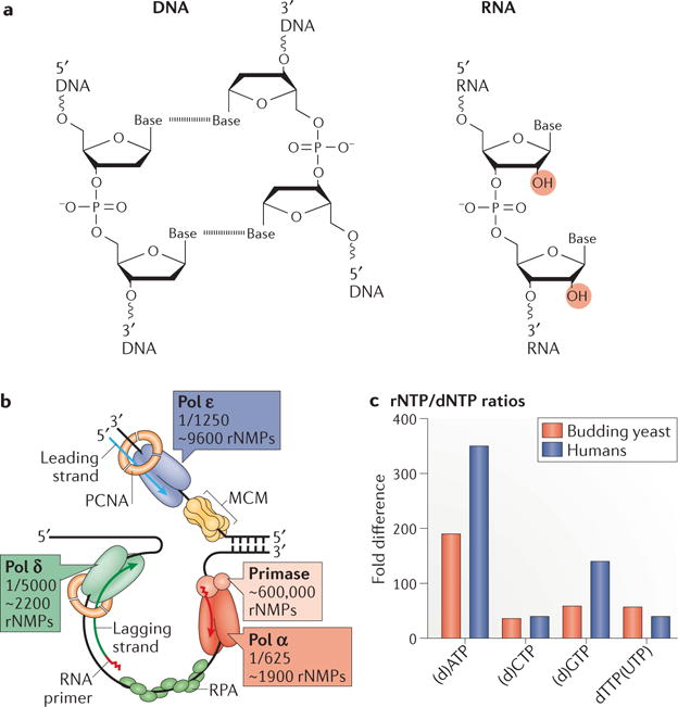
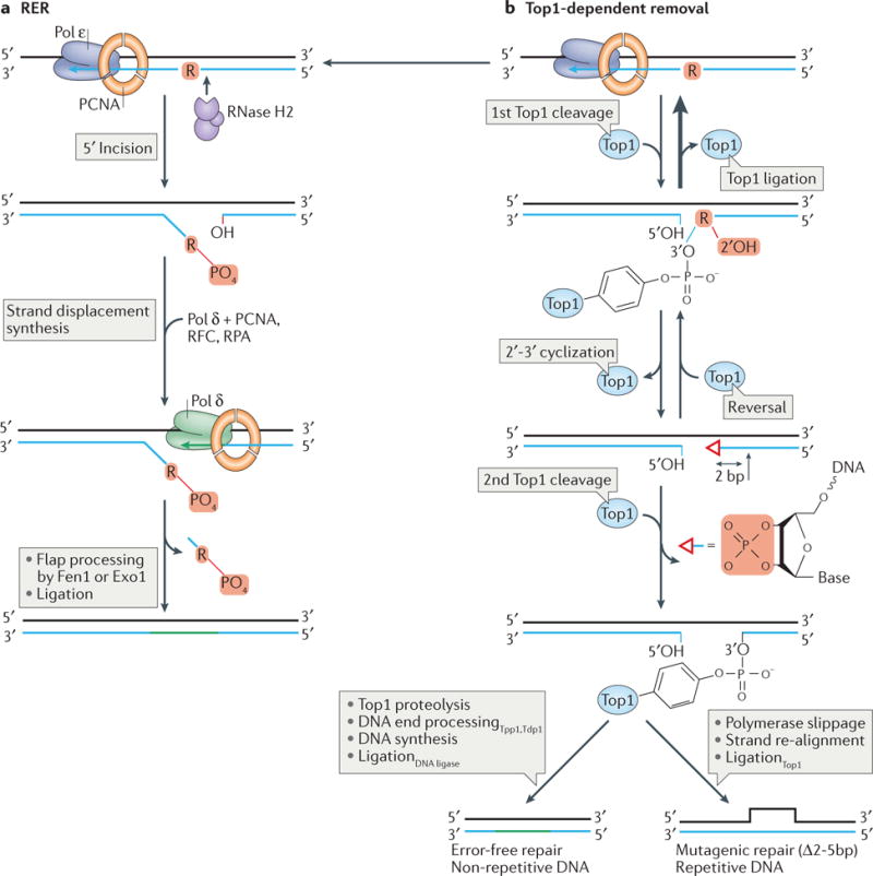
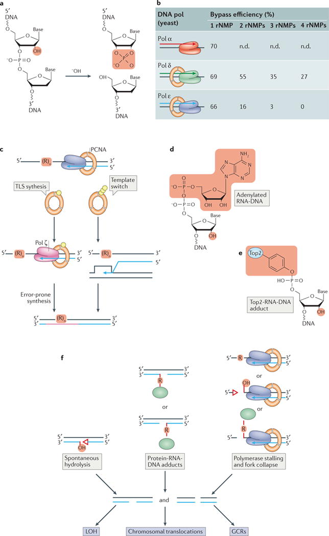
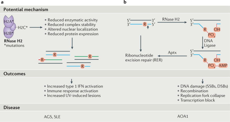
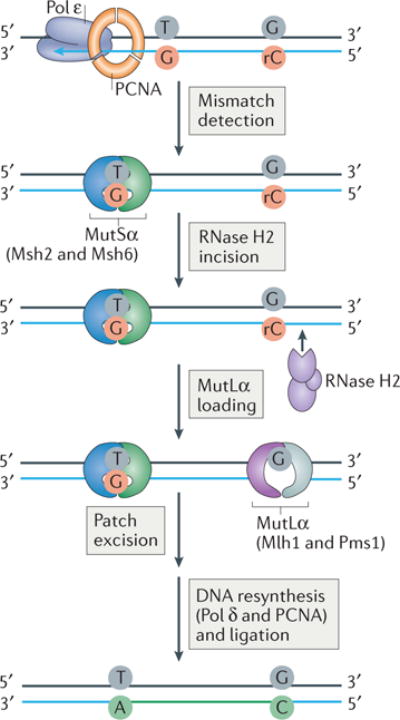
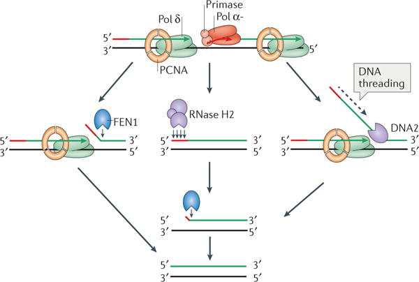
The information encoded in DNA is influenced by the presence of non-canonical nucleotides, the most frequent of which are ribonucleotides. In this Review, we discuss recent discoveries about ribonucleotide incorporation into DNA during replication by the three major eukaryotic replicases, DNA polymerases α, δ and ε. The presence of ribonucleotides in DNA causes short deletion mutations and may result in the generation of single- and double-strand DNA breaks, leading to genome instability. We describe how these ribonucleotides are removed from DNA through ribonucleotide excision repair and by topoisomerase I. We discuss the biological consequences and the physiological roles of ribonucleotides in DNA, and consider how deficiencies in their removal from DNA may be important in the aetiology of disease.


Conflict of interest statement

Competing interests statement

The authors declare no competing interests.

Figures

Figure 1
Figure 1. Ribonucleotide incorporation into DNA by polymerases
a | A comparison of the chemical structures of DNA and RNA. The dashed line between bases indicates hydrogen bonding in the double stranded DNA structure. The presence of a 2′-OH group on the ribose ring (highlighted in red) is the distinguishing feature that renders RNA less stable than DNA. b | A model of the division of labor at the budding yeast replication fork. RNA primase initiates synthesis of both strands by laying down a short RNA primer which is extended by the major leading strand polymerase, Polymerase ε (Pol ε), in a continuous fashion on the leading strand. On the lagging strand, the polymerase activity of Pol α takes over for a short stretch which is then extended by Pol δ to synthesize Okazaki fragments which are subsequently processed and joined during Okazaki fragment maturation. Both in vitro and in vivo studies have demonstrated the propensity of DNA polymerases to incorporate a significant number of ribonucleotides during synthesis. The fraction represents one ribonucleotide incorporated per x number of deoxyribonucleotides. The estimates of the total number of ribonucleotides (as ribonucleoside monophosphates (rNMPs)) incorporated into the 12 million base pair S. cerevisiae genome are indicated for leading and lagging strands. PCNA, proliferating cell nuclear antigen, a sliding clamp that binds to Pols δ and ε and enhances their processivity; RPA, replication protein A, single-strand DNA binding protein; MCM, minichromosome maintenance protein that serves as the eukaryotic replicative helicase complex. c | The nucleotide pool imbalances present in asynchronous cycling S. cerevisiae and human cells. Displayed is the ribonucleotide (rNTP) to deoxyribonucleotide (dNTP) ratio for each.
Figure 2
Figure 2. Ribonucleotide removal during ribonucleotide excision repair (RER) or Topoisomerase 1 (Top1)-processing
a | RER is initiated when RNase H2 incises 5′ to an embedded ribonucleotide. This is followed by a nick-translation reaction during which the nicked strand is displaced, allowing Pol δ (or Pol ε) in complex with proliferating cell nuclear antigen (PCNA) to bind and fill the gap. Replication factor C (RFC) is an ATP-dependent PCNA clamp-loader and RPA is a single-strand DNA binding protein. The flap generated following strand displacement is then nucleolytically-processed by the flap structure-specific endonuclease 1 (Fen1) or exonuclease 1 (Exo1). Although it is not depicted here, RER is important for removal of ribonucleotides incorporated during both leading and lagging strand DNA synthesis. b | In addition to RNase H2, Top1 is also able to cleave at genomic ribonucleotides. Top1 can incise the DNA on the 3′ side of a ribonucleotide, creating a nick that is typically reversed by Top1-mediated ligation. This reversal generates the original ribonucleotide-containing substrate and allows for another repair opportunity via RER (panel a). Alternatively, as occurs in cells that are RER-deficient, nucleophilic attack by the 2′-OH group on the ribose generates a 2′-3′-cyclic-PO4 (depicted by the open red triangle). This cyclic product can either be reversed through the activity of Top1 or efficiently removed following a second, this time irreversible Top1 cleavage reaction two-nucleotides upstream (5′) of the first cleavage site to generate a small gap. When the ribonucleotide is located in non-repetitive DNA, repair can proceed in an error-free fashion through the action of Top1-proteolysis and DNA end-processing enzymes (tyrosyl-DNA phosphodiesterase 1 (Tdp1), which hydrolyses the DNA-tyrosyl bond, and three prime phosphatase 1 (Tpp1), which processes the 3′ end) together with DNA polymerase(s) and DNA ligase to ultimately seal the nick. When the ribonucleotide is incorporated into a region containing repetitive DNA, mutagenic repair can occur to create a deletion of 2–5 base pairs (Δ2–5bp) in size, depending on the size of the repeat unit. This occurs as a result of re-alignment of the DNA strands followed by Top1-mediated ligation across the gap to generate a deletion of one of the repeat units. Not depicted here is the ability of Exo1 nuclease and the Srs2 helicase to enlarge this gap to prevent deletion mutations.
Figure 3
Figure 3. Consequences of unrepaired ribonucleotides in DNA
a | Hydrolysis at a ribonucleotide (occurring spontaneously or induced by alkali treatment) generates a ‘dirty’ DNA single strand break containing unligatable 2′–3′-cyclic-PO4 and 5′-OH termini. Not shown is the other intact DNA strand in the duplex molecule. b | DNA polymerases can bypass ribonucleotides in template DNA with varying proficiencies. This is dependent on the polymerase and the number of consecutive ribonucleotides present in the DNA as well as the identity of the base and the sequence context (not included in the table); n.d., not determined. c | In the absence of RNase H activity, the presence of ribonucleotides in template DNA causes DNA polymerase stalling and activates post-replication DNA repair (PRR) through ubiquitylation (yellow spheres) of proliferating cell nuclear antigen (PCNA). Through poly-ubiquitylation of PCNA ribonucleotide bypass can be achieved by a template switch mechanism. This is a form of DNA damage-tolerance in which a switch in template strands occurs during replication in order to bypass DNA damage and ensure completion of replication (right panel). Monoubiquitylation of PCNA initiates translesion DNA synthesis (TLS, left panel). This involves the recruitment of TLS polymerases such as Polymerase ζ (Pol ζ), which can bypass the lesion. However, Pol ζ lacks proofreading activity and has relatively low fidelity, causing this synthesis to be error-prone and thus mutagenic. d and e | Formation of compound DNA lesions is promoted by unrepaired genomic ribonucleotides. d | An abortive ligation lesion, adenylated RNA–DNA (5′-AMP-RNA-DNA), is formed following failure of DNA ligase to seal an RNase H2-generated nick. Highlighted in red are the adenylate group (top) and the ribose 2′-OH group (bottom). e | An irreversible topoisomerase 2 (Top2)–RNA–DNA adduct is formed when Top2 incises 5′ to a ribonucleotide. Highlighted in red are the covalently-linked Top2 adduct (top) and the ribose 2′-OH group (bottom). f | Failure of RNase H2-dependent repair can lead to spontaneous DNA hydrolysis (left), formation of protein–RNA–DNA adducts (green ellipses in the middle; see also panel e) or DNA (or RNA) polymerase stalling and fork collapse when encountering a ribonucleotide or ribonucleotide-provoked lesion (right). These events can trigger the formation of DNA single- (SSBs) and double-strand breaks (DSBs) that promote recombination and various genome rearrangements observed in yeast and mouse cells deficient in RNase H2. LOH, loss of heterozygosity; GCRs, gross chromosomal rearrangements.
Figure 4
Figure 4. The connections between genomic ribonucleotides and human disease
A schematic diagram illustrating the potential mechanisms, outcomes, and diseases associated with failure of ribonucleotide removal or processing. a | Mutations in RNase H2 are associated with Aicardi Goutières Syndrome (AGS) and Systemic Lupus Erythematosus (SLE). Patients suffering from AGS display increased type 1 interferon (IFN) activation, an immune response potentially related to the accumulation of aberrant ribonucleotide-containing nucleic acid species upon reduction of RNase H2-dependent repair. This reduced RNase H2 function may be related to the fact that these mutations in RNase H2 perturb enzymatic activity, decrease protein expression, affect localization to replication and repair sites and alter complex assembly and stability,,. Photosensitivity and skin disease are common in SLE patients, and the demonstration of an increase in ultraviolet (UV)-induced lesions in both AGS and SLE patient cells raises the possibility that unrepaired genomic ribonucleotides renders DNA more susceptible to UV-induced DNA damage. b | Mutations in Aprataxin (Aptx) are associated with the neurological disease, Ataxia with Oculomotor Apraxia 1 (AOA1). Following RNase H2 incision at a ribonucleotide in DNA to initiate ribonucleotide excision repair (RER), premature engagement of this RNA-DNA substrate by DNA ligase triggers abortive ligation and transfer of an adenylate (AMP) group from DNA ligase to the RNA-DNA junction to generate a compound 5′-AMP-RNA-DNA lesion. Failure of removal of this bulky adenylate group by Aprataxin causes accumulation of adenylated RNA-DNA and may lead to the formation of DNA breaks, affect processes such as replication and transcription, and contribute to AOA1.
Figure 5
Figure 5. Ribonucleotides act as a strand-discrimination signal during DNA mismatch repair (MMR)
A model depicting how ribonucleotides incorporated into DNA by Polymerase ε (Pol ε) during leading strand synthesis may provide a signal for MMR in yeast. MMR involves mismatch recognition by MutSα, a heterodimer of Msh2 and Msh6, which are homologs of the bacterial MutS protein required for MMR. This recognition is followed by mismatch removal by MutLα (comprised of a heterodimer of Mlh1 and (yeast) Pms1), which are homologs of bacterial MutL.An entry point for MutLα is provided by RNase H2-mediated incision at a ribonucleotide incorporated into DNA by the replicative polymerase (Pol ε) during DNA replication to create a nick. Following MutLα loading at the incision site, excision of the mismatch and correct re-synthesis of the DNA by Pol δ or ε is followed by ligation to complete repair. Thus, the incorporated ribonucleotide serves to distinguish the newly synthesized mismatch-containing strand from the template strand.
None

References

    1. Kuchta RD, Stengel G. Mechanism and evolution of DNA primases. Biochim Biophys Acta. 2010;1804:1180–1189. - PMC - PubMed
    1. Martinez-Jimenez MI, et al. Alternative solutions and new scenarios for translesion DNA synthesis by human PrimPol. DNA Repair (Amst) 2015;29:127–138. - PubMed
    1. Goodman MF, Woodgate R. Translesion DNA polymerases. Cold Spring Harb Perspect Biol. 2013;5:a010363. - PMC - PubMed
    1. Boiteux S, Jinks-Robertson S. DNA repair mechanisms and the bypass of DNA damage in Saccharomyces cerevisiae. Genetics. 2013;193:1025–1064. - PMC - PubMed
    1. Sollier J, Cimprich KA. Breaking bad: R-loops and genome integrity. Trends Cell Biol. 2015;25:514–522. - PMC - PubMed

LinkOut - more resources