. 2016 Apr 19;7(2):e01388-15.
doi: 10.1128/mBio.01388-15.

The Eng1 β-Glucanase Enhances Histoplasma Virulence by Reducing β-Glucan Exposure

Affiliations

The Eng1 β-Glucanase Enhances Histoplasma Virulence by Reducing β-Glucan Exposure

Andrew L Garfoot et al. mBio. .

Abstract

The fungal pathogen Histoplasma capsulatum parasitizes host phagocytes. To avoid antimicrobial immune responses, Histoplasma yeasts must minimize their detection by host receptors while simultaneously interacting with the phagocyte. Pathogenic Histoplasma yeast cells, but not avirulent mycelial cells, secrete the Eng1 protein, which is a member of the glycosylhydrolase 81 (GH81) family. We show that Histoplasma Eng1 is a glucanase that hydrolyzes β-(1,3)-glycosyl linkages but is not required for Histoplasma growth in vitro or for cell separation. However, Histoplasma yeasts lacking Eng1 function have attenuated virulence in vivo, particularly during the cell-mediated immunity stage. Histoplasma yeasts deficient for Eng1 show increased exposure of cell wall β-glucans, which results in enhanced binding to the Dectin-1 β-glucan receptor. Consistent with this, Eng1-deficient yeasts trigger increased tumor necrosis factor alpha (TNF-α) and interleukin-6 (IL-6) cytokine production from macrophages and dendritic cells. While not responsible for large-scale cell wall structure and function, the secreted Eng1 reduces levels of exposed β-glucans at the yeast cell wall, thereby diminishing potential recognition by Dectin-1 and proinflammatory cytokine production by phagocytes. In α-glucan-producing Histoplasma strains, Eng1 acts in concert with α-glucan to minimize β-glucan exposure: α-glucan provides a masking function by covering the β-glucan-rich cell wall, while Eng1 removes any remaining exposed β-glucans. Thus, Histoplasma Eng1 has evolved a specialized pathogenesis function to remove exposed β-glucans, thereby enhancing the ability of yeasts to escape detection by host phagocytes.

Importance: The success of Histoplasma capsulatum as an intracellular pathogen results, in part, from an ability to minimize its detection by receptors on phagocytic cells of the immune system. In this study, we showed that Histoplasma pathogenic yeast cells, but not avirulent mycelia, secrete a β-glucanase, Eng1, which reduces recognition of fungal cell wall β-glucans. We demonstrated that the Eng1 β-glucanase promotes Histoplasma virulence by reducing levels of surface-exposed β-glucans on yeast cells, thereby enabling Histoplasma yeasts to escape detection by the host β-glucan receptor, Dectin-1. As a consequence, phagocyte recognition of Histoplasma yeasts is reduced, leading to less proinflammatory cytokine production by phagocytes and less control of Histoplasma infection in vivo Thus, Histoplasma yeasts express two mechanisms to avoid phagocyte detection: masking of cell wall β-glucans by α-glucan and enzymatic removal of exposed β-glucans by the Eng1 β-glucanase.


Figures

FIG 1
FIG 1
Histoplasma ENG1 expression is enriched in cells in the pathogenic phase. Relative expression of putative Histoplasma endoglucanase genes in the virulent (yeast) and avirulent (mycelia) phases are indicated. Gene expression levels were determined by qRT-PCR of RNA harvested from wild-type G186A yeast-phase or mycelial-phase Histoplasma cultures. Gene expression levels were normalized to the level seen with the constitutively expressed TEF1 gene, and the fold change in specific mRNA levels (yeast phase relative to mycelial phase) was determined. Yeast-phase enrichment was confirmed by expression of the CBP1 yeast-phase-specific gene. Error bars indicate the standard deviations of results from biological replicates (n = 3).
FIG 2
FIG 2
Depletion of Eng1 does not impair cell separation. Data represent distributions of the number of yeast cells per cluster for Eng1-producing (ENG1) and Eng1-deficient (ENG1-RNAi) yeasts from the G217B background in late-exponential-stage growth. The numbers of yeasts per cluster were quantified by microscopy (n >150 clusters per strain). Data represent the average percentage of each class, and error bars represent the standard deviations of results from biological replicates for each line (n = 3).
FIG 3
FIG 3
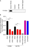
Eng1 is an extracellular β-(1,3)-glucanase. (A) Immunoblot of protein fractions representing yeast culture filtrate, cytosol, SDS extract of the cell wall (“cell wall”), and proteins solubilized by zymolyase digestion of the cell wall (“cell wall digest”). Protein fractions were prepared from wild-type Histoplasma yeasts expressing a FLAG epitope-tagged Eng1 protein, and the Eng1 protein was detected by anti-FLAG epitope immunoblotting. (B) Total β-(1,3)-glucanase activity in culture filtrates from Eng1-producing (ENG1; black bars) and Eng1-deficient (ENG1-RNAi; red bars) yeasts and purified Eng1 protein (purple bar). Glucanase activity was determined by incubating culture filtrates or protein with a β-(1,3)-glucan substrate (laminarin) and quantification of the released reducing saccharides. Reaction controls include zymolyase [a β-(1,3)-glucanase; blue bar] and heat-inactivated Eng1-producing culture filtrate (gray bar). The dashed line indicates the limit of detection. Data represent average glucanase activity ± standard deviations of results from replicates (n = 3). Asterisks denote the statistical significance of results of comparisons between Eng1-producing and Eng1-deficient yeasts as determined by one-tailed Student’s t test (***, P < 0.001).
FIG 4
FIG 4
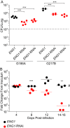
Eng1 promotes Histoplasma virulence in vivo. Wild-type C57BL/6 mice were infected intranasally with 1 × 10⁴ Eng1-expressing (ENG1; black data points) or Eng1-deficient (ENG1-RNAi; red data points) yeast cells, and the fungal burden (CFU) in lungs was determined by plating of lung tissue homogenates. (A) Histoplasma burden in lungs 8 days postinfection with yeasts of the G186A or G217B genetic background. The dashed line indicates the inoculum level, and data points represent the Histoplasma CFU counts from each mouse (n = 4 to 5). (B) Kinetics of lung infection by ENG1-expressing and ENG1-deficient Histoplasma yeasts of the G217B background determined at 4, 8, 12, 14, or 16 days postinfection. Mice infected with Eng1-expressing yeasts were moribund at 14 days postinfection (# symbol), at which point lung tissue was harvested. Mice infected with Eng1-deficient Histoplasma remained alive, and lung tissue was harvested at 16 days postinfection († symbol). The data represent the fold change in CFU from the initial inoculum at each time point (n = 3 mice per strain). Horizontal bars represent means, and asterisks represent statistically significant differences between infections with Eng1-expressing and Eng1-deficient strains as determined by one-tailed Student’s t test (**, P < 0.01; ***, P < 0.001).
FIG 5
FIG 5
Eng1 decreases Dectin-1 recognition of Histoplasma yeasts. (A) Relative levels of binding of Eng1-expressing (ENG1; gray bar) and Eng1-deficient (ENG1-RNAi; red bars) yeasts to Dectin-1-expressing cells. Uvitex-labeled yeasts were added to Dectin-1-expressing 3T3 fibroblast cells. Adherent yeasts were released by lysis of the fibroblasts and the yeasts quantified by Uvitex fluorescence. Laminarin was added to the Dectin-1-expressing 3T3 cells as a competitive inhibitor (black bars) prior to addition of yeast cells. All data were normalized to the average level of binding of Eng1-expressing yeasts (ENG1) in the absence of laminarin. (B) Ability of Eng1-containing culture filtrates to reduce Dectin-1 recognition of yeasts. Eng1-deficient yeasts were treated with saline solution (PBS; black bar), Eng1-containing culture filtrate (ENG1 CF; gray bar), Eng1-deficient culture filtrate (ENG1-RNAi CF; red bars), the β-glucanase zymolyase (blue bar), or purified Eng1 (green bar). Treated yeast cells were added to 3T3–Dectin-1 cells and the bound yeasts quantified. Data represent the relative amounts of bound yeasts after normalization to yeast cells treated with Eng1-containing culture filtrate. Error bars represent the standard deviations of results from replicate assays (n = 3), and asterisks represent statistically significant differences between Eng1-expressing and Eng1-deficient conditions as determined by one-tailed Student’s t test (n = 3; ***, P < 0.001). (C) Representative images of Histoplasma yeasts showing surface-exposed β-glucans. Yeasts were fixed and exposed β-glucans detected by immunofluorescence following incubation with a soluble Dectin-1 molecule (FcDectin-1). Yeasts were visualized at ×600 magnification by differential interference contrast (DIC), Uvitex fluorescence (blue), and FcDectin-1 immunofluorescence (green) microscopy.
FIG 6
FIG 6
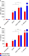
Eng1 activity decreases macrophage recognition and response to Histoplasma yeasts. Data represent cytokine production by phagocytes infected with Eng1-producing (ENG1) or Eng1-deficient (ENG1-RNAi) Histoplasma yeasts. Murine peritoneal macrophages (red bars) or bone marrow-derived dendritic cells (blue bars) were infected with Histoplasma or Candida albicans yeast cells for 8 h at an MOI of 0.5:1, and production of TNF-α (A) and IL-6 (B) was quantified by cytokine-specific ELISA of culture supernatants. Data indicate the average cytokine levels, and error bars represent the standard deviations of results from replicate infections (n = 3). Asterisks indicate statistically significant differences in cytokine stimulation between infections with Eng1-expressing and Eng1-deficient yeasts as determined by one-tailed Student’s t test (*, P < 0.05; ***, P < 0.001).
FIG 7
FIG 7
Dectin-1 mediates control of Eng1-deficient yeasts in vivo. Wild-type C57BL/6 (Dectin-1 +/+; circles) or Dectin-1 knockout (Dectin-1 −/−; squares) mice were infected intranasally with 1 × 10⁴ Eng1-expressing (ENG1; black data points) or Eng1-deficient (ENG1-RNAi; red data points) yeast cells, and the fungal burden in lungs (CFU) (A) or spleens (B) was determined by plating of lung tissue homogenates. Data points represent the Histoplasma CFU counts from each mouse (n = 4 to 5) at 8 days postinfection. The dashed line indicates the inoculum level, and horizontal bars represent the mean CFU recovered. Asterisks represent statistically significant differences between infections with Eng1-expressing and Eng1-deficient strains as determined by one-tailed Student’s t test (***, P < 0.001) ns, not significant.
FIG 8
FIG 8
Eng1 and α-glucan combine to reduce yeast β-glucan exposure. Data represent Dectin-1 recognition of G186A-background yeasts lacking Eng1 function (ENG1-RNAi; red bar) or α-glucan (ags1Δ; green bar) or both factors (ags1Δ ENG1-RNAi; purple bar). Uvitex-labeled yeasts were added to Dectin-1-expressing 3T3-fibroblasts, and adherent yeasts were quantified by Uvitex fluorescence. Data indicate the average number of yeasts bound by Dectin-1 relative to the number of bound wild-type yeasts (WT; gray bar). Error bars represent the standard deviations of results from replicates (n = 3). Asterisks represent statistically significant differences in recognition as determined by one-tailed Student’s t test (*, P < 0.05; **, P < 0.01; ***, P < 0.001).

Comment in

References

    1. Garfoot AL, Rappleye CA. 2016. Histoplasma capsulatum surmounts obstacles to intracellular pathogenesis. FEBS J. 283:619–633. doi:10.1111/febs.13389. - DOI - PMC - PubMed
    1. Wu-Hsieh B, Howard DH. 1984. Inhibition of growth of Histoplasma capsulatum by lymphokine-stimulated macrophages. J Immunol 132:2593–2597. - PubMed
    1. Newman SL, Gootee L, Bucher C, Bullock WE. 1991. Inhibition of intracellular growth of Histoplasma capsulatum yeast cells by cytokine-activated human monocytes and macrophages. Infect Immun 59:737–741. - PMC - PubMed
    1. Bullock WE, Wright SD. 1987. Role of the adherence-promoting receptors, CR3, LFA-1, and p150,95, in binding of Histoplasma capsulatum by human macrophages. J Exp Med 165:195–210. - PMC - PubMed
    1. Newman SL, Bucher C, Rhodes J, Bullock WE. 1990. Phagocytosis of Histoplasma capsulatum yeasts and microconidia by human cultured macrophages and alveolar macrophages. Cellular cytoskeleton requirement for attachment and ingestion. J Clin Invest 85:223–230. doi:10.1172/JCI114416. - DOI - PMC - PubMed

Publication types