Review
. 2016 Jun;27(6):392-403.
doi: 10.1016/j.tem.2016.03.016. Epub 2016 Apr 16.

Marrow Adipose Tissue: Trimming the Fat

Affiliations
Review

Marrow Adipose Tissue: Trimming the Fat

Erica L Scheller et al. Trends Endocrinol Metab. 2016 Jun.

Abstract

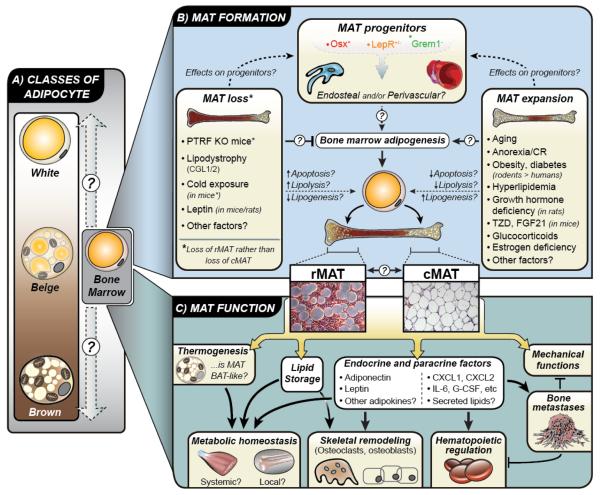
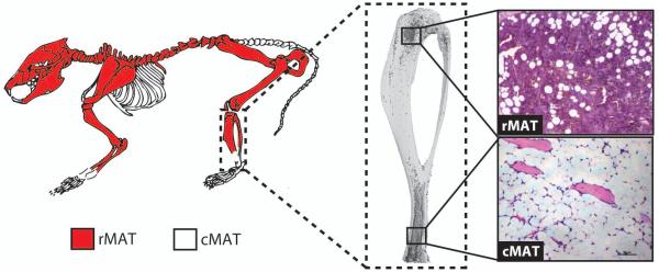
Marrow adipose tissue (MAT) is a unique fat depot, located in the skeleton, that has the potential to contribute to both local and systemic metabolic processes. In this review we highlight several recent conceptual developments pertaining to the origin and function of MAT adipocytes; consider the relationship of MAT to beige, brown, and white adipose depots; explore MAT expansion and turnover in humans and rodents; and discuss future directions for MAT research in the context of endocrine function and metabolic disease. MAT has the potential to exert both local and systemic effects on metabolic homeostasis, skeletal remodeling, hematopoiesis, and the development of bone metastases. The diversity of these functions highlights the breadth of the potential impact of MAT on health and disease.

Keywords: adiponectin; adipose tissue; anorexia; beige fat; marrow fat; obesity.


Figures

Figure 1
Figure 1. KEY FIGURE. The knowns and unknowns of MAT
(A) Adipose tissue is typically classified as white, brown, or beige. Bone marrow adipocytes are morphologically similar to white adipocytes; however, it is unclear where they fall on this ‘white-beige-brown’ spectrum, if at all. (B) Lineage tracing studies demonstrate that bone marrow adipocyte progenitors express osterix (Osx), but not Grem1. Some marrow adipocytes are also derived from progenitors that express the leptin receptor (LepR). Based on these findings it is unclear if marrow adipocyte progenitors are endosteal and/or perivascular in origin, although they are clearly distinct to progenitors for white and brown adipocytes. Also unclear is how these progenitors are driven toward adipogenesis to generate bone marrow adipocytes, which can be classed as two distinct subtypes: regulated (rMAT) and constitutive (cMAT). Do these subtypes derive from distinct progenitors, and can rMAT and cMAT interconvert? In addition to physiological MAT formation, various conditions are associated with MAT loss or MAT expansion, predominantly in rMAT. However, the mechanisms linking these conditions to MAT loss or gain remain largely uncertain. (C) The function of MAT is also yet to be firmly established. Some reports suggest that MAT has BAT-like properties, though this remains controversial. Instead, MAT may have more WAT-like properties such as lipid storage and endocrine functions. It is now clear that MAT can release adipokines such as adiponectin and leptin, as well as paracrine factors such as cytokines and lipids. These secreted factors may allow MAT to exert both local and systemic effects on metabolic homeostasis, skeletal remodeling, hematopoiesis, and development of bone metastases. The diversity of these functions highlights the breadth of MAT’s potential impact on health and disease.
BOX 1 Figure I
BOX 1 Figure I. Regulated and constitutive marrow adipose tissue (MAT) in the mouse
(A) Proposed distribution of regulated MAT (rMAT) and constitutive MAT (cMAT) in the mouse skeleton when marrow is present. Regulated MAT is found in the more proximal regions including the mid- to proximal-tibia, femur, and lumbar vertebrae. Constitutive MAT is found in the most distal portion of the tibia and tail vertebrae. (B) Three-dimensional reconstruction of an osmium-stained mouse tibia. (C) Representative histology of rMAT and cMAT adipocytes within the bone marrow.

Similar articles

Cited by

References

    1. Ng M, et al. Global, regional, and national prevalence of overweight and obesity in children and adults during 1980-2013: a systematic analysis for the Global Burden of Disease Study 2013. The Lancet. 2014;384:766–781. - PMC - PubMed
    1. Scheller EL, Rosen CJ. What’s the matter with MAT? Marrow adipose tissue, metabolism, and skeletal health. Ann N Y Acad Sci. 2014;1311:14–30. - PMC - PubMed
    1. Kricun ME. Red-yellow marrow conversion: its effect on the location of some solitary bone lesions. Skeletal Radiol. 1985;14:10–19. - PubMed
    1. Cawthorn WP, et al. Expansion of Bone Marrow Adipose Tissue During Caloric Restriction Is Associated With Increased Circulating Glucocorticoids and Not With Hypoleptinemia. Endocrinology. 2016;157:508–521. - PMC - PubMed
    1. Scheller EL, et al. Region-specific variation in the properties of skeletal adipocytes reveals regulated and constitutive marrow adipose tissues. Nat Commun. 2015;6:7808. - PMC - PubMed

Publication types

LinkOut - more resources