

3D rotational fluoroscopy for intraoperative clip control in patients with intracranial aneurysms--assessment of feasibility and image quality
- PMID: 27094510
- PMCID: PMC4837534
- DOI: 10.1186/s12880-016-0133-0
3D rotational fluoroscopy for intraoperative clip control in patients with intracranial aneurysms--assessment of feasibility and image quality
Abstract
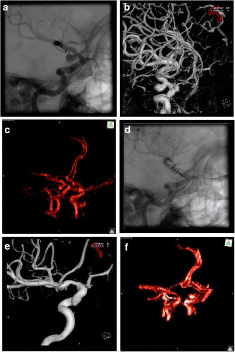
Background: Mobile 3D fluoroscopes have become increasingly available in neurosurgical operating rooms. In this series, the image quality and value of intraoperative 3D fluoroscopy with intravenous contrast agent for the evaluation of aneurysm occlusion and vessel patency after clip placement was assessed in patients who underwent surgery for intracranial aneurysms.
Materials and methods: Twelve patients were included in this retrospective analysis. Prior to surgery, a 360° rotational fluoroscopy scan was performed without contrast agent followed by another scan with 50 ml of intravenous iodine contrast agent. The image files of both scans were transferred to an Apple PowerMac® workstation, subtracted and reconstructed using OsiriX® free software. The procedure was repeated after clip placement. Both image sets were compared for assessment of aneurysm occlusion and vessel patency.
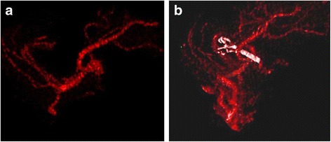
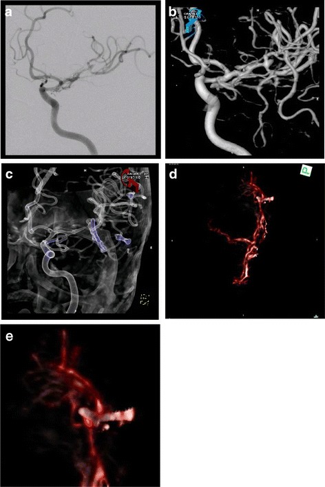
Results: Image acquisition and contrast administration caused no adverse effects. Image quality was sufficient to follow the patency of the vessels distal to the clip. Metal artifacts reduce the assessability of the immediate vicinity of the clip. Precise image subtraction and post-processing can reduce metal artifacts and make the clip-site assessable and depict larger neck-remnants.
Conclusion: This technique quickly supplies images at adequate quality to evaluate distal vessel patency after aneurysm clipping. Significant aneurysm remnants may be depicted as well. As it does not require visual control of all vessels that are supposed to be evaluated intraoperatively, this technique may be complementary to other intraoperative tools like indocyanine green videoangiography and micro-Doppler, especially for the assessment of larger aneurysms. At the momentary state of this technology, it cannot replace postoperative conventional angiography. However, 3D fluoroscopy and image post-processing are young technologies. Further technical developments are likely to result in improved image quality.
Keywords: 3D fluoroscopy; Aneurysm surgery; Angiography; Clip control; Contrast; Image quality; Intraoperative; Post-processing; Vessel patency.
Figures
References
-
- Zhang H1, Hou C, Zhou Z, Zhang H, Zhou G, Zhang G. Evaluating of small intracranial aneurysms by 64-detector CT Angiography: a comparison with 3-dimensional rotation DSA or surgical findings. J Neuroimaging. 2014;24(2):137-43. - PubMed
-
- Raabe A, Beck J, Gerlach R, Zimmermann M, Seifert V. Near-infrared indocyanine green video angiography: a new method for intraoperative assessment of vascular flow. Neurosurgery. 2003;52(1):132–139. - PubMed
-
- Westermaier T, Linsenmann T, Keler AF, Stetter C, Willner N, Solymosi L, Ernestus RI, Vince GH. Intraoperative cerebral angiography by intravenous contrast administration with 3-dimensional rotational fluoroscopy in patients with intracranial aneurysms: a feasibility study. Neurosurgery. 2015;11(Suppl 2):119–126. - PubMed
Publication types
MeSH terms
Substances
LinkOut - more resources
Full Text Sources
Other Literature Sources
Medical
Research Materials
