. 2016 Apr 18:12:1744806916644927.
doi: 10.1177/1744806916644927. Print 2016.

Interferon-gamma potentiates NMDA receptor signaling in spinal dorsal horn neurons via microglia-neuron interaction

Affiliations

Interferon-gamma potentiates NMDA receptor signaling in spinal dorsal horn neurons via microglia-neuron interaction

Mayumi Sonekatsu et al. Mol Pain. .

Abstract

Background: Glia-neuron interactions play an important role in the development of neuropathic pain. Expression of the pro-inflammatory cytokne →cytokine Interferon-gamma (IFNγ) is upregulated in the dorsal horn after peripheral nerve injury, and intrathecal IFNγ administration induces mechanical allodynia in rats. A growing body of evidence suggests that IFNγ might be involved in the mechanisms of neuropathic pain, but its effects on the spinal dorsal horn are unclear. We performed blind whole-cell patch-clamp recording to investigate the effect of IFNγ on postsynaptic glutamate-induced currents in the substantia gelatinosa neurons of spinal cord slices from adult male rats.

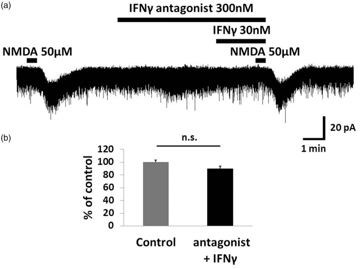
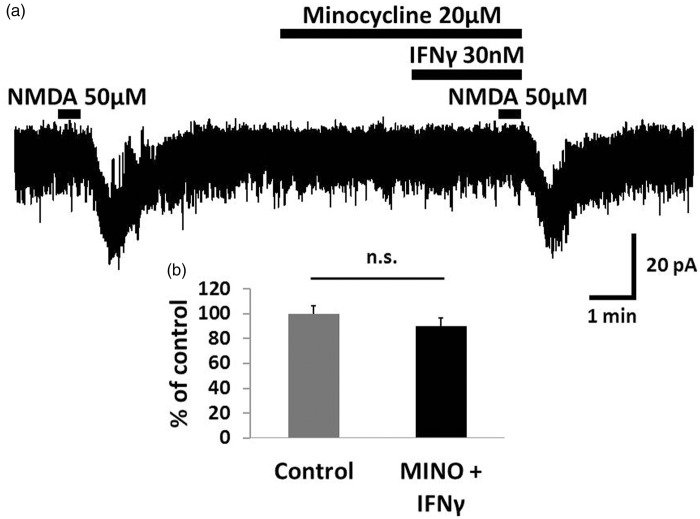
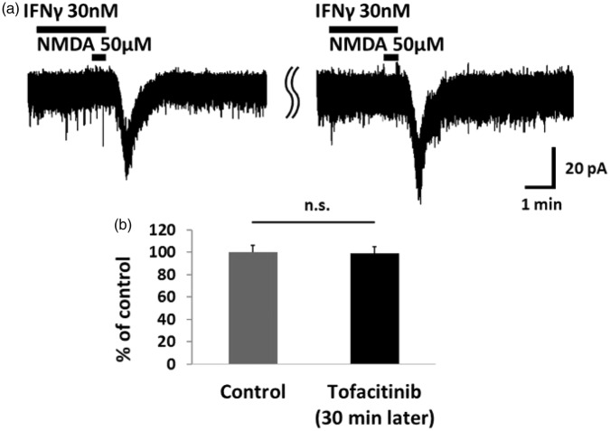
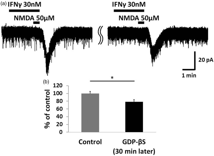
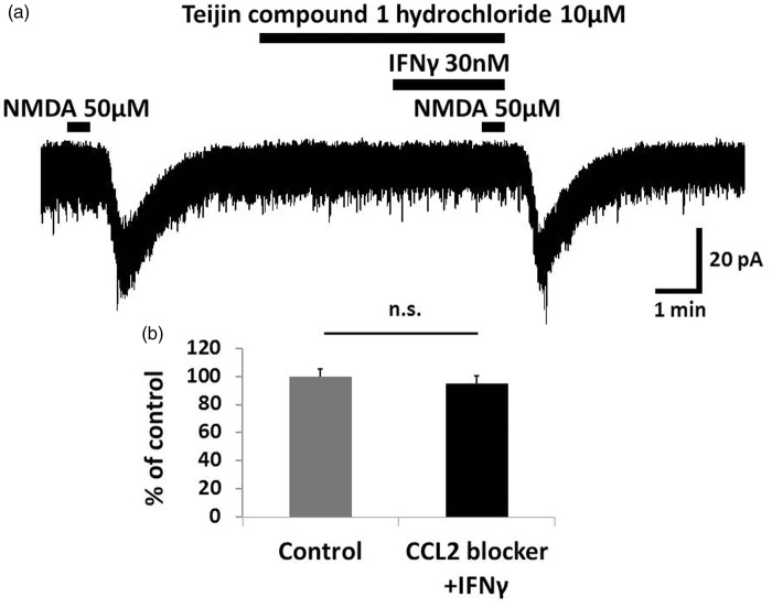
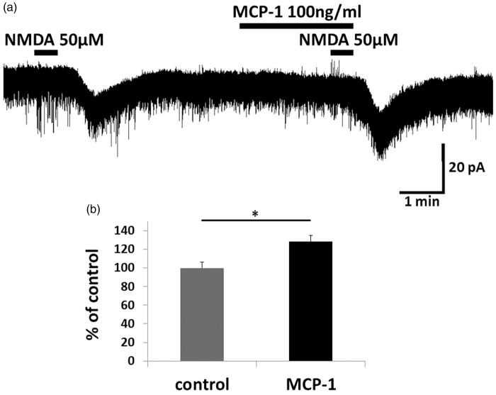
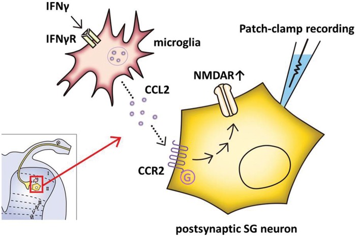
Results: IFNγ perfusion significantly enhanced the amplitude of NMDA-induced inward currents in substantia gelatinosa neurons, but did not affect AMPA-induced currents. The facilitation of NMDA-induced current by IFNγ was inhibited by bath application of an IFNγ receptor-selective antagonist. Adding the Janus activated kinase inhibitor tofacitinib to the pipette solution did not affect the IFNγ-induced facilitation of NMDA-induced currents. However, the facilitatory effect of IFNγ on NMDA-induced currents was inhibited by perfusion of the microglial inhibitor minocycline. These results suggest that IFNγ binds the microglial IFNγ receptor and enhances NMDA receptor activity in substantia gelatinosa neurons. Next, to identify the effector of signal transmission from microglia to dorsal horn neurons, we added an inhibitor of G proteins, GDP-β-S, to the pipette solution. In a GDP-β-S-containing pipette solution, IFNγ-induced potentiation of the NMDA current was significantly suppressed after 30 min. In addition, IFNγ-induced potentiation of NMDA currents was blocked by application of a selective antagonist of CCR2, and its ligand CCL2 increased NMDA-induced currents.

Conclusion: Our findings suggest that IFNγ enhance the amplitude of NMDA-induced inward currents in substantia gelatinosa neurons via microglial IFNγ receptors and CCL2/CCR2 signaling. This mechanism might be partially responsible for the development of persistent neuropathic pain.

Keywords: CCR2; NMDA receptor; Neuropathic pain; dorsal horn; interferon-gamma; microglia; patch-clamp recording; spinal cord.


Figures

Figure 1.
Figure 1.
AMPA- and NMDA-induced currents under bath application of IFNγ in adult rat SG neurons. (a) Recordings of AMPA-induced inward currents at a holding potential of −70 mV. Perfusion of IFNγ (30 nM, 2.5 min) did not result in any significant change in AMPA-induced currents. (b) Effects of IFNγ on NMDA-induced inward currents at a holding potential of −50 mV. NMDA-induced current was significantly increased by IFNγ (30 nM, 2.5 min). (c) The peak amplitude of the AMPA-induced currents after IFNγ treatment was 100.0 ± 3.7% of baseline. (d) The peak amplitude of the NMDA-induced currents after IFNγ treatment was 147.3 ± 10.7% of baseline. In Figure 1(b) and subsequent figures, NMDA responses were recorded in voltage-clamp mode (VH = −50 mV). n.s.: not significant. * p < 0.05. AMPA: α-amino-3-hydroxy-5-methyl-4-Isoxazole-4-propionic acid; IFNγ: interferon-gamma; NMDA: N-methyl-d-aspartate.
Figure 2.
Figure 2.
An IFNγ receptor selective antagonist inhibited IFNγ-induced facilitation of the NMDA current. (a) Perfusion of the IFNγ receptor-selective antagonist (300 nM, 5.5 min) inhibited the IFNγ-induced facilitation of NMDA current. (b) Bar graphs show the peak amplitude of NMDA-induced current compared to baseline when the IFNγ receptor-selective antagonist and IFNγ are perfused simultaneously. Therefore, IFNγ likely binds to IFNγ receptors and activates NMDA receptors in SG neurons. IFNγ: interferon-gamma; NMDA: N-methyl-d-aspartate.
Figure 3.
Figure 3.
The potentiation of NMDA-induced currents by IFNγ was inhibited by minocycline. (a) Minocycline (20 μM, 5.5 min), an inhibitor of microglia activation, inhibited the IFNγ-induced facilitation of the NMDA current. (b) Bar graphs show the peak amplitude of the NMDA-induced currents compared with baseline when minocycline and IFNγ were perfused simultaneously. Therefore, IFNγ likely binds to microglial IFNγ receptors and enhances the activity of NMDA receptors in dorsal horn neurons. IFNγ: interferon-gamma; NMDA: N-methyl-d-aspartate.
Figure 4.
Figure 4.
The JAK inhibitor tofacitinib had no effect on IFNγ-induced NMDA currents. (a) Tofacitinib (1 mM) addition to the pipette solution did not affect the IFNγ-induced NMDA current. In this experimental system, the control corresponds to the IFNγ-induced NMDA currents recorded immediately after patch clamping. (b) Bar graphs show the average rate of NMDA current enhancement by IFNγ at the start of patch-clamp recording and 30 min later. The peak amplitude of the IFNγ-induced NMDA currents did not change compared with the control recording. This confirmed that IFNγ enhances neuronal NMDA-induced inward current via the microglial IFNγ receptor. IFNγ: interferon-gamma; NMDA: N-methyl-d-aspartate; n.s.: not significant.
Figure 5.
Figure 5.
The G protein inhibitor GDP-β-S significantly inhibited IFNγ-induced NMDA currents. (a) In a potassium gluconate pipette solution containing GDP-β-S (1 mM), the IFNγ-induced NMDA current after 30 min of recording was significantly lower than the control recording of IFNγ-induced NMDA currents recorded at the start of patch clamping. (b) Bar graphs show the average rate of IFNγ-induced enhancement of NMDA current in the same neuron at the beginning of the recording and after 30 min. The value of the right bar is 78.7 ± 5.1% of the left bar. This finding suggested that the IFNγ-induced enhancement of NMDA currents involves the activation of G protein-coupled receptors. *p < 0.05. IFNγ: interferon-gamma; NMDA: N-methyl-d-aspartate.
Figure 6.
Figure 6.
The enhancement of NMDA-induced inward current by IFNγ was mediated by CCR2. (a) Bath application of the CCR2 antagonist Teijin compound 1 hydrochloride (10 μM, 5.5 min) blocked the IFNγ-induced enhancement of NMDA currents. (b) The peak amplitude of the NMDA-induced currents after simultaneous application of Teijin compound 1 hydrochloride and IFNγ was 95.0 ± 5.3% that of baseline. IFNγ likely increases the NMDA-induced inward current in SG neurons via CCR2. IFNγ: interferon-gamma; NMDA: N-methyl-d-aspartate; n.s.: not significant.
Figure 7.
Figure 7.
Bath application of CCL2 enhanced NMDA-induced inward current. (a) Perfusion of spinal cord slices with CCL2 (100 ng/ml, 2.5 min) increased NMDA-induced inward currents. (b) The peak amplitude of the NMDA-induced currents after simultaneous CCL2 application is 128.3 ± 6.4% compared with baseline. This suggests that IFNγ increases the NMDA-induced inward current in SG neurons via CCL2/CCR2 signaling. * p < 0.05. MCP-1: monocyte chemoattractant protein 1; NMDA: N-methyl-d-aspartate.
Figure 8.
Figure 8.
Schematic overview. When nerve injury occurs, IFNγ levels are increased in the dorsal horn of the spinal cord and binds to IFNγ receptors on microglia. CCL2 subsequently enhances the neuronal NMDA-induced inward current in lamina II neurons via CCR2. This potentiation of NMDA receptors likely contributes to the development of neuropathic pain. CCL2: chemokine ligand 2; CCR2: C-C chemokine receptor type 2; IFNγ: interferon-gamma; SG: substantia gelatinosa.

Similar articles

Cited by

References

    1. Costigan M, Scholz J, Woolf CJ. Neuropathic pain: a maladaptive response of the nervous system to damage. Annu Rev Neurosci 2009; 32: 1–32. - PMC - PubMed
    1. Cohen SP, Mao J. Neuropathic pain: mechanisms and their clinical implications. BMJ 2014; 348: 1–12. - PubMed
    1. Latremoliere A, Woolf CJ. Central sensitization: a generator of pain hypersensitivity by central neural plasticity. J Pain 2009; 10: 895–926. - PMC - PubMed
    1. Scholz J, Woolf CJ. The neuropathic pain triad: neurons, immune cells and glia. Nat Neurosci 2007; 10: 1361–1368. - PubMed
    1. Ren K, Dubner R. Interactions between the immune and nervous systems in pain. Nat Med 2010; 16: 1267–1276. - PMC - PubMed

Publication types

MeSH terms

LinkOut - more resources