. 2016 Apr 20:6:24083.
doi: 10.1038/srep24083.

Mutations in human C2CD3 cause skeletal dysplasia and provide new insights into phenotypic and cellular consequences of altered C2CD3 function

Affiliations

Mutations in human C2CD3 cause skeletal dysplasia and provide new insights into phenotypic and cellular consequences of altered C2CD3 function

Claudio R Cortés et al. Sci Rep. .

Abstract

Ciliopathies are a group of genetic disorders caused by defective assembly or dysfunction of the primary cilium, a microtubule-based cellular organelle that plays a key role in developmental signalling. Ciliopathies are clinically grouped in a large number of overlapping disorders, including the orofaciodigital syndromes (OFDS), the short rib polydactyly syndromes and Jeune asphyxiating thoracic dystrophy. Recently, mutations in the gene encoding the centriolar protein C2CD3 have been described in two families with a new sub-type of OFDS (OFD14), with microcephaly and cerebral malformations. Here we describe a third family with novel compound heterozygous C2CD3 mutations in two fetuses with a different clinical presentation, dominated by skeletal dysplasia with no microcephaly. Analysis of fibroblast cultures derived from one of these fetuses revealed a reduced ability to form cilia, consistent with previous studies in C2cd3-mutant mouse and chicken cells. More detailed analyses support a role for C2CD3 in basal body maturation; but in contrast to previous mouse studies the normal recruitment of the distal appendage protein CEP164 suggests that this protein is not sufficient for efficient basal body maturation and subsequent axonemal extension in a C2CD3-defective background.


Figures

Figure 1
Figure 1. Clinical features of the two subjects in this study.
(a–g) G2P1; (h–k) G3P1. Common features include short ribs, shortened long bones, micrognathia (arrow, b,e,i) and trident acetubula (arrow, a,h). G2P1 displayed preaxial polydactyly (duplicated hallux) of the feet (arrow, c,g), and no polydactyly of the hands, although digits appear short (d,f). G3P1 displayed preaxial polydactyly (triplicated hallux) of the feet (arrow j), and pre- (arrow, k) and postaxial (arrowhead, k) polydactyly of the hands. The tibiae in G3P1 were hypoplastic (arrowhead, h) but appeared normal in G2P1.
Figure 2
Figure 2. Segregation of C2CD3 mutations, and protein ideogram indicating location of variants.
(a) C2CD3 variants present in the parents and affected individuals in the reported family. Sequence reads highlight genotype for variants identified by Sanger sequencing under each individual. Arrows denote missense mutation, dotted lines denote deletion site. Note that the sequence scan for the c.195G > C mutation shows the reverse strand sequence; the c.1429delA mutation is shown on the forward strand. (b) Human C2CD3 protein schematic showing mutations detected in the present study (red lines, top), mutations reported in previously reported OFDS cases (dotted lines, bottom) and mouse C2cd3 alleles C2Cd3Gt (Gt) and Hearty (Hty) (dotted lines, bottom). Note the C2cd3Gt mutation is predicted to result in a truncated protein encoding the N-terminal 161 amino acids only; aberrant splicing in Hty leads to proteins either truncated at amino acid 235, or with small in-frame deletions in the same region. C2: canonical C2 domains, C2CD3N: non-canonical globular C2 domain.
Figure 3
Figure 3. Ciliogenesis and IFT protein recruitment is impaired in fibroblasts isolated from G3P1.
(a,b) Images showing a lower percentage of cilia (as marked by Arl13b staining) relative to DAPI-stained nuclei in serum-starved C2CD3-mutant fibroblast cultures (G3P1), compared to control juvenile dermal fibroblasts. Scale bar = 10 μm. Quantification is shown in (c). (d–i) Representative images of IFT88 staining (green) in non-ciliated cells (indicated by lack of red Arl13b staining along the axoneme) in serum-starved cultures. IFT88 localises to the centrosome (stained with gamma-tubulin in red) in control fibroblasts (d–f) whereas in fibroblasts isolated from G3P1, IFT88 is absent from most centrosomes (g–i). Scale bar = 5 μm. (j) Quantification of percentage of non-ciliated cells with IFT88-postive centrosomes in cultured C2CD3-mutant fibroblasts, relative to normal juvenile fibroblasts. ***p < 0.0001, error bars show SEM. All ciliated cells show normal localisation of IFT88 in both control and mutant cultures, reflecting the requirement for IFT88 in ciliogenesis.
Figure 4
Figure 4. Centriolar appendage and basal body maturation in C2CD3-mutant fibroblasts.
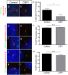
(a,b) Images of CP110 staining (green) in serum starved control and G3P1 fibroblasts. Note that the image in panel (b) with CP110 at both centrioles is representative of approximately 77% of cells in G3P1cultures (23% of cells have CP110 removed from the mother centriole, i.e. show staining at just one centriole). This quantification is shown graphically in (c) where random cells across both mutant and control cultures with 2 distinct centriolar spots were scored for removal of CP110 from the mother centriole. CP110 is removed in fewer mutant cells relative to control cells. *p < 0.05. (d–g) Representative images of staining for the distal appendage marker CEP164 (green) in non-ciliated (d,e) and ciliated cells (f,g). (h) Quantification of cells with CEP164-positive mother centriole or basal body. (i–l) Representative images of staining for the sub-distal appendage marker ODF2 (green) in non-ciliated (i–j) and ciliated cells (k,l). (m) Quantification of cells with ODF2-positive mother centriole or basal body. Scale bar = 5 μm. In all cases the centrosome is marked with gamma-tubulin and the axoneme with Arl13b, both in red. n.s. not significant, error bars show SEM.
Figure 5
Figure 5. Schematic model for C2CD3-deficient mouse and human cells.
(a) In normal conditions, the centrosome moves to the cell surface and the mother centriole with subdistal and distal appendage proteins docks to the membrane. Prior to docking CP110 is removed from the distal end of the mother centriole, a step essential for axonemal extension. (b) In C2CD3-deficient mouse cells, the mother centriole does not form distal appendages or anchor to the plasma membrane, and shows defective removal of CP110. (c) In C2CD3-deficient human G3P1 cells, the subdistal and distal appendage proteins are localised normally at the mother centriole and basal body, but CP110 is not efficiently removed, hence blocking axonemal extension and ciliogenesis.

References

    1. Delling M., DeCaen P. G., Doerner J. F., Febvay S. & Clapham D. E. Primary cilia are specialized calcium signalling organelles. Nature 504, 311–314 (2013). - PMC - PubMed
    1. Huangfu D. et al. Hedgehog signalling in the mouse requires intraflagellar transport proteins. Nature 426, 83–87 (2003). - PubMed
    1. Jones C. et al. Ciliary proteins link basal body polarization to planar cell polarity regulation. Nat. Genet. 40, 69–77 (2008). - PubMed
    1. Lancaster M. A., Schroth J. & Gleeson J. G. Subcellular spatial regulation of canonical Wnt signalling at the primary cilium. Nat. Cell Biol. 13, 700–707 (2011). - PMC - PubMed
    1. Liem K. F. et al. The IFT-A complex regulates Shh signaling through cilia structure and membrane protein trafficking. J. Cell Biol. 197, 789–800 (2012). - PMC - PubMed

Publication types

Substances

LinkOut - more resources