. 2016 Apr 20:6:24712.
doi: 10.1038/srep24712.

Chromatin folding and DNA replication inhibition mediated by a highly antitumor-active tetrazolato-bridged dinuclear platinum(II) complex

Affiliations

Chromatin folding and DNA replication inhibition mediated by a highly antitumor-active tetrazolato-bridged dinuclear platinum(II) complex

Ryosuke Imai et al. Sci Rep. .

Abstract

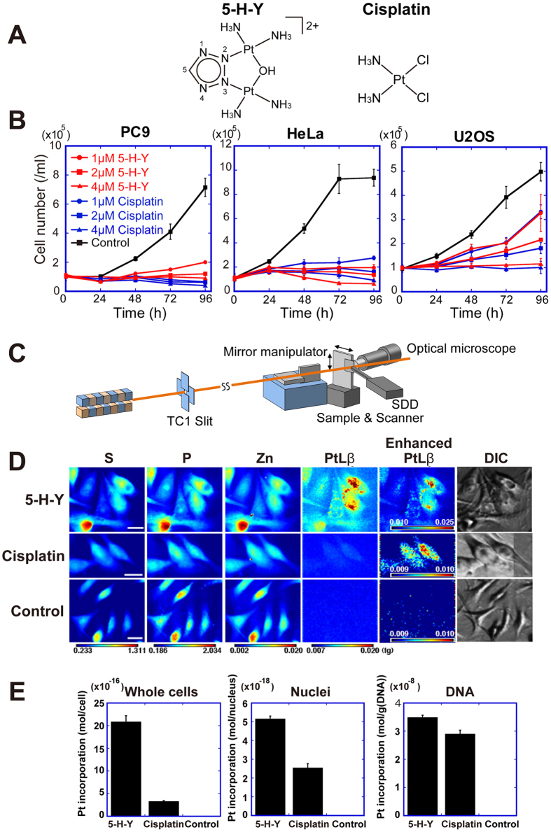
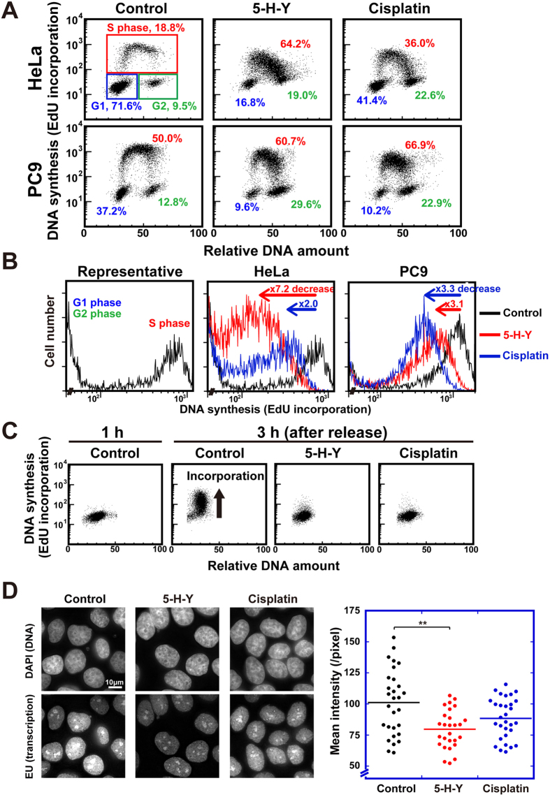
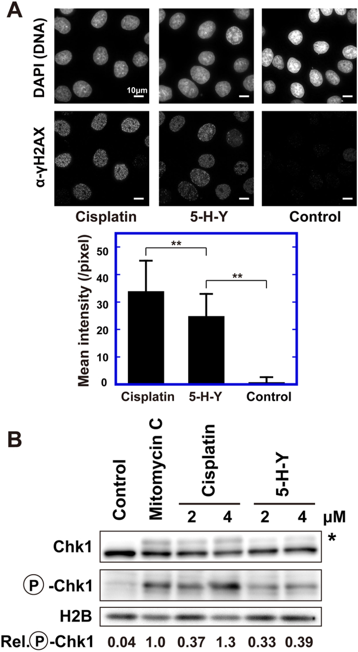
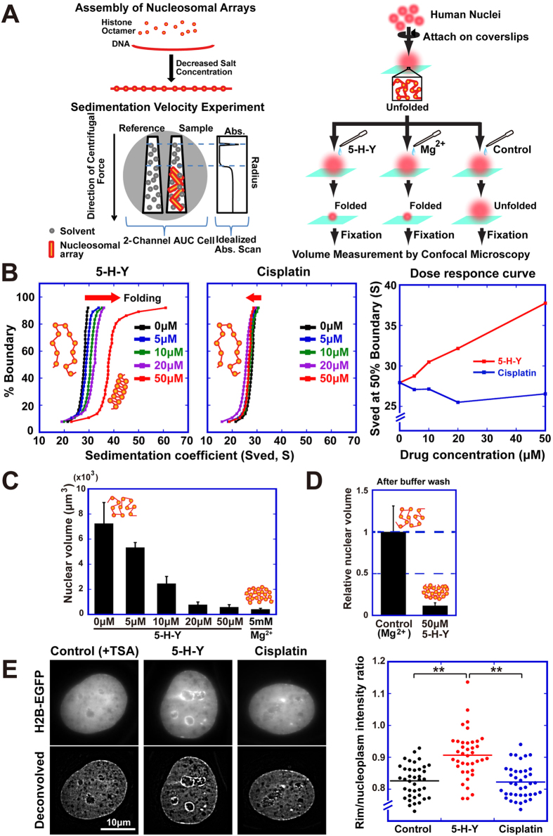
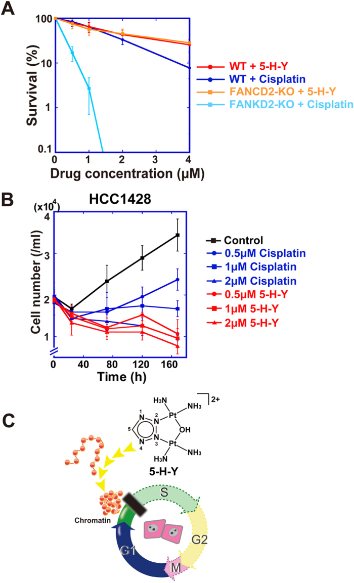
Chromatin DNA must be read out for various cellular functions, and copied for the next cell division. These processes are targets of many anticancer agents. Platinum-based drugs, such as cisplatin, have been used extensively in cancer chemotherapy. The drug-DNA interaction causes DNA crosslinks and subsequent cytotoxicity. Recently, it was reported that an azolato-bridged dinuclear platinum(II) complex, 5-H-Y, exhibits a different anticancer spectrum from cisplatin. Here, using an interdisciplinary approach, we reveal that the cytotoxic mechanism of 5-H-Y is distinct from that of cisplatin. 5-H-Y inhibits DNA replication and also RNA transcription, arresting cells in the S/G2 phase, and are effective against cisplatin-resistant cancer cells. Moreover, it causes much less DNA crosslinking than cisplatin, and induces chromatin folding. 5-H-Y will expand the clinical applications for the treatment of chemotherapy-insensitive cancers.


Figures

Figure 1
Figure 1. 5-H-Y inhibits cell proliferation and is incorporated into cell nuclei.
(A) Chemical structures of [{cis-Pt(NH3)2}2(μ-OH)(μ-tetrazolato-N2,N3)]2+ (5-H-Y) (left) and cis-diamminedichloridoplatinum(II) (cisplatin) (right). (B) Cell proliferation assays with 5-H-Y or cisplatin treatment. Four human cell lines (PC9, HeLa, U2OS, and TIG-1) were treated with the indicated concentrations of 5-H-Y or cisplatin, and the cell numbers were monitored from 0 to 96 h for human cells (for TIG-1, see also Fig. S1A). (C) Schematic view of scanning X-ray fluorescence microscopy. The X-ray beam, highly focused by a set of mirrors (KB-mirror) was focused on the cells refs and . Then X-ray fluorescence was detected by the silicon drift detector (SDD). (D) SXFM analysis after drug treatment. Cell morphologies obtained by Nomarski (DIC). Brighter colors indicate a higher signal intensity of each element. Representative results are shown. Results are shown for 5-H-Y (top) and cisplatin (middle), untreated control PC9 cells (bottom). Note the high intensity of Pt in 5-H-Y treated cells. Pt, platinum signal, P, phosphorus, S, sulfur, Zn, zinc. Color bars indicate elemental content, expressed in fg/μm2. The phosphorus- and zinc -rich regions in the cells seem to be nuclei. Bars show 10 μm. (E) Amounts of platinum in PC9 whole cells, nuclei, and DNA fractions of 5-H-Y- and cisplatin-treated cells.
Figure 2
Figure 2. 5-H-Y inhibits DNA replication and RNA transcription, arresting the cells in the S/G2 phase.
(A) Flow cytometry results for HeLa cells (upper row) and PC9 cells (lower row) with/without 5-H-Y (2 μM) or cisplatin (2 μM). Vertical and horizontal axes show DNA synthesis activity (EdU incorporation) and DNA amount, respectively. Each dot represents a single cell and results using 10,000 cells are plotted. In the plot of control HeLa, the corresponding cell cycle stages are indicated. Percentages of each cell cycle population are indicated. See also Fig. S2. (B) EdU incorporation versus cell numbers plots of (A). Left panel shows a representative plot (control HeLa). Note that EdU incorporation was high in the S-phase. Fold decreases in EdU incorporation upon 5-H-Y (red) or cisplatin (blue) treatment are indicated in the plots of HeLa (middle) and PC9 (right). Note the several-fold decreases in EdU incorporation in the 5-H-Y (red) or cisplatin (blue) treated cells. (C) Results for HeLa cells synchronized at G1/S by nocodazole-thymidine block with/without 5-H-Y (2 μM) or cisplatin (2 μM). 5-H-Y and cisplatin both inhibit very early phases of DNA replication. (D) Effect of 5-H-Y on RNA transcription in vivo. (Left) Fluorescence microscopy images of 5-H-Y or cisplatin-treated cells. DNA stain, upper; EU fluorescent labeling, lower. (Right) Dot plot of the mean intensity of EU fluorescence in each nucleus (each group, n = 27–30). **p < 0.01, Student’s t-test.
Figure 3
Figure 3. DNA damage response in 5-H-Y treated cells.
(A) γH2AX foci formation in 5-H-Y or cisplatin-treated HeLa cells. DNA stain, upper; anti-γH2AX antibody staining, lower. Scale bars are 10 μm. The bar graph indicates quantification of the γH2AX signal intensity averaged from ~50 nuclei. Note that the signal in 5-H-Y-treated cells was significantly lower than in cisplatin-treated cells. **p < 0.01, Student’s t-test. (B) Chk1 activation on drug treatment. Western blotting analysis of cell lysates using anti-Chk1 (1st row) and anti-phospho-Chk1 (P-Chk1) (2nd row) antibody. In the 1st row, the position of phosphorylated (activated) Chk1 is marked by the asterisk. Control, no treatment; mitomycin C, mitomycin C treatment for efficient DNA crosslinking. The third row is a loading control using H2B. The values at the bottom indicate quantification of the phosphorylated Chk1 signal intensity. Note that the relative intensity of phosphorylated signal in 5-H-Y-treated cells was considerably lower than that in cisplatin-treated cells. The blots were cropped at the positions of the proteins for clarity and space considerations.
Figure 4
Figure 4. Much lower interstrand crosslinking (ICL) activity of 5-H-Y.
(A) Covalent binding of cisplatin (blue square) and 5-H-Y (red square) to calf-thymus DNA (n = 4). The rb value is defined as the molar ratio of platinum complex bound per nucleotide. (B) Interstrand crosslinking of drug-treated plasmid DNA. Two types of plasmid DNAs, pUC19 (left) and pBluescript II (pBSII) (right), were treated with no drug (Control), cisplatin, or 5-H-Y for 24 h (upper) or 48 h (lower). The treated plasmid DNAs were electrophoresed on alkaline agarose gels. The gels with EtBr staining are shown. The positions of dsDNA, representing interstrand crosslinks, and ssDNA, including no crosslinks and intrastrand crosslinks, are shown. Values below the gels indicate intensities of dsDNA normalized by that of cisplatin. Note that there is much more dsDNA in cisplatin-treated DNA than in 5-H-Y-treated DNA. The two DNA templates, pUC19 and pBSII, produced similar results. (C) Experimental scheme of PCR amplification. DNA templates were treated with cisplatin or 5-H-Y. If inter-strand (middle) or intrastrand (bottom) crosslinks occur in the template DNA, DNA amplification by PCR is inhibited. (D) PCR results. Two types of plasmid DNAs, pUC19 (left) and pBluescript II (pBSII) (right), were used as PCR templates. They were treated with no drug (Control), cisplatin, or 5-H-Y for 24 h or 48 h. The PCR products (marked with arrow) on the agarose gel after electrophoresis are shown. Values below the gel indicate the fluorescent intensities of the PCR product normalized by that of control. In the “Cisplatin + Control” lanes, PCR was performed using mixed templates of cisplatin-treated and no-treated plasmids. Note that PCR using cisplatin-treated template produced much less product.
Figure 5
Figure 5. 5-H-Y binds to chromatin DNA tightly and folds chromatin in vitro and in vivo.
(A) Experimental scheme of the two chromatin folding assays: ultracentrifuge assay (left) and nuclear volume assay (right). (B) 5-H-Y induces chromatin folding. Samples of reconstituted nucleosome fibers were exposed to the indicated concentrations of 5-H-Y (left) or cisplatin (center) and analyzed by sedimentation velocity analytical ultracentrifugation. The integral distribution of diffusion-corrected sedimentation coefficients obtained after analysis of the data by the method of Demeler and van Holde are shown. (Right) Summary of analytical ultracentrifuge-SV results. Values at the 50% boundary are displayed as a function of drug concentration added 5-H-Y (red) or cisplatin (blue). (C) Nuclear volume was decreased by 5-H-Y in a dose-dependent manner. Nuclei treated with 50 μM 5-H-Y showed a 12-fold decrease in the volume. This indicates that 5-H-Y induces chromatin folding. The nuclei treated with 5 mM Mg2+ were prepared as a control for the nuclei with highly folded chromatin. The error bars represent the standard deviation. For each point, n = ~100. (D) Volumes between Mg2+-pretreated and 5-H-Y-pretreated nuclei after buffer washing. When the volume was normalized by Mg2+-pretreated nuclei, although Mg2+-pretreated nuclei became large after the washing (relative nuclear volume = 1), 5-H-Y-pretreated nuclei did not change (~0.1). 5-H-Y seems to bind tightly to chromatin DNA, in contrast to Mg2+. The error bars represent the standard deviation. (E) 5-H-Y induces chromatin condensation in vivo. HeLa cells were treated with TSA to decondense chromatin and then with 5-H-Y. 5-H-Y induced enrichment of chromatin at nuclear periphery and nucleoli (left) although we cannot exclude the possibility that condensation by 5-H-Y only occur around nucleoli and nuclear periphery. Right plot shows the intensity quantification of nuclear periphery chromatin. **p < 0.01, Chi-square test.
Figure 6
Figure 6. DNA damage by 5-H-Y is repaired primarily by different pathways from ICL repair.
(A) Sensitivity assay to the 5-H-Y or cisplatin in wtDT40 cells and FUNCD2-KO cells using a colony formation assay. Mean ± SD of three independent experiments is shown. See also Fig. S7. (B) Cell proliferation assay of cisplatin-resistant HCC1428 cells upon 5-H-Y or cisplatin treatment. HCC1428 cells were treated with the indicated concentrations of 5-H-Y or cisplatin, and cell numbers were monitored. 5-H-Y was effective even in this cisplatin-resistant cell line. (C) A model figure. This study demonstrated that 5-H-Y inhibited DNA replication, and arrests the treated cells in S/G2 phase. 5-H-Y binds tightly to chromatin DNA and induces chromatin folding.

Similar articles

Cited by

References

    1. Watson J. D. et al. Molecular Biology of the Gene, 7/E. (Benjamin Cummings, 2013).
    1. Davey G. E. & Davey C. A. Chromatin - a new, old drug target? Chem Biol Drug Des 72, 165–170 (2008). - PubMed
    1. Rosenberg B. & VanCamp L. The successful regression of large solid sarcoma 180 tumors by platinum compounds. Cancer Res 30, 1799–1802 (1970). - PubMed
    1. Kociba R. J., Sleight S. D. & Rosenberg B. Inhibition of Dunning asc itic leukemia and Walker 256 carcinosarcoma with cis-diamminedichloroplatinum (NSC-119875). Cancer Chemother Rep 54, 325–328 (1970). - PubMed
    1. Mansy S., Rosenberg B. & Thomson A. J. Binding of cis- and trans-dichlorodiammineplatinum(II) to nucleosides. I. Location of the binding sites. J Am Chem Soc 95, 1633–1640 (1973). - PubMed

Publication types

MeSH terms

LinkOut - more resources