. 2016 Jun;22(6):905-19.
doi: 10.1261/rna.055731.115. Epub 2016 Apr 19.

Packaging of Mason-Pfizer monkey virus (MPMV) genomic RNA depends upon conserved long-range interactions (LRIs) between U5 and gag sequences

Affiliations

Packaging of Mason-Pfizer monkey virus (MPMV) genomic RNA depends upon conserved long-range interactions (LRIs) between U5 and gag sequences

Rawan M Kalloush et al. RNA. 2016 Jun.

Abstract

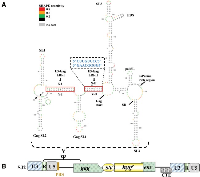
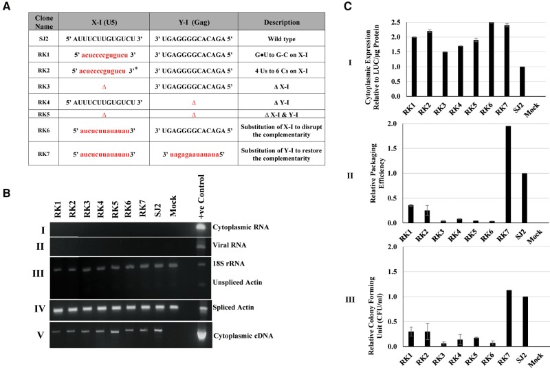
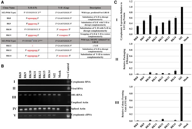
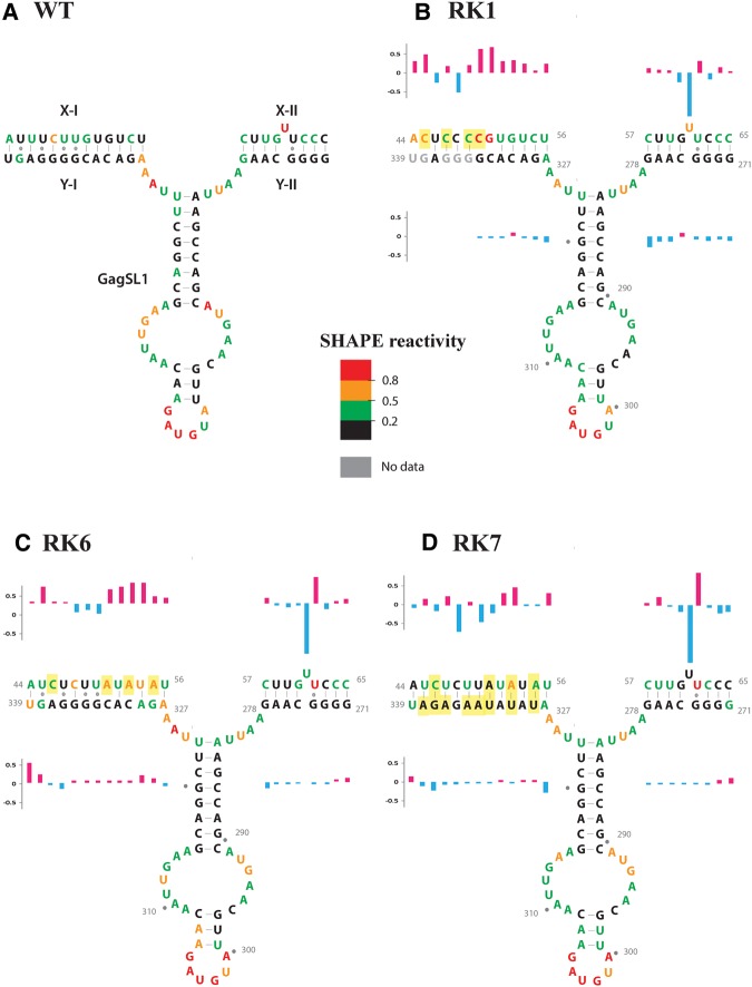
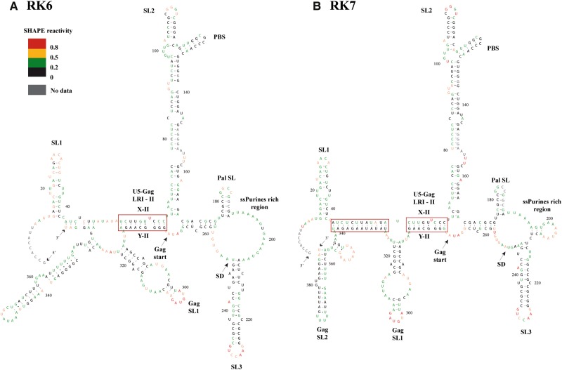
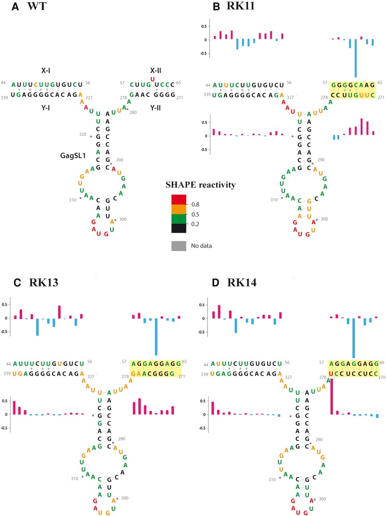
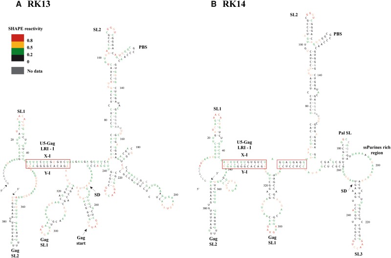
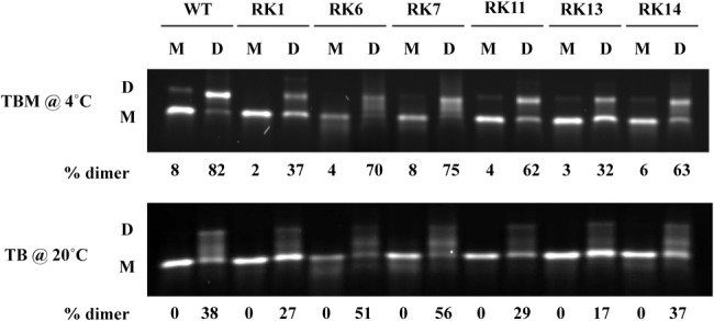
MPMV has great potential for development as a vector for gene therapy. In this respect, precisely defining the sequences and structural motifs that are important for dimerization and packaging of its genomic RNA (gRNA) are of utmost importance. A distinguishing feature of the MPMV gRNA packaging signal is two phylogenetically conserved long-range interactions (LRIs) between U5 and gag complementary sequences, LRI-I and LRI-II. To test their biological significance in the MPMV life cycle, we introduced mutations into these structural motifs and tested their effects on MPMV gRNA packaging and propagation. Furthermore, we probed the structure of key mutants using SHAPE (selective 2'hydroxyl acylation analyzed by primer extension). Disrupting base-pairing of the LRIs affected gRNA packaging and propagation, demonstrating their significance to the MPMV life cycle. A double mutant restoring a heterologous LRI-I was fully functional, whereas a similar LRI-II mutant failed to restore gRNA packaging and propagation. These results demonstrate that while LRI-I acts at the structural level, maintaining base-pairing is not sufficient for LRI-II function. In addition, in vitro RNA dimerization assays indicated that the loss of RNA packaging in LRI mutants could not be attributed to the defects in dimerization. Our findings suggest that U5-gag LRIs play an important architectural role in maintaining the structure of the 5' region of the MPMV gRNA, expanding the crucial role of LRIs to the nonlentiviral group of retroviruses.

Keywords: Mason-Pfizer monkey virus (MPMV); RNA packaging and dimerization; RNA secondary structure; SHAPE (selective 2′hydroxyl acylation analyzed by primer extension); long-range interactions (LRI); retroviruses.


Figures

FIGURE 1.
FIGURE 1.
SHAPE-validated structure of MPMV packaging signal RNA. (A) RNA structure of MPMV packaging signal after applying SHAPE constraints. Nucleotides are color-coded as per the SHAPE reactivity key. Arrows point to important structural elements of the virus genome. The solid boxed regions show the two long-range interactions (LRIs), LRI-I and LRI-II, observed in the structure. The SHAPE-validated structure corroborates well with the predicted structure except for subtle differences in LRI-II (the predicted LRI-II structure shown in the enlarged dashed box). A uridine residue (U62) is unpaired and forms a bulge in the SHAPE-validated, but not in the predicted LRI-II structure, while C65 is base-paired in the SHAPE-validated structure, but unpaired in the predicted structure (figure adapted with permission from Aktar et al. 2013). (B) Schematic representation of MPMV subgenomic transfer vector, SJ2 (Jaballah et al. 2010), in which region between R and 120 nt of Gag that has been shown to be important for MPMV RNA packaging and dimerization is demarcated. The same region was used to predict and validate the RNA secondary structure.
FIGURE 2.
FIGURE 2.
Effect of substitution/deletion mutations in U5-gag LRIs on MPMV gRNA packaging and propagation. (A) Table outlining the substitution/deletion mutations introduced into the U5-gag sequences of LRI-I and LRI-II. Sequences of the mutations introduced are represented in lower case and in red. In RK2, (*) denotes that only part of the U5 sequence for both LRIs is shown due to space limitations. (B) Representative gel images of the controls needed for validating different aspects of the three-plasmid in vivo packaging and propagation assay: (I and II) PCR amplification of DNase-treated RNA from the cytoplasmic and viral RNA preparations, respectively, with MPMV-specific vector primers; (III) multiplex amplification of unspliced β-actin mRNA and 18S rRNA; (IV) PCR amplification of spliced β-actin mRNA to check for the nucleocytoplasmic fractionation technique; and (V) PCR amplification of transfer vector cytoplasmic cDNAs using MPMV vector-specific primers. (C) Bar graphs represent: (I) the relative cytoplasmic expression of transfer vector RNAs in 293T cells relative to the wild-type (SJ2 vector) after normalization with the β-actin endogenous control and luciferase expression; (II) packaging efficiency of the mutant transfer vector RNAs relative to SJ2 after normalization with β-actin and luciferase expression; and (III) the relative propagation of MMTV transfer vector RNAs as measured by the luciferase-normalized hygromycin-resistant colony-forming units (CFU)/mL for mutant transfer vectors compared to the wild-type SJ2 vector. The data represented in the histograms correspond to the mean of samples tested in triplicates (±SD) following transfection and infection experiments.
FIGURE 3.
FIGURE 3.
Effect of deletion/substitution mutations introduced into U5-gag LRI-II on MPMV gRNA packaging and propagation. (A) Table outlining the deletion/substitution mutations introduced into the U5-gag sequences of LRI-II. Sequences of the introduced mutations are represented in lower case and in red. Two sets of mutants are outlined in the table. The first set of mutations were introduced into the Mfold-predicted 8-nt LRI-II structure, while the second set describes mutations introduced into the SHAPE-validated 9-nt LRI-II structure. (*) Denotes the 8-nt U5 sequence of the predicted LRI-II. (**) Denotes the 9-nt U5 sequence of the SHAPE-validated LRI-II. (B) Representative gel images of the controls needed for validating different aspects of the three-plasmid in vivo packaging and propagation assay: (I and II) PCR amplification of DNase-treated RNA from the cytoplasmic and viral RNA preparations, respectively, with MPMV-specific vector primers; (III) multiplex amplification of unspliced β-actin mRNA and 18S rRNA; (IV) PCR amplification of spliced β-actin mRNA to check for the nucleocytoplasmic fractionation technique; and (V) PCR amplification of transfer vector cytoplasmic cDNAs using MPMV vector-specific primers. (C) Bar graphs represent: (I) the relative cytoplasmic expression of transfer vector RNAs in 293T cells relative to the wild-type (SJ2 vector) after normalization with the β-actin endogenous control and luciferase expression; (II) packaging efficiency of the mutant transfer vector RNAs relative to SJ2 after normalization with β-actin and luciferase expression; and (III) the relative propagation of MMTV transfer vector RNAs as measured by the luciferase-normalized hygromycin resistant colony-forming units (CFU)/mL for mutant transfer vectors compared to the wild-type SJ2 vector. The data represented in the histograms correspond to the mean of samples tested in triplicates (±SD) following transfection and infection experiments.
FIGURE 4.
FIGURE 4.
SHAPE reactivity of the region corresponding to LRI-I/II and Gag SL1 of wild-type and mutant (RK1, RK6, and RK7) gRNAs. The SHAPE reactivity is color-coded on the SHAPE-validated structure of wild-type and mutant RNAs. (AD) The SHAPE reactivity is reported on the wild-type structure for the sake of comparison. For mutants RK1, RK6, and RK7, a structure similar to the wild-type structure is drawn to facilitate comparison of the SHAPE reactivity, but this structure does not necessarily reflect the actual structure (see text and Fig. 5 for details). The differences in reactivity in LRI-I and LRI-II in the mutants compared to wild-type are indicated by histograms (red and blue correspond to increase and decrease in reactivity, respectively). Mutations are highlighted in yellow.
FIGURE 5.
FIGURE 5.
SHAPE-validated RNA secondary structural models of mutant RK6 designed to disrupt LRI-I (A) and RK7 designed to restore LRI-I (B). Nucleotides are color-coded as per the SHAPE reactivity key. Major structural elements that also exist in wild-type gRNA are labeled as in Figure 1.
FIGURE 6.
FIGURE 6.
SHAPE reactivity of the region corresponding to LRI-I/II and Gag SL1 of wild-type gRNA and mutants RK11, RK13, and RK14. The SHAPE reactivity is color-coded on the SHAPE-validated structure of wild-type and mutant RNAs. (AD) The SHAPE reactivity is reported on the wild-type structure for the sake of comparison. For these mutants, RK11, RK13, and RK14, a structure similar to the wild-type structure is drawn to facilitate comparison of the SHAPE reactivity, but this structure does not necessarily reflect the actual structure (see text and Fig. 7 for details). The differences in reactivity in LRI-I and LRI-II in the mutants compared to wild-type are indicated by histograms (red and blue correspond to increase and decrease in reactivity, respectively). Mutations are highlighted in yellow.
FIGURE 7.
FIGURE 7.
SHAPE-validated RNA secondary structure models of mutant RK13 designed to disrupt the complementarity of LRI-II (A) and RK14 designed to restore a heterologous LRI-II (B). Nucleotides are color-coded as per the SHAPE reactivity key. Structural elements that also exist in wild-type gRNA are labeled as in Figure 1.
FIGURE 8.
FIGURE 8.
In vitro RNA dimerization on SHAPE-interrogated LRI mutants. Wild-type and mutant RNAs were incubated in monomer (M) or dimer (D) buffer and analyzed by electrophoresis on 1% agarose gel either in TBM buffer at 4°C (top panel) or in TB buffer at 20°C (lower panel). Experiments were performed in duplicates and the mean dimerization percent is indicated below each lane.

Similar articles

Cited by

References

    1. Abbink TE, Berkhout B. 2003. A novel long distance base-pairing interaction in human immunodeficiency virus type 1 RNA occludes the Gag start codon. J Biol Chem 278: 11601–11611. - PubMed
    1. Abd El-Wahab EW, Smyth RP, Mailler E, Bernacchi S, Vivet-Boudou V, Hijnen M, Jossinet F, Mak J, Paillart JC, Marquet R. 2014. Specific recognition of the HIV-1 genomic RNA by the Gag precursor. Nat Commun 5: 4304. - PubMed
    1. Aktar SJ, Jabeen A, Ali LM, Vivet-Boudou V, Marquet R, Rizvi TA. 2013. SHAPE analysis of the 5′ end of the Mason-Pfizer monkey virus (MPMV) genomic RNA reveals structural elements required for genome dimerization. RNA 19: 1648–1658. - PMC - PubMed
    1. Aktar SJ, Vivet-Boudou V, Ali LM, Jabeen A, Kalloush RM, Richer D, Mustafa F, Marquet R, Rizvi TA. 2014. Structural basis of genomic RNA (gRNA) dimerization and packaging determinants of mouse mammary tumor virus (MMTV). Retrovirology 11: 96. - PMC - PubMed
    1. Al Dhaheri NS, Phillip PS, Ghazawi A, Ali J, Beebi E, Jaballah SA, Rizvi TA. 2009. Cross-packaging of genetically distinct mouse and primate retroviral RNAs. Retrovirology 6: 66. - PMC - PubMed

Publication types

LinkOut - more resources