

Complementarity of medium-throughput in situ RNA hybridization and tissue-specific transcriptomics: case study of Arabidopsis seed development kinetics
- PMID: 27095274
- PMCID: PMC4837347
- DOI: 10.1038/srep24644
Complementarity of medium-throughput in situ RNA hybridization and tissue-specific transcriptomics: case study of Arabidopsis seed development kinetics
Abstract
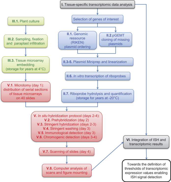
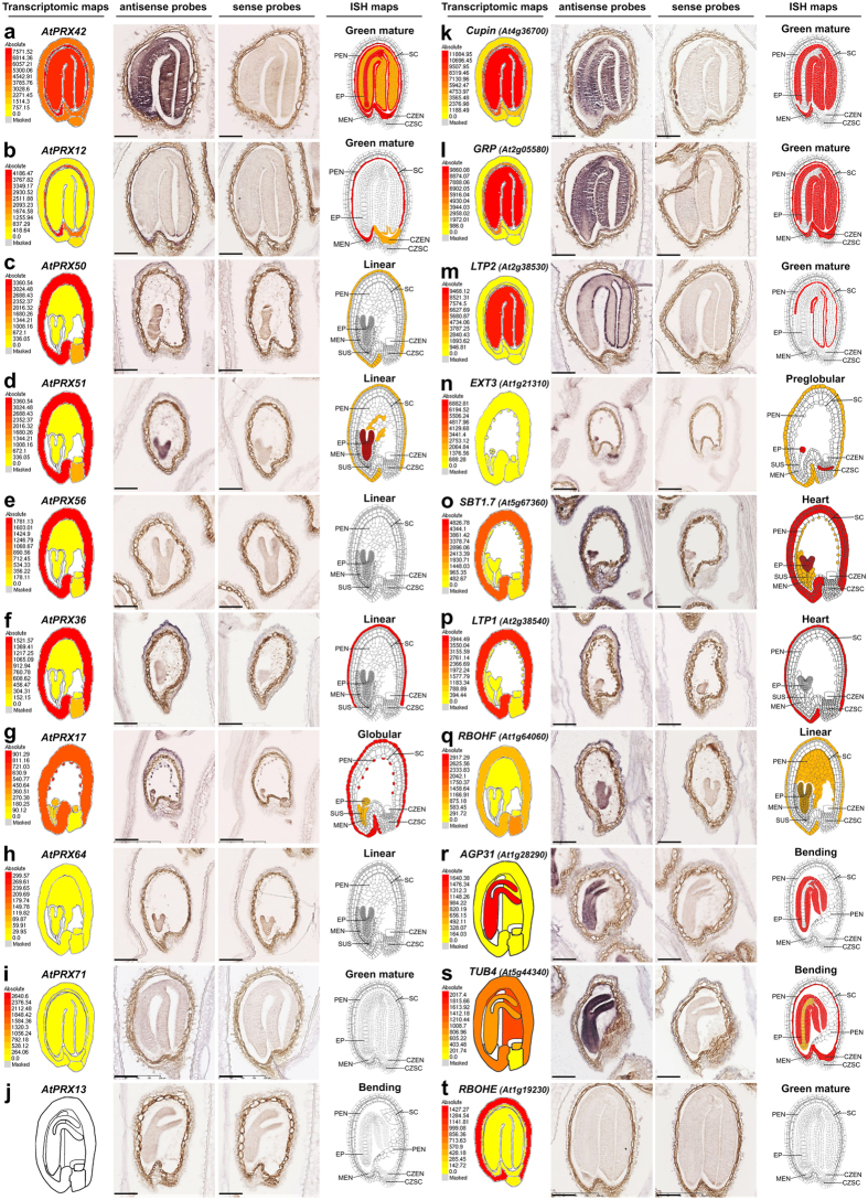
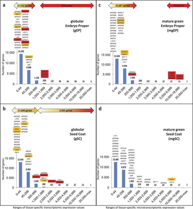
The rationale of this study is to compare and integrate two heterologous datasets intended to unravel the spatiotemporal specificities of gene expression in a rapidly growing and complex organ. We implemented medium-throughput RNA in situ hybridization (ISH) for 39 genes mainly corresponding to cell wall proteins for which we have particular interest, selected (i) on their sequence identity (24 class III peroxidase multigenic family members and 15 additional genes used as positive controls) and (ii) on their expression levels in a publicly available Arabidopsis thaliana seed tissue-specific transcriptomics study. The specificity of the hybridization signals was carefully studied, and ISH results obtained for the 39 selected genes were systematically compared with tissue-specific transcriptomics for 5 seed developmental stages. Integration of results illustrates the complementarity of both datasets. The tissue-specific transcriptomics provides high-throughput possibilities whereas ISH provides high spatial resolution. Moreover, depending on the tissues and the developmental stages considered, one or the other technique appears more sensitive than the other. For each tissue/developmental stage, we finally determined tissue-specific transcriptomic threshold values compatible with the spatiotemporally-specific detection limits of ISH for lists of hundreds to tens-of-thousands of genes.
Figures
Similar articles
-
Medium-Throughput RNA In Situ Hybridization of Serial Sections from Paraffin-Embedded Tissue Microarrays.Methods Mol Biol. 2019;1933:99-130. doi: 10.1007/978-1-4939-9045-0_6. Methods Mol Biol. 2019. PMID: 30945181
-
Computational and experimental analysis identifies Arabidopsis genes specifically expressed during early seed development.BMC Genomics. 2006 Feb 28;7:38. doi: 10.1186/1471-2164-7-38. BMC Genomics. 2006. PMID: 16504176 Free PMC article.
-
Combining association mapping and transcriptomics identify HD2B histone deacetylase as a genetic factor associated with seed dormancy in Arabidopsis thaliana.Plant J. 2013 Jun;74(5):815-28. doi: 10.1111/tpj.12167. Epub 2013 Apr 4. Plant J. 2013. PMID: 23464703
-
Transcriptomics approaches in the early Arabidopsis embryo.Trends Plant Sci. 2013 Sep;18(9):514-21. doi: 10.1016/j.tplants.2013.04.011. Epub 2013 May 29. Trends Plant Sci. 2013. PMID: 23726727 Review.
-
A primer for generating and using transcriptome data and gene sets.Development. 2020 Dec 23;147(24):dev193854. doi: 10.1242/dev.193854. Development. 2020. PMID: 33361089 Free PMC article. Review.
Cited by
-
Immunochemical Identification of the Main Cell Wall Polysaccharides of the Early Land Plant Marchantia polymorpha.Cells. 2023 Jul 12;12(14):1833. doi: 10.3390/cells12141833. Cells. 2023. PMID: 37508498 Free PMC article.
-
A Comparative Study of Sample Preparation for Staining and Immunodetection of Plant Cell Walls by Light Microscopy.Front Plant Sci. 2017 Aug 29;8:1505. doi: 10.3389/fpls.2017.01505. eCollection 2017. Front Plant Sci. 2017. PMID: 28900439 Free PMC article.
-
An integrative Study Showing the Adaptation to Sub-Optimal Growth Conditions of Natural Populations of Arabidopsis thaliana: A Focus on Cell Wall Changes.Cells. 2020 Oct 7;9(10):2249. doi: 10.3390/cells9102249. Cells. 2020. PMID: 33036444 Free PMC article.
-
TBL38 atypical homogalacturonan-acetylesterase activity and cell wall microdomain localization in Arabidopsis seed mucilage secretory cells.iScience. 2024 Apr 5;27(5):109666. doi: 10.1016/j.isci.2024.109666. eCollection 2024 May 17. iScience. 2024. PMID: 38665206 Free PMC article.
-
Demystifying biotrophs: FISHing for mRNAs to decipher plant and algal pathogen-host interaction at the single cell level.Sci Rep. 2020 Aug 31;10(1):14269. doi: 10.1038/s41598-020-70884-4. Sci Rep. 2020. PMID: 32868853 Free PMC article.
References
Publication types
MeSH terms
LinkOut - more resources
Full Text Sources
Other Literature Sources
Research Materials
