. 2016 May 27:622:30-6.
doi: 10.1016/j.neulet.2016.04.033. Epub 2016 Apr 16.

Confounding effect of EEG implantation surgery: Inadequacy of surgical control in a two hit model of temporal lobe epilepsy

Affiliations

Confounding effect of EEG implantation surgery: Inadequacy of surgical control in a two hit model of temporal lobe epilepsy

Irena Balzekas et al. Neurosci Lett. .

Abstract

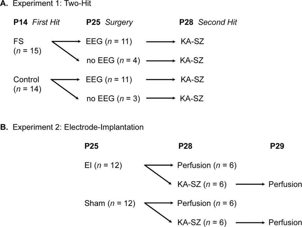
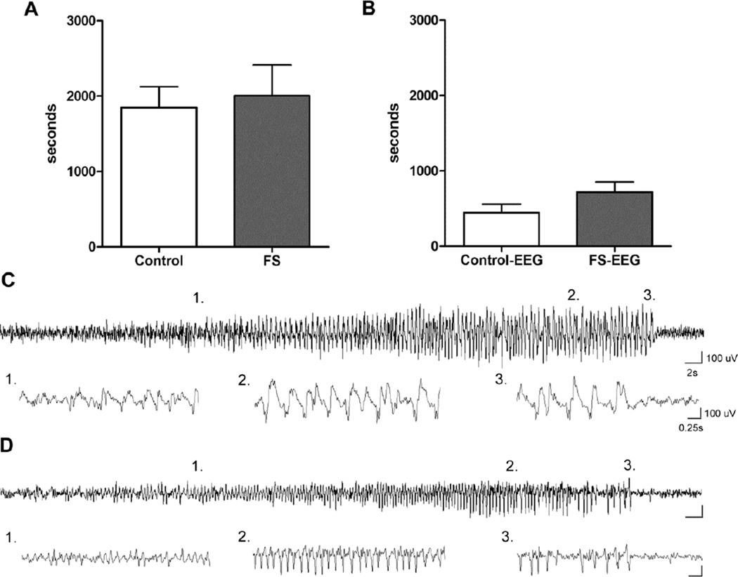
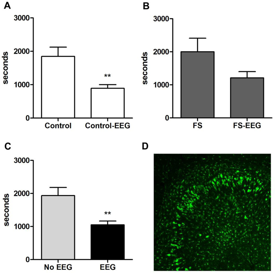
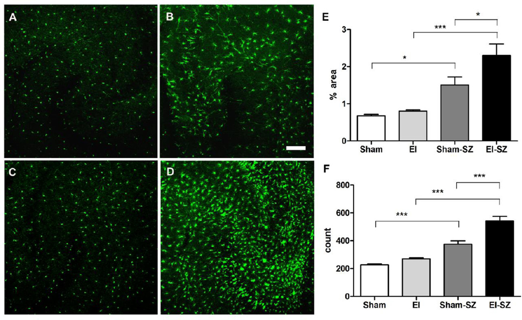
In rodent models of epilepsy, EEG implantation surgery is an essential modality to evaluate electrographic seizures. The inflammatory consequences of EEG electrode-implantation and their resultant effects on seizure susceptibility are unclear. We evaluated electrode-implantation in a two-hit model of epileptogenesis in C57BL/6 mice that included brief, recurrent febrile seizures (FS) at P14 and kainic acid induced seizures (KA-SZ) at P28. During KA-SZ, latencies to first electrographic and behavioral seizures, seizure severity, and KA dose sensitivity were measured. Mice that received subdural screw electrode implants at P25 for EEG monitoring at P28 had significantly shorter latencies to seizures than sham mice, regardless of early life seizure experience. Electrode-implanted mice were sensitive to low dose KA as shown by high mortality rate at KA doses above 10mg/kg. We then directly compared electrode-implantation and KA-SZ in seizure naive CX3CR1(GFP/+) transgenic C57BL/6 mice, wherein microglia express green fluorescent protein (GFP), to determine if microglia activation related to surgery was associated with the increased seizure susceptibility in electrode-implanted mice from the two-hit model. Hippocampal microglia activation, as demonstrated by percent area GFP signal and GFP positive cell counts, prior to seizures was indistinguishable between electrode-implanted mice and controls, but was significantly greater in electrode-implanted mice following seizures. Electrode-implantation had a confounding priming effect on the inflammatory response to subsequent seizures.

Keywords: EEG; Epilepsy; Inflammation; Kainic acid; Microglia; Surgery.


Figures

Fig. 1
Fig. 1
Experimental Design.
Fig. 2
Fig. 2
Lack of effect of brief, recurrent FS on later life seizure susceptibility. A. Latency to onset of behavioral seizures (GIII) during KA-SZ at P28 in mice with prior exposure to febrile seizures at P14 and their control littermates (n = 4; 3, p = 0.78). These mice had no EEG implants and underwent only sham surgery at P25. B. Latency to first electrographic seizures was also similar between FS and control (n = 11/groups, p = 0.13). C. Example of first electrographic seizure detected and accompanied by subtle behavioral changes: comparison between control mouse (top panel) and D. FS mouse (bottom panel).
Fig. 3
Fig. 3
Significant increase in susceptibility to KA-SZ and marked seizure-induced microglia activation after EEG implantation. A. Latency to first behavioral seizures (GIII) in sham-operated control mice (Control-sham) compared to EEG-implanted control mice (Control-EEG) (n = 3 vs n = 11, p = 0.0027). Neither group experienced early-life FS. B. Latency to first behavioral seizures (GIII) in sham-operated FS mice (FS-sham) compared to EEG-implanted FS mice (FS-EEG) (n = 4 vs n = 11, p = 0.065) C. Pooled behavioral seizure latency data comparing all mice (FS and controls) with versus without EEG implants (1054 ± 111.7 seconds vs 1937 ± 243.9 seconds; n = 22 vs n = 7, p = 0.001). D. Example of activated microglia in hippocampus of EEG-implanted control CX3CR1GFP/+ mouse. The mouse experienced the first hit control condition, EEG implantation, and second hit KA-SZ. GFP positive cells were enlarged and abundant near the CA3 hippocampal subfields where prolonged KA-SZ are known to cause neuronal death.
Fig. 4
Fig. 4
Significant increase in inflammation after KA-SZ with electrode implantation. A-D: Representative confocal images of hippocampi and microglia morphology at P28 A. Hippocampus of sham mouse (Sham); B. Hippocampus of sham mouse 24 hours after KA-SZ (Sham-SZ); C. Hippocampus of electrode-implanted mouse 3 days after surgery (EI); D. Hippocampus of electrode-implanted mouse 3 days after surgery and 24 hours after KA-SZ (EI-SZ); E. Percent area green fluorescent signal in CA3 region of sham mice (Sham), electrode-implanted mice (EI), sham mice after KA (Sham-SZ), and electrode-implanted mice after KA (EISZ), n = 6/group, p < 0.0001, One-way ANOVA with post-hoc Tukey test (* p<0.05, ***p < 0.001). F. GFP positive cell count in CA3 region, n = 6/group, p = 0.001, One-way ANOVA with post-hoc Tukey test (***p < 0.001).

References

    1. Abraham J, Fox PD, Condello C, Bartolini A, Koh S. Minocycline attenuates microglia activation and blocks the long-term epileptogenic effects of early-life seizures. Neurobiol Dis. 2012;46:425–430. - PMC - PubMed
    1. Aihara N, Hall JJ, Pitts LH, Fukuda K, Noble LJ. Altered immunoexpression of microglia and macrophages after mild head injury. J Neurotrauma. 1995;12:53–63. - PubMed
    1. Annegers JF, Hauser WA, Shirts SB, Kurland LT. Factors prognostic of unprovoked seizures after febrile convulsions. N Engl J Med. 1987;316:493–498. - PubMed
    1. Benson MJ, Manzanero S, Borges K. Complex alterations in microglial M1/M2 markers during the development of epilepsy in two mouse models. Epilepsia. 2015;56:895–905. - PubMed
    1. Dube C, Richichi C, Bender RA, Chung G, Litt B, Baram TZ. Temporal lobe epilepsy after experimental prolonged febrile seizures: prospective analysis. Brain. 2006;129:911–922. - PMC - PubMed

Publication types

MeSH terms

LinkOut - more resources