Review
. 2016 Jun 9;127(23):2814-23.
doi: 10.1182/blood-2016-03-378588. Epub 2016 Apr 19.

Inherited platelet disorders: toward DNA-based diagnosis

Affiliations
Review

Inherited platelet disorders: toward DNA-based diagnosis

Claire Lentaigne et al. Blood. .

Abstract

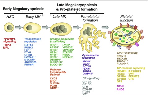
Variations in platelet number, volume, and function are largely genetically controlled, and many loci associated with platelet traits have been identified by genome-wide association studies (GWASs).(1) The genome also contains a large number of rare variants, of which a tiny fraction underlies the inherited diseases of humans. Research over the last 3 decades has led to the discovery of 51 genes harboring variants responsible for inherited platelet disorders (IPDs). However, the majority of patients with an IPD still do not receive a molecular diagnosis. Alongside the scientific interest, molecular or genetic diagnosis is important for patients. There is increasing recognition that a number of IPDs are associated with severe pathologies, including an increased risk of malignancy, and a definitive diagnosis can inform prognosis and care. In this review, we give an overview of these disorders grouped according to their effect on platelet biology and their clinical characteristics. We also discuss the challenge of identifying candidate genes and causal variants therein, how IPDs have been historically diagnosed, and how this is changing with the introduction of high-throughput sequencing. Finally, we describe how integration of large genomic, epigenomic, and phenotypic datasets, including whole genome sequencing data, GWASs, epigenomic profiling, protein-protein interaction networks, and standardized clinical phenotype coding, will drive the discovery of novel mechanisms of disease in the near future to improve patient diagnosis and management.


Figures

Figure 1
Figure 1
The 51 genes underlying IPDs. The cartoon depicts the process of megakaryopoiesis and platelet formation. Each of the 51 known IPD genes are indicated and categorized according to their effect on megakaryocyte and platelet biology. *IPDs typically associated with phenotypes outside of the blood system. HSC, hematopoietic stem cell.
Figure 2
Figure 2
Expression levels of 51 genes underlying IPDs across hematopoietic stem and progenitor cells. High relative expression is shown in red and low relative expression in blue. The expression of each gene is normalized relative to the mean expression across all samples. Genes are ordered and color-coded according to their predicted effect on platelet biology (as in Fig. 1). Information about the levels of transcripts for the 51 genes determined by RNA-seq was retrieved from Chen et al. HSC, hematopoietic stem cell; MPP, multipotent progenitor; CLP, common lymphoid progenitor; CMP, common myeloid progenitor; GMP, granulocyte-monocyte progenitor; MEP, megakaryocyte-erythrocyte precursor; EB, erythroblast; MK, megakaryocyte.
Figure 3
Figure 3
Genomic location of the 51 genes underlying IPDs. Circos diagram illustrating the location of known IPD genes across human chromosomes. Track 1: Cytoband with chromosome name with centromeres in blue. Track 2: Genomic location of 51 established IPD genes and the year in which variants in the gene were first identified as a cause of IPD in humans in brackets. Gene names in red represent genes identified by HTS. Track 3: Log10 of the number of amino acids encoded by the reference CCDS transcript. Log10 scale is indicated at 12 o’clock. Track 4: Log10 of the number of rare variants predicted to affect amino acid sequence observed in 6390 individuals enrolled to the NIHR BioResource–Rare Diseases. Log10 scale is indicated at 12 o’clock.
Figure 4
Figure 4
Protein–protein interaction network reflecting the molecules and pathways implicated in megakaryopoiesis, the formation of platelets, thrombosis, and hemostasis. (A) PPIN of 1684 nodes (proteins) connected by 5360 edges (biochemical reactions). The 1517 first-order interacting nodes and all but 24 of the 5360 edges were obtained from the Reactome (n = 3625) and IntAct (n = 1711) databases. The 24 edges were added on the basis of manual literature curation. (B) The 200 baits are colored as per the Venn diagram, except for the 8 baits present in >1 category, which are pink, and the 8 prototype proteins involved in the synthesis of thromboxane and signaling via the thromboxane receptor (Tbxa2r) pathway. The Venn diagram shows the 3 gene sets in ochre, blue, and purple for the ThromboGenomics HTS test platform gene set, the platelet volume and count GWAS gene set, or the gene set identified by ChIP-seq in human megakaryocytes and showing binding of all 5 transcription factors (Fli1, Gata1, Gata2, Runx1, and Tal1) at their promoter, respectively. (C-D) Subnetworks retrieved from the PPIN in A. (C) A subnetwork of 156 nodes and 874 edges obtained by retrieving the first-order interactors of Tbxa2r, the receptor for thromboxane. (D) A subnetwork of 26 nodes and 42 edges involved in the synthesis of thromboxane and obtained by selecting the first-order interactors of Tbxas1 (thromboxane synthase 1) and Pla2g4a (phospholipase A2). The red nodes in C and D are a set of prototype proteins related to thromboxane synthesis and signaling and the other colored nodes are baits. The surface areas of the red colored nodes in A and all colored nodes in C and D reflect their transcript level determined by sequencing of RNA from human megakaryocytes (data retrieved from Chen et al). An interactive version of the network, containing gene expression levels and other annotation features, is available for download in Cytoscape format from the supplemental Data.

References

    1. Gieger C, Radhakrishnan A, Cvejic A, et al. New gene functions in megakaryopoiesis and platelet formation. Nature. 2011;480(7376):201–208. - PMC - PubMed
    1. Westbury SK, Turro E, Greene D, et al. BRIDGE-BPD Consortium. Human phenotype ontology annotation and cluster analysis to unravel genetic defects in 707 cases with unexplained bleeding and platelet disorders. Genome Med. 2015;7(1):36. - PMC - PubMed
    1. Dasouki MJ, Rafi SK, Olm-Shipman AJ, et al. Exome sequencing reveals a thrombopoietin ligand mutation in a Micronesian family with autosomal recessive aplastic anemia. Blood. 2013;122(20):3440–3449. - PMC - PubMed
    1. Dasouki M, Roberts J, Santiago A, Saadi I, Hovanes K. Confirmation and further delineation of the 3q26.33-3q27.2 microdeletion syndrome. Eur J Med Genet. 2014;57(2-3):76–80. - PubMed
    1. Ihara K, Ishii E, Eguchi M, et al. Identification of mutations in the c-mpl gene in congenital amegakaryocytic thrombocytopenia. Proc Natl Acad Sci USA. 1999;96(6):3132–3136. - PMC - PubMed

Publication types

MeSH terms