

Obliquity of the Stapes in Otosclerosis: A New Radiological Sign
- PMID: 27096011
- PMCID: PMC4835337
- DOI: 10.1055/s-0036-1579743
Obliquity of the Stapes in Otosclerosis: A New Radiological Sign
Abstract
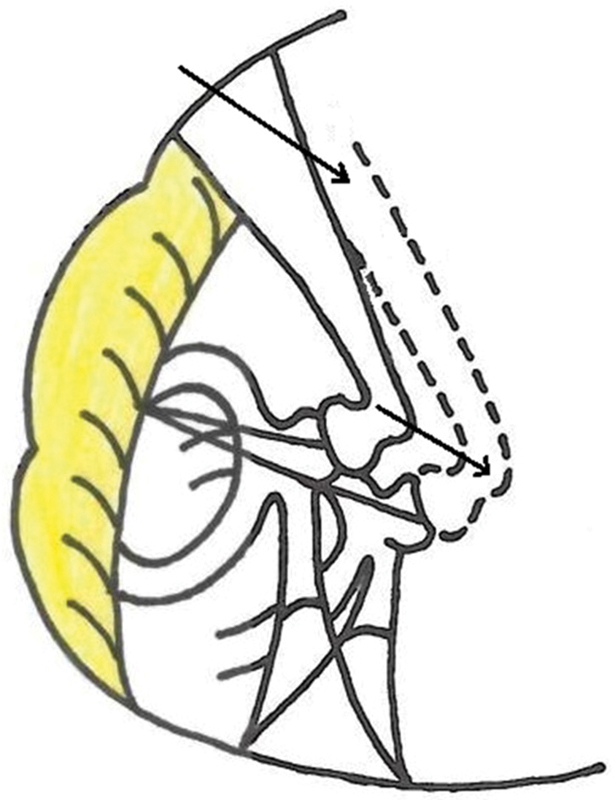
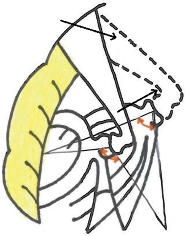
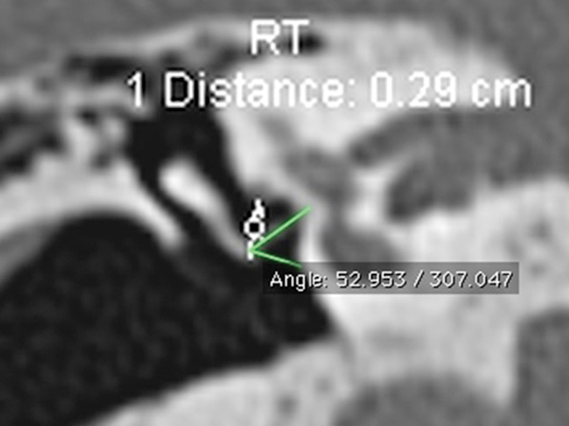
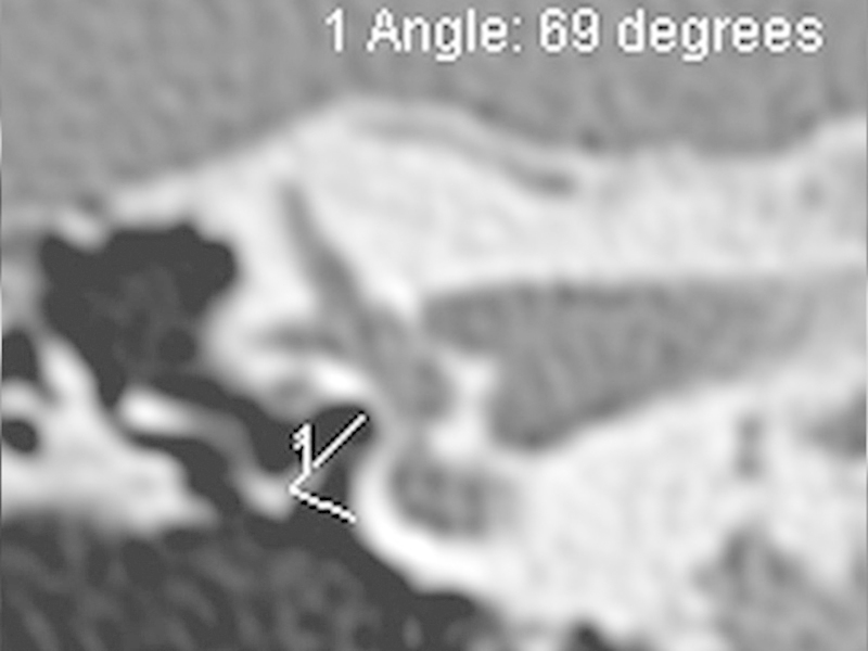
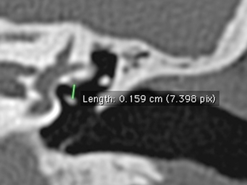
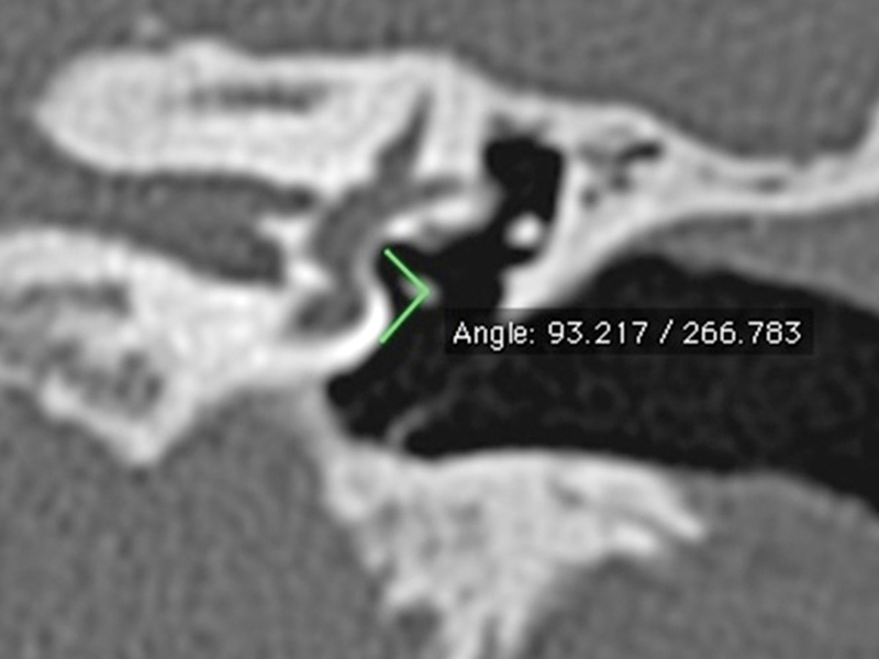
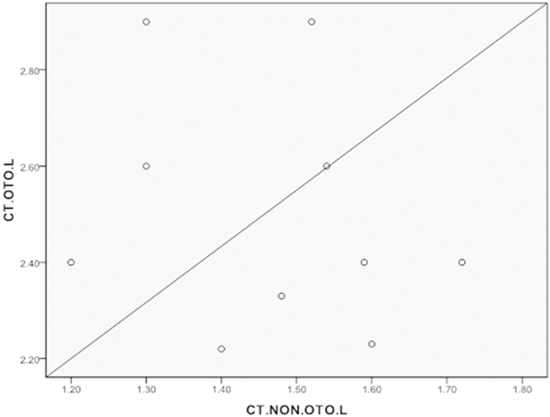
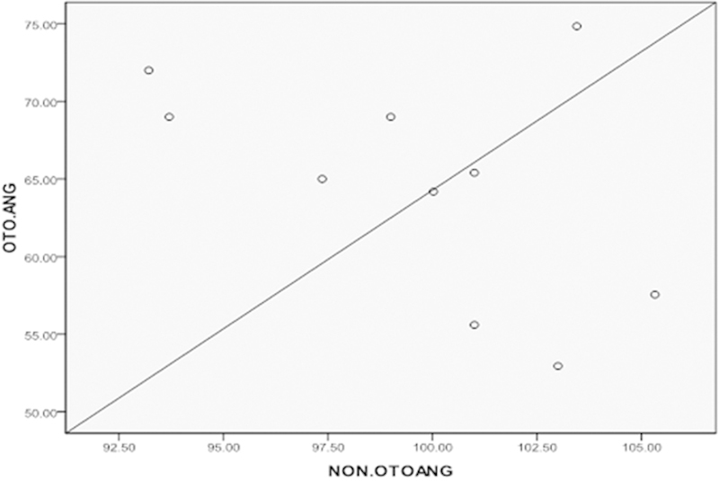
Introduction Observing the obliquity of stapes by closely scrutinizing the HRCT temporal bone in otosclerosis revealed a reliable and consistent finding. This finding can add to the existing radiological criteria in diagnosis of otosclerosis. Objective The objective of this study is to establish the obliquity of stapes in otosclerosis by radiological measurements using HRCT temporal bone by comparing: (a) the distance between the horizontal (tympanic) segment of facial nerve and stapes head in otosclerotic ears (study group) with non-otosclerotic ears (control group); and (b) the angle subtended by stapes with promontory in the study and control groups. Methods This is a prospective study performed after the institutional Ethics Committee clearance (IEC 3/2013). Results An increased mean distance between the horizontal segment of facial nerve and stapes head in otosclerotic patients (i.e., 2.49mm +/- 0.24mm SD), when compared with the non-otosclerotic patients (i.e., 1.46mm +/- 0.16mm SD) is noted. There is a change in angle (i.e., 64.550 +/- 7.190 SD) subtended by the stapes toward the promontory in otosclerotic ears when compared with that of controls (i.e., 99.700 +/- 40 SD). We applied the Mann-Whitney U non-parametric test and considered p value of < 0.0001 highly significant. Conclusions Obliquity of stapes in otosclerosis referred to as a "Pisa" sign by the senior author has diagnostic value as a new radiological sign in imaging of otosclerosis. This obliquity explains the torsional effect of otosclerosis on the ossicular chain. The findings correlate with late complications and failures in stapes surgery.
Keywords: obliquity; otosclerosis; stapes; temporal bone.
Conflict of interest statement
Figures
References
-
- Politzer A. Uber primare Erkrankung der Knocheren Labyrinth-Kapsel. Johrenheilk. 1894;25:309–327.
-
- Anson B J, Cauldwell E W, Bast T H. The fissula ante fenestram of the human otic capsule; developmental and normal adult structure. Ann Otol Rhinol Laryngol. 1947;56(4):957–985. - PubMed
-
- Bast T H. Development of otic capsule. Residual cartilages and defective ossification and their relation to otosclerotic foci. Arch Otolaryngol. 1940;32:771–782.
-
- Anson B J, Cauldwell E W, Bast T H. The fissula ante fenestram of the human otic capsule; aberrant form and contents. Ann Otol Rhinol Laryngol. 1948;57(1):103–128. - PubMed
-
- Swartz D J, Loevener A L. New York, USA: Thieme; 2009. Imaging of the Temporal bone. 4th ed.
LinkOut - more resources
Full Text Sources
Other Literature Sources