. 2016 Jun 2;15(11):1450-61.
doi: 10.1080/15384101.2016.1175256. Epub 2016 Apr 20.

PP2A regulates kinetochore-microtubule attachment during meiosis I in oocyte

Affiliations

PP2A regulates kinetochore-microtubule attachment during meiosis I in oocyte

An Tang et al. Cell Cycle. .

Abstract

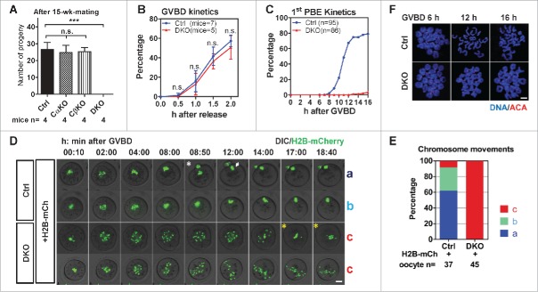
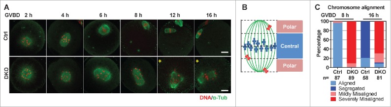
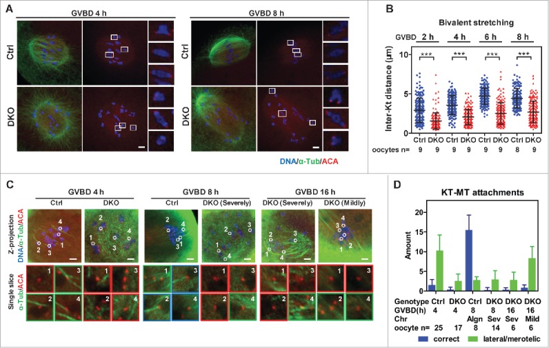
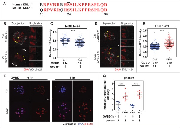
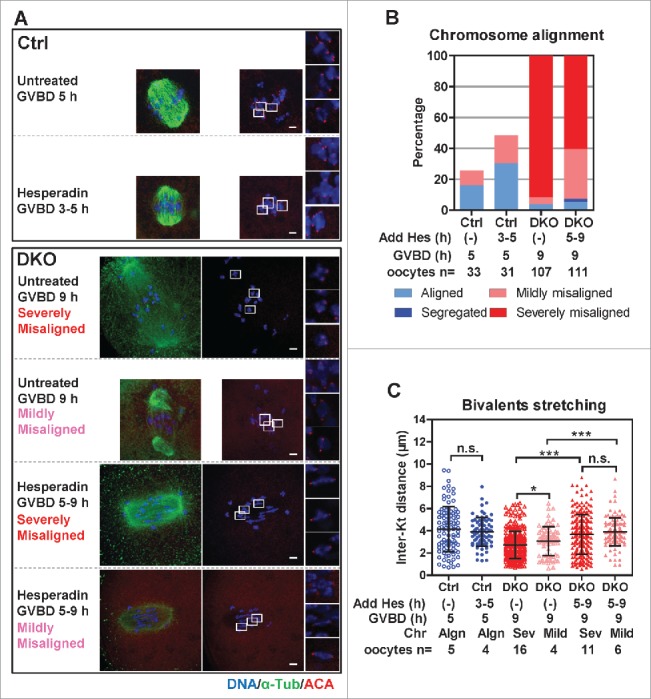
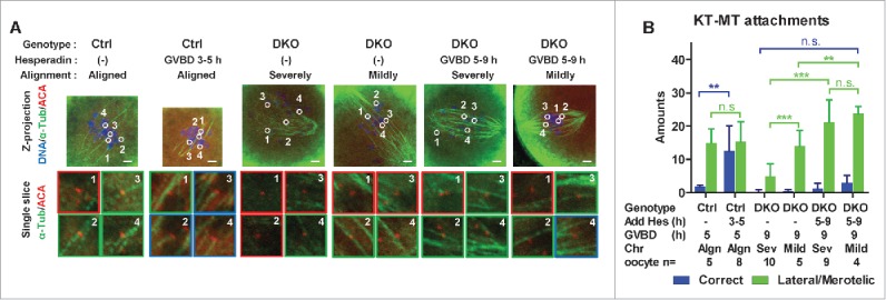
Studies using in vitro cultured oocytes have indicated that the protein phosphatase 2A (PP2A), a major serine/threonine protein phosphatase, participates in multiple steps of meiosis. Details of oocyte maturation regulation by PP2A remain unclear and an in vivo model can provide more convincing information. Here, we inactivated PP2A by mutating genes encoding for its catalytic subunits (PP2Acs) in mouse oocytes. We found that eliminating both PP2Acs caused female infertility. Oocytes lacking PP2Acs failed to complete 1(st) meiotic division due to chromosome misalignment and abnormal spindle assembly. In mitosis, PP2A counteracts Aurora kinase B/C (AurkB/C) to facilitate correct kinetochore-microtubule (KT-MT) attachment. In meiosis I in oocyte, we found that PP2Ac deficiency destabilized KT-MT attachments. Chemical inhibition of AurkB/C in PP2Ac-null oocytes partly restored the formation of lateral/merotelic KT-MT attachments but not correct KT-MT attachments. Taken together, our findings demonstrate that PP2Acs are essential for chromosome alignments and regulate the formation of correct KT-MT attachments in meiosis I in oocytes.

Keywords: KT-MT attachment; PP2A; fertility; meiosis; oocyte.


Figures

Figure 1.
Figure 1.
Loss of PP2Acs in oocytes caused female infertility and bivalent segregation failure in meiosis I. (A) 6-wk-old female mice were mated with wild-type male C57BL/6J mice for 15 wks. Cumulative number of pups between Ctrl, Ppp2caf/f:Zp3-Cre (Cα KO), Ppp2cbf/f:Zp3-Cre (Cβ KO) and DKO females were compared (mean ± SD). Experiments were repeated twice and representative result is shown. Numbers of female mice were indicated. (B) DKO oocytes had normal GVBD kinetics. The percentage of oocytes that had undergone GVBD was scored every 0.5 h after release from IBMX (mean ± SD). Experiments were repeated 3 times and a representative result is shown. The numbers of mice are indicated. (C) Oocytes from DKO mice could not extrude 1st PBs. Kinetics of 1st PBE after GVBD are shown. Oocytes that had undergone GVBD within 1.5 h after release into IBMX-free medium were selected for further culture. 1st PBE was scored every 1 h. Data were collected from 3 independent experiments. The numbers of oocytes are indicated. (D) Chromosomes failed to segregate in DKO oocytes. Still images with times from representative live imaging series are shown. The DIC (differential interference contrast) channel and H2B-mCherry channel were merged together. Genotypes of oocytes and injected mRNA (H2B-mCh for H2B-mCherry) are indicated. Types of chromosome movements are labeled on the right side of each series. Time is shown in h:min after GVBD. The white asterisk indicates the chromosome segregation in Ctrl oocytes; the white arrow indicates the metaphase II plate; yellow asterisks indicate unstable chromosome alignment in DKO oocytes. Bar = 20 μm. (E) Comparison of the frequencies of different types of chromosome segregation in oocytes. The numbers of ooyctes used for analysis are indicated. Data were collected from 3 independent experiments. (F) Bivalents failed to separate in DKO oocytes after GVBD. Chromosome spreads were prepared from the indicated time after GVBD and immunostained for DNA and centromeres (ACA, anti-centromere antibodies). Bar = 5 μm. Statistics: n.s., no significant difference; ***, P < 0.001.
Figure 2.
Figure 2.
Bivalents were severely misaligned in DKO oocytes. (A) Bivalents in DKO oocytes were misaligned after GVBD. Oocytes were fixed at the indicated times after GVBD and immunostained for DNA and α-tubulin. Representative z-projected confocal images are shown. The yellow asterisks indicate the DKO oocytes containing multipolar spindles. Bar = 20 μm. (B) Cartoon showing the possible chromosome distribution in a spindle during meiosis I. Spindle area was 1:2:1 divided into a central region and the 2 flanking polar regions. Four different Chromosome alignments were distinguished: chromosomes aligned inside the central region (defined as ‘aligned’); no more than 6 chromosomes distributed in the polar regions (defined as ‘mildly misaligned’); more than 6 chromosomes distributed in the polar regions (defined as ‘severely misaligned’); chromosomes were separated (defined as ‘segregated’). (C) Chromosomes in the majority of DKO oocytes were severely misaligned after GVBD. The frequencies of 4 types of chromosome alignments in Ctrl and DKO oocytes at GVBD 8 h and GVBD 16 h are shown. The numbers of oocytes used for analysis are indicated. Data were collected from 3 independent experiments
Figure 3.
Figure 3.
Lack of bivalent stretching and stable KT-MT attachments in DKO oocytes. (A) Oocytes were immunostained for DNA, α-tubulin and KTs (Anti-centromere antibodies, ACA) to determine bivalent biorientation and stretching. Representative z-projected confocal images of Ctrl and DKO oocytes at GVBD 4 h and GVBD 8 h are shown. Magnified views for 3 representative bivalents in each oocyte are shown in the right side of each z-projected image. Bar = 5 μm. (B) Inter-KT distances of bivalents in DKO and Ctrl oocytes at indicated times after GVBD were plotted (mean ± SD are shown). The numbers of oocytes used for analysis are indicated. Representative result from 3 independent experiments. (C) Oocytes at indicated time points were ice-treated and immunostained for DNA, α-tubulin and KTs (ACA) to determine KT-MT attachments. Representative z-projected images of the whole spindles are shown in the upper row. Magnified views of the KT-MT attachments, indicated by circles and numbers in the upper row, from a single slice, are shown in the lower rows. The types of misalignment of DKO oocytes are indicated. Red panes indicate no attachment; Green panes indicate lateral/merotelic attachments; blue panes indicate correct attachments. Bar = 5 μm. (D) KT-MT attachments in oocytes at times after GVBD were analyzed. Correct attachments (blue column) and lateral/merotelic attachments (green column) were scored (mean ± SD). Chromosome alignments (Algn, aligned; Sev, severely misaligned; Mild, mildly misaligned), numbers of oocytes used for analysis are indicated. Representative result from 3 independent experiments. Statistics: ***, P < 0.001.
Figure 4.
Figure 4.
Phosphorylation of hKNL1-ser24 and pH3s10 were increased DKO oocytes. A polyclonal antibody recognizing the phosphorylated ser24 in hKNL1 protein (hKNL1-ser24) of KMN network has been developed based on the human protein sequences by Iain M.Cheeseman's group. (A) Similarity of the immunogenic peptide sequences of hKNL1-ser24 was compared between human and mouse. Identical residues are indicated in red font. The black box marks the Serine 24 phosphorylation site. (B) Feasibility of the antibody was evaluated in Ctrl oocytes. Ctrl oocytes were fixed at 2 h or 8 h after GVBD and immunostained for DNA and hKNL1-ser24. Representative z-projected images are shown in the left. Magnified views of the single chromosomes, indicated by the yellow arrows, from single slices are shown in the right panels. Note that the anti-hKNL1-ser24 antibody also targeted in the area of MTOCs. Bar = 5 μm. (C) The anti-hKNL1-ser24 antibody is feasible for immunostaining mouse oocytes. Intensities of hKNL1-ser24 signals on KTs were normalized to the average of Ctrl oocytes at GVBD 2 h and plotted (mean ± SD bars are shown). Numbers of oocytes used for analysis are indicated. Representative result from 3 independent experiments. (D) Oocytes were fixed at 6 h after GVBD and immunostained for DNA and hKNL1-ser24. Z-projected images of all the chromosomes are shown in the left panel. Magnified views of the representative single chromosomes, indicated by the yellow arrows, from single slices are shown in the right panels. Bar = 5 μm. (E) The intensities of the hKNL1-ser24 signal on KTs were normalized to the average intensity of Ctrl oocytes at GVBD 6 h and plotted (mean ± SD bars are shown). The numbers of oocytes used for analysis are indicated. Representative result from 3 independent experiments. (F) Oocytes were immunostained for DNA and pH3s10 at 4 h and 8 h after GVBD. Z-projected images of all the chromosomes are shown. Bar = 5 μm. (G) The intensities of pH3s10 signal on Chromosome clusters were normalized to the average intensity of Ctrl oocytes at GVBD 4 h and plotted (mean ± SD bars are shown). Representative result from 2 independent experiments. Statistics: ***, P < 0.001.
Figure 5.
Figure 5.
Hesperadin treatment partly recovered bivalent alignment and stretching in DKO oocytes. (A) Ctrl and DKO oocytes were treated with or without hesperadin (0.25 μm) during indicated times after GVBD and then immunostained for DNA, α-tubulin and KTs (ACA). Genotypes of oocytes, drug treatments and types of chromosome alignment are indicated. Representative z-projected images are shown, magnified views for 3 representative bivalents in each oocyte are shown in the right. Bar = 5 μm. (B) After hesperadin treatments, oocytes were immunostained and analyzed. Chromosome alignments were classified according to Figure 2B. The histogram shows the frequencies of different types of chromomsome alignment. Genotypes of oocytes, drug treatments and oocyte numbers are indicated. Data were collected from 3 independent experiments. (C) The inter-KT distances of bivalents in Ctrl and DKO oocytes after hesperadin treatments were plotted (mean ± SD are shown). Genotypes of oocytes, drug treatments, chromosome alignments (Algn, aligned; Sev, severely misaligned; Mild, mildly misaligned) and oocyte numbers are indicated. Representative result from 3 independent experiments. Statistics: n.s., no significant difference; *, P < 0.05; ***, P < 0.001.
Figure 6.
Figure 6.
Hesperadin treatment partly restored lateral/merotelic KT-MT attachments in DKO oocytes. (A) After hesperadin treatment, oocytes were ice-treated and immunostained for DNA, α-tubulin and KTs (ACA). Representative z-projected images of the whole spindles are shown in the upper row. Magnified views of the KT-MT attachments, indicated by circles and numbers in the upper panels, from a single slice, are shown in the lower rows. The types of misalignment of DKO oocytes are indicated. Green panes indicate the stable KT-MT attachments; red panes indicate the incorrect attachments. Genotypes, treatments and alignment types are indicated. Bar = 5 μm. (B) KT-MT attachments in oocytes after hesperadin treatment were analyzed. Correct attachments (blue column) and lateral/merotelic attachments (green column) were scored (mean ± SD). Chromosome alignments (Algn, aligned; Sev, severely misaligned; Mild, mildly misaligned), numbers of oocytes used for analysis are indicated. Representative result from 3 independent experiments. Statistics: n.s., no significant difference; **, P < 0.01; ***, P < 0.001.

Comment in

  • PP2A in meiotic oocytes.
    Yan X, Zhu X. Yan X, et al. Cell Cycle. 2016 Aug 2;15(15):1950-1. doi: 10.1080/15384101.2016.1188604. Epub 2016 May 21. Cell Cycle. 2016. PMID: 27210114 Free PMC article. No abstract available.

References

    1. Holt JE, Tran SM, Stewart JL, Minahan K, Garcia-Higuera I, Moreno S, Jones KT. The APC/C activator FZR1 coordinates the timing of meiotic resumption during prophase I arrest in mammalian oocytes. Development 2011; 138:905-13; PMID:21270054; http://dx.doi.org/10.1242/dev.059022 - DOI - PubMed
    1. Jessberger R. Age-related aneuploidy through cohesion exhaustion. EMBO Rep 2012; 13:539-46; PMID:22565322; http://dx.doi.org/10.1038/embor.2012.54 - DOI - PMC - PubMed
    1. Tachibana-Konwalski K, Godwin J, van der Weyden L, Champion L, Kudo NR, Adams DJ, Nasmyth K. Rec8-containing cohesin maintains bivalents without turnover during the growing phase of mouse oocytes. Genes Dev 2010; 24:2505-16; PMID:20971813; http://dx.doi.org/10.1101/gad.605910 - DOI - PMC - PubMed
    1. Lister LM, Kouznetsova A, Hyslop LA, Kalleas D, Pace SL, Barel JC, Nathan A, Floros V, Adelfalk C, Watanabe Y, et al.. Age-related meiotic segregation errors in mammalian oocytes are preceded by depletion of cohesin and Sgo2. Curr Biol 2010; 20:1511-21; PMID:20817533; http://dx.doi.org/10.1016/j.cub.2010.08.023 - DOI - PubMed
    1. Kitajima TS, Ohsugi M, Ellenberg J. Complete kinetochore tracking reveals error-prone homologous chromosome biorientation in mammalian oocytes. Cell 2011; 146:568-81; PMID:21854982; http://dx.doi.org/10.1016/j.cell.2011.07.031 - DOI - PubMed

Publication types

MeSH terms