. 2017 Feb;219(2):441-452.
doi: 10.1111/apha.12697. Epub 2016 May 17.

Increased microvascular permeability in mice lacking Epac1 (Rapgef3)

Affiliations

Increased microvascular permeability in mice lacking Epac1 (Rapgef3)

R K Kopperud et al. Acta Physiol (Oxf). 2017 Feb.

Abstract

Aim: Maintenance of the blood and extracellular volume requires tight control of endothelial macromolecule permeability, which is regulated by cAMP signalling. This study probes the role of the cAMP mediators rap guanine nucleotide exchange factor 3 and 4 (Epac1 and Epac2) for in vivo control of microvascular macromolecule permeability under basal conditions.

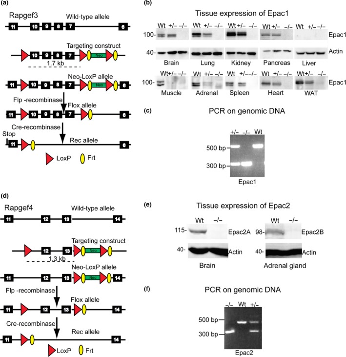
Methods: Epac1-/- and Epac2-/- C57BL/6J mice were produced and compared with wild-type mice for transvascular flux of radio-labelled albumin in skin, adipose tissue, intestine, heart and skeletal muscle. The transvascular leakage was also studied by dynamic contrast-enhanced magnetic resonance imaging (DCE-MRI) using the MRI contrast agent Gadomer-17 as probe.

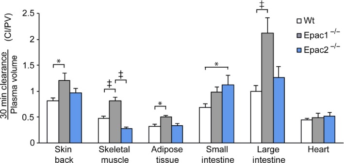
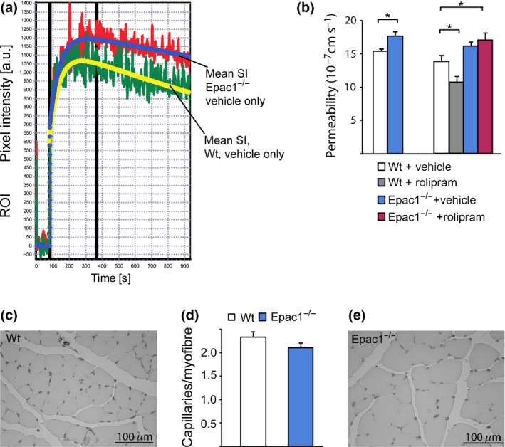
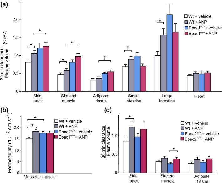
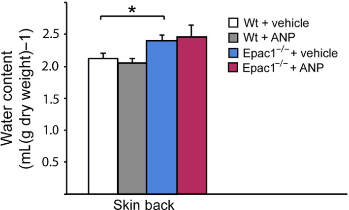
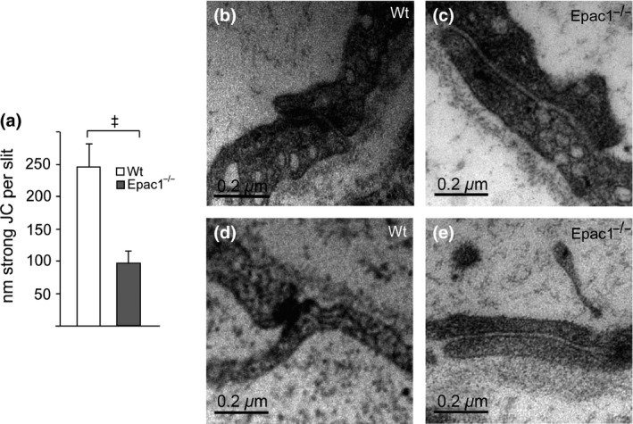
Results: Epac1-/- mice had constitutively increased transvascular macromolecule transport, indicating Epac1-dependent restriction of baseline permeability. In addition, Epac1-/- mice showed little or no enhancement of vascular permeability in response to atrial natriuretic peptide (ANP), whether probed with labelled albumin or Gadomer-17. Epac2-/- and wild-type mice had similar basal and ANP-stimulated clearances. Ultrastructure analysis revealed that Epac1-/- microvascular interendothelial junctions had constitutively less junctional complex.

Conclusion: Epac1 exerts a tonic inhibition of in vivo basal microvascular permeability. The loss of this tonic action increases baseline permeability, presumably by reducing the interendothelial permeability resistance. Part of the action of ANP to increase permeability in wild-type microvessels may involve inhibition of the basal Epac1-dependent activity.

Keywords: Epac deletion (mouse); Rapgef; atrial natriuretic peptide; cAMP; endothelial junction; microvascular permeability (in vivo).


Figures

Figure 1
Figure 1
Generation of the Epac1−/− and Epac2−/− mouse strains. (a) The Epac1 (Rapgef3) gene targeting construct and deletion strategy. (b) Anti‐Epac1 immunoblots showing lack of Epac1 protein in Epac1−/− mouse tissue and decreased expression in Epac1−/+ compared to Wt tissue. (c) PCR genotyping demonstrates the sizes of PCR products predicted for the Epac1 deletion (see Methods for details; WAT is white adipose tissue). (d) The Epac2 (Rapgef4) gene targeting construct and the deletion strategy. The deletion includes the functional cAMP‐binding domain common for all known Epac2 isoforms. (e) Anti‐Epac2 immunoblots showing lack of Epac2 protein in Epac2−/− mouse organs (brain, adrenal cortex) with high expression of Epac2. (f) PCR genotyping validation of the deletion.
Figure 2
Figure 2
The normalized albumin clearance is increased in skin, skeletal muscle, adipose tissue and large intestine of Epac1−/−, but not Epac2−/− mice. The normalized clearance (NC) in tissue samples of back skin, skeletal muscle, white adipose tissue, small and large intestine, and heart is shown mL g−1 dry weight (average ± SEM). The NC is the ratio of the clearance (based on relative tissue sample content of 125I‐HSA infused 35 min before euthanasia) and the local tissue plasma volume (relative content of 131I‐HSA, infused 5 min before euthanasia). P‐values < 0.05 (*) and <0.001 (±) are shown. The plasma volume (mL g−1 dry weight) was, with the exception of jejunum from the Epac2−/− mice, similar between Wt (n = 19), Epac1−/− (n = 9) and Epac2−/− (n = 9) mouse tissues: Skin: Wt: 0.023 ± 0.002, Epac1−/−: 0.020 ± 0.002, Epac2−/−: 0.027 ± 0.003; quadriceps muscle (corresponding values): 0.022 ± 0.003, 0.014 ± 0.001, and 0.018 ± 0.002; inguinal fat: 0.024 ± 0.001, 0.023 ± 0.002 and 0.028 ± 0.003; jejunum: 0.101 ± 0.008, 0.116 ± 0.017 and 0.064 ± 0.006; colon: 0.042 ± 0.004, 0.033 ± 0.002 and 0.032 ± 0.003; heart: 0.229 ± 0.013, 0.215 ± 0.019, and 0.205 ± 0.015. HSA, human serum albumin.
Figure 3
Figure 3
The constitutive Gadomer‐17 clearance is increased in masseter muscle of Epac1−/− compared to Wt mice. (a) Typical magnetic resonance imaging (MRI) signal intensity (SI) obtained upon continuous scanning of a masseter region from Epac1−/− (red) or Wt (green) mouse before and after Gadomer (Gd)‐17 injection. The solid lines show fits based on the calculated permeability coefficient. (b) The Gd 17 permeability coefficient calculated from the scanning data obtained before and after infusion of vehicle or rolipram, each animal serving as its own control. P‐values < 0.05 are shown (n = 10). (c–e) The endothelium of transverse sections of M. masseter from Epac1−/− (c) or Wt (e) mice was visualized by anti‐CD31 staining and the capillary/myofibre ratio determined (d). Error bars are SEM. P‐values < 0.05 (*) are shown.
Figure 4
Figure 4
The effect of atrial natriuretic peptide (ANP) on macromolecule microvascular permeability in tissue from Wt, Epac1−/− and Epac2−/− mice. (a) The effect of ANP on the normalized albumin clearance is shown for tissues from Wt and Epac1−/− mice. The mice were given vehicle or ANP 30 minutes before euthanasia, and the apparent plasma volume and albumin clearance determined by the [131I]albumin/[125I]albumin method as for the experiment shown in Figure 2. (b) The effect of ANP on the microvascular permeability of Gadomer‐17 is shown for masseter muscle from Wt and Epac1−/− mice. (c) The effect of ANP on the normalized albumin clearance for tissues from Wt and Epac2−/− mice. Error bars are SEM (n = 10). P‐values < 0.05 (*) and <0.01 (+) are shown.
Figure 5
Figure 5
The water content of back skin from Wt and Epac1−/− mice. The water content (mL g−1 dry weight) of back skin from Epac1−/− mice and Wt mice injected with vehicle or atrial natriuretic peptide (ANP) was determined in the tissue samples used for the experiments on albumin clearance shown in Figures 2 and 4a, as described in the experimental section. Error bars are SEM (n = 10). P‐value < 0.05 (*) is shown.
Figure 6
Figure 6
The endothelial junctional density of Epac1−/− and Wt capillaries. (a) The average length of each endothelial slit found associated with strong junctional complex (JC) in Wt and Epac1−/− M. masseter. Error bars are SEM. The P‐value for the difference between Epac1−/− and Wt slits was <0.00002 by the Mann–Whitney two‐tailed U‐test and <0.0003 by the two‐tailed t‐test. (b–e) Examples of junctions from Wt and Epac1−/− mice. The two Wt junctions shown (b, c) had more abundant JC than found in any of the 48 Epac1−/− junctions studied. The two Epac1−/− junctions shown (d, e) had less abundant JC than any of 57 Wt junctions studied.

Comment in

Similar articles

Cited by

References

    1. Birukova, A.A. , Zagranichnaya, T. , Alekseeva, E. , Bokoch, G.M. & Birukov, K.G. 2008. Epac/Rap and PKA are novel mechanisms of ANP‐induced Rac‐mediated pulmonary endothelial barrier protection. J Cell Physiol 215, 715–724. - PMC - PubMed
    1. Chen, W. , Gassner, B. , Borner, S. , Nikolaev, V.O. , Schlegel, N. , Waschke, J. , Steinbronn, N. , Strasser, R. & Kuhn, M. 2012. Atrial natriuretic peptide enhances microvascular albumin permeability by the caveolae‐mediated transcellular pathway. Cardiovasc Res 93, 141–151. - PMC - PubMed
    1. Chen, H. , Ding, C. , Wild, C. , Liu, H. , Wang, T. , White, M.A. , Cheng, X. & Zhou, J. 2013. Efficient synthesis of ESI‐09, a novel non‐cyclic nucleotide EPAC antagonist. Tetrahedron Lett 54, 1546–1549. - PMC - PubMed
    1. Christensen, A.E. , Selheim, F. , de Rooij, J. , Dremier, S. , Schwede, F. , Dao, K.K. , Martinez, A. , Maenhaut, C. , Bos, J.L. , Genieser, H.G. & Doskeland, S.O. 2003. cAMP analog mapping of Epac1 and cAMP kinase. Discriminating analogs demonstrate that Epac and cAMP kinase act synergistically to promote PC‐12 cell neurite extension. J Biol Chem 278, 35394–35402. - PubMed
    1. Cullere, X. , Shaw, S.K. , Andersson, L. , Hirahashi, J. , Luscinskas, F.W. & Mayadas, T.N. 2005. Regulation of vascular endothelial barrier function by Epac, a cAMP‐activated exchange factor for Rap GTPase. Blood 105, 1950–1955. - PubMed

Publication types

Substances

LinkOut - more resources