

Crowding-Induced Mixing Behavior of Lipid Bilayers: Examination of Mixing Energy, Phase, Packing Geometry, and Reversibility
- PMID: 27096947
- PMCID: PMC5519306
- DOI: 10.1021/acs.langmuir.6b00831
Crowding-Induced Mixing Behavior of Lipid Bilayers: Examination of Mixing Energy, Phase, Packing Geometry, and Reversibility
Abstract
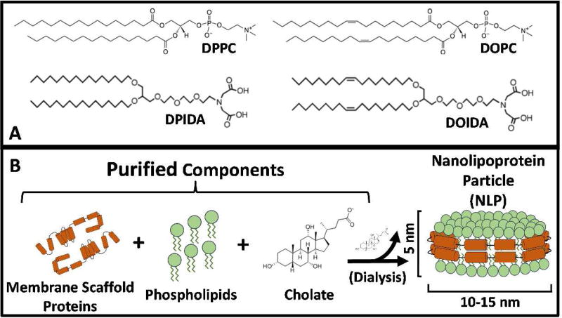
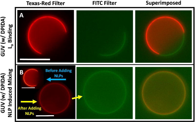
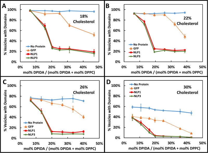
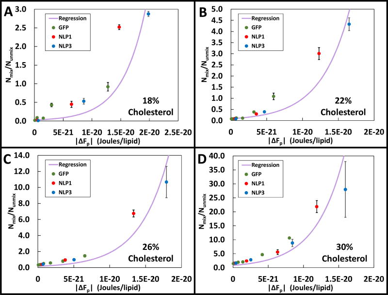
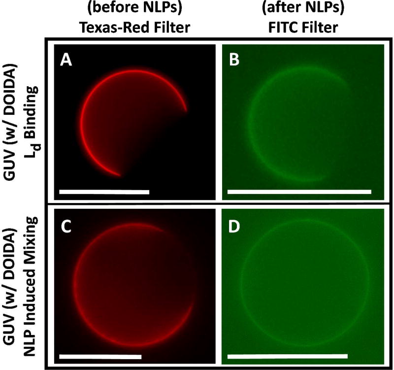
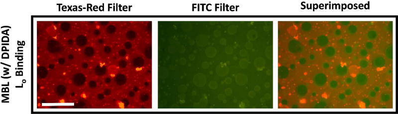
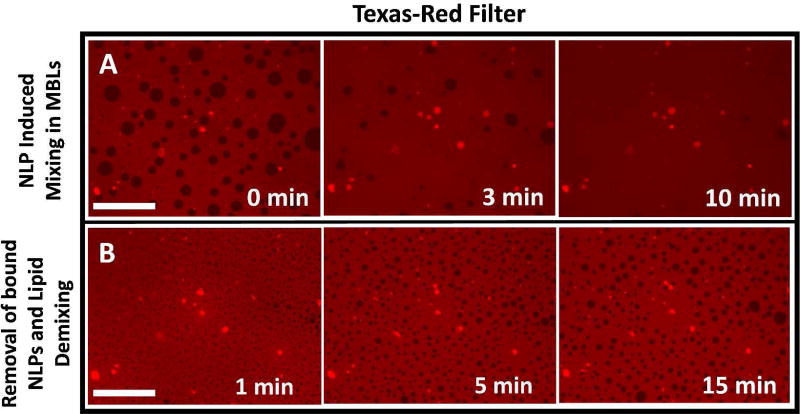
In an effort to develop a general thermodynamic model from first-principles to describe the mixing behavior of lipid membranes, we examined lipid mixing induced by targeted binding of small (Green Fluorescent Protein (GFP)) and large (nanolipoprotein particles (NLPs)) structures to specific phases of phase-separated lipid bilayers. Phases were targeted by incorporation of phase-partitioning iminodiacetic acid (IDA)-functionalized lipids into ternary lipid mixtures consisting of DPPC, DOPC, and cholesterol. GFP and NLPs, containing histidine tags, bound the IDA portion of these lipids via a metal, Cu(2+), chelating mechanism. In giant unilamellar vesicles (GUVs), GFP and NLPs bound to the Lo domains of bilayers containing DPIDA, and bound to the Ld region of bilayers containing DOIDA. At sufficiently large concentrations of DPIDA or DOIDA, lipid mixing was induced by bound GFP and NLPs. The validity of the thermodynamic model was confirmed when it was found that the statistical mixing distribution as a function of crowding energy for smaller GFP and larger NLPs collapsed to the same trend line for each GUV composition. Moreover, results of this analysis show that the free energy of mixing for a ternary lipid bilayer consisting of DOPC, DPPC, and cholesterol varied from 7.9 × 10(-22) to 1.5 × 10(-20) J/lipid at the compositions observed, decreasing as the relative cholesterol concentration was increased. It was discovered that there appears to be a maximum packing density, and associated maximum crowding pressure, of the NLPs, suggestive of circular packing. A similarity in mixing induced by NLP1 and NLP3 despite large difference in projected areas was analytically consistent with monovalent (one histidine tag) versus divalent (two histidine tags) surface interactions, respectively. In addition to GUVs, binding and induced mixing behavior of NLPs was also observed on planar, supported lipid multibilayers. The mixing process was reversible, with Lo domains reappearing after addition of EDTA for NLP removal.
Figures
Similar articles
-
Dynamics of Crowding-Induced Mixing in Phase Separated Lipid Bilayers.J Phys Chem B. 2016 Nov 3;120(43):11180-11190. doi: 10.1021/acs.jpcb.6b07119. Epub 2016 Oct 25. J Phys Chem B. 2016. PMID: 27723342 Free PMC article.
-
Incorporation of Triacylglycerol and Cholesteryl Ester Droplets in Phase-Separated Giant Unilamellar Vesicles.Langmuir. 2025 Mar 11;41(9):6113-6123. doi: 10.1021/acs.langmuir.4c05063. Epub 2025 Feb 26. Langmuir. 2025. PMID: 40009743
-
Targeting proteins to liquid-ordered domains in lipid membranes.Langmuir. 2011 Feb 15;27(4):1457-62. doi: 10.1021/la1041458. Epub 2010 Dec 14. Langmuir. 2011. PMID: 21155607
-
Preparing giant unilamellar vesicles (GUVs) of complex lipid mixtures on demand: Mixing small unilamellar vesicles of compositionally heterogeneous mixtures.Biochim Biophys Acta. 2015 Dec;1848(12):3175-80. doi: 10.1016/j.bbamem.2015.09.020. Epub 2015 Sep 28. Biochim Biophys Acta. 2015. PMID: 26417657
-
Exploring the raft-hypothesis by probing planar bilayer patches of free-standing giant vesicles at nanoscale resolution, with and without Na,K-ATPase.Biochim Biophys Acta. 2016 Dec;1858(12):3041-3049. doi: 10.1016/j.bbamem.2016.09.001. Epub 2016 Sep 9. Biochim Biophys Acta. 2016. PMID: 27616046 Review.
Cited by
-
Functional lipid pairs as building blocks of phase-separated membranes.Proc Natl Acad Sci U S A. 2020 Mar 3;117(9):4749-4757. doi: 10.1073/pnas.1919264117. Epub 2020 Feb 18. Proc Natl Acad Sci U S A. 2020. PMID: 32071249 Free PMC article.
-
Structure Versus Stochasticity-The Role of Molecular Crowding and Intrinsic Disorder in Membrane Fission.J Mol Biol. 2018 Aug 3;430(16):2293-2308. doi: 10.1016/j.jmb.2018.03.024. Epub 2018 Apr 5. J Mol Biol. 2018. PMID: 29627460 Free PMC article. Review.
-
Liquid-like protein interactions catalyse assembly of endocytic vesicles.Nat Cell Biol. 2021 Apr;23(4):366-376. doi: 10.1038/s41556-021-00646-5. Epub 2021 Apr 5. Nat Cell Biol. 2021. PMID: 33820972 Free PMC article.
-
Lipid and Protein Transfer between Nanolipoprotein Particles and Supported Lipid Bilayers.Langmuir. 2019 Sep 17;35(37):12071-12078. doi: 10.1021/acs.langmuir.9b01288. Epub 2019 Sep 6. Langmuir. 2019. PMID: 31442053 Free PMC article.
-
Interactions of HIV gp41's membrane-proximal external region and transmembrane domain with phospholipid membranes from 31P NMR.Biochim Biophys Acta Biomembr. 2021 Nov 1;1863(11):183723. doi: 10.1016/j.bbamem.2021.183723. Epub 2021 Aug 2. Biochim Biophys Acta Biomembr. 2021. PMID: 34352242 Free PMC article.
References
-
- Lingwood D, Simons K. Lipid Rafts as a Membrane-Organizing Principle. Science (Washington, DC, US.) 2010;327:46–50. - PubMed
-
- Juhasz J, Sharom FJ, Davis JH. Quantitative Characterization of Coexisting Phases in Dopc/Dppc/Cholesterol Mixtures: Comparing Confocal Fluorescence Microscopy and Deuterium Nuclear Magnetic Resonance. Biochim. Biophys. Acta. 2009;1788:2541–2552. - PubMed
-
- Marsh D. Cholesterol-Induced Fluid Membrane Domains: A Compendium of Lipid-Raft Ternary Phase Diagrams. Biochim. Biophys. Acta. 2009;1788:2114–2123. - PubMed
-
- Veatch SL, Keller SL. Seeing Spots: Complex Phase Behavior in Simple Membranes. Biochim. Biophys. Acta. 2005;1746:172–185. - PubMed
Publication types
MeSH terms
Substances
Grants and funding
LinkOut - more resources
Full Text Sources
Other Literature Sources
Research Materials
