

Optical and nuclear imaging of glioblastoma with phosphatidylserine-targeted nanovesicles
- PMID: 27096954
- PMCID: PMC5078058
- DOI: 10.18632/oncotarget.8763
Optical and nuclear imaging of glioblastoma with phosphatidylserine-targeted nanovesicles
Abstract
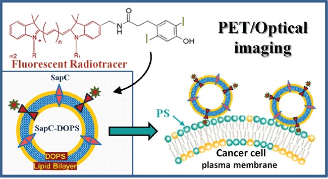
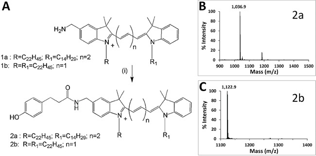
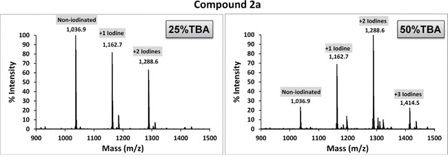
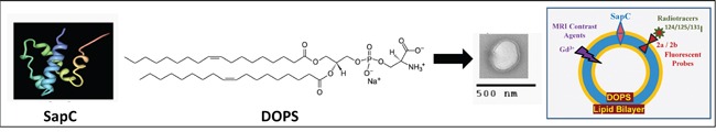
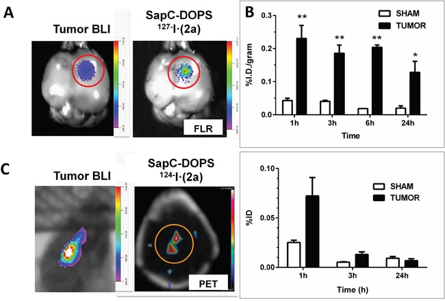
Multimodal tumor imaging with targeted nanoparticles potentially offers both enhanced specificity and sensitivity, leading to more precise cancer diagnosis and monitoring. We describe the synthesis and characterization of phenol-substituted, lipophilic orange and far-red fluorescent dyes and a simple radioiodination procedure to generate a dual (optical and nuclear) imaging probe. MALDI-ToF analyses revealed high iodination efficiency of the lipophilic reporters, achieved by electrophilic aromatic substitution using the chloramide 1,3,4,6-tetrachloro-3α,6α-diphenyl glycoluril (Iodogen) as the oxidizing agent in an organic/aqueous co-solvent mixture. Upon conjugation of iodine-127 or iodine-124-labeled reporters to tumor-targeting SapC-DOPS nanovesicles, optical (fluorescent) and PET imaging was performed in mice bearing intracranial glioblastomas. In addition, tumor vs non-tumor (normal brain) uptake was compared using iodine-125. These data provide proof-of-principle for the potential value of SapC-DOPS for multimodal imaging of glioblastoma, the most aggressive primary brain tumor.
Keywords: PET; SapC-DOPS; glioblastoma; liposome; optical imaging.
Conflict of interest statement
Patents applications are in progress for the intellectual property disclosed in this manuscript between University of Cincinnati and Molecular Targeting Technologies, Inc. (MTTI). X. Qi is listed as an inventor on the patent for SapC-DOPS technology that is the subject of this research. Consistent with current Cincinnati Children's Hospital Medical Center policies, the development and commercialization of this technology has been licensed to Bexion Pharmaceuticals, LLC, in which Dr. Qi, holds a minor (< 5%) equity interest. Dr. Gray is an employee and Dr. Pak is a shareholder of MTTI. The other authors declared no conflict of interest.
Figures

Similar articles
-
Detection of cancer cells using SapC-DOPS nanovesicles.Mol Cancer. 2016 May 10;15(1):33. doi: 10.1186/s12943-016-0519-1. Mol Cancer. 2016. PMID: 27160923 Free PMC article. Review.
-
Phosphatidylserine-selective targeting and anticancer effects of SapC-DOPS nanovesicles on brain tumors.Oncotarget. 2014 Aug 30;5(16):7105-18. doi: 10.18632/oncotarget.2214. Oncotarget. 2014. PMID: 25051370 Free PMC article.
-
In vivo optical imaging of brain tumors and arthritis using fluorescent SapC-DOPS nanovesicles.J Vis Exp. 2014 May 2;(87):51187. doi: 10.3791/51187. J Vis Exp. 2014. PMID: 24837630 Free PMC article.
-
Systemic delivery of SapC-DOPS has antiangiogenic and antitumor effects against glioblastoma.Mol Ther. 2013 Aug;21(8):1517-25. doi: 10.1038/mt.2013.114. Epub 2013 Jun 4. Mol Ther. 2013. PMID: 23732993 Free PMC article.
-
SapC-DOPS - a Phosphatidylserine-targeted Nanovesicle for selective Cancer therapy.Cell Commun Signal. 2020 Jan 9;18(1):6. doi: 10.1186/s12964-019-0476-6. Cell Commun Signal. 2020. PMID: 31918715 Free PMC article. Review.
Cited by
-
Medical Imaging Technology and Imaging Agents.Adv Exp Med Biol. 2023;1199:15-38. doi: 10.1007/978-981-32-9902-3_2. Adv Exp Med Biol. 2023. PMID: 37460725
-
Phosphatidylserine: The Unique Dual-Role Biomarker for Cancer Imaging and Therapy.Cancers (Basel). 2022 May 21;14(10):2536. doi: 10.3390/cancers14102536. Cancers (Basel). 2022. PMID: 35626139 Free PMC article. Review.
-
Detection of cancer cells using SapC-DOPS nanovesicles.Mol Cancer. 2016 May 10;15(1):33. doi: 10.1186/s12943-016-0519-1. Mol Cancer. 2016. PMID: 27160923 Free PMC article. Review.
-
Glioblastoma Treatments: An Account of Recent Industrial Developments.Front Pharmacol. 2018 Sep 13;9:879. doi: 10.3389/fphar.2018.00879. eCollection 2018. Front Pharmacol. 2018. PMID: 30271342 Free PMC article. Review.
-
Real-time glioblastoma tumor microenvironment assessment by SpiderMass for improved patient management.Cell Rep Med. 2024 Apr 16;5(4):101482. doi: 10.1016/j.xcrm.2024.101482. Epub 2024 Mar 28. Cell Rep Med. 2024. PMID: 38552622 Free PMC article.
References
-
- Culver J, Akers W, Achilefu S. Multimodality molecular imaging with combined optical and SPECT/PET modalities. Journal of Nuclear Medicine. 2008;49:169–172. - PubMed
-
- Jennings LE, Long NJ. ‘Two is better than one’-probes for dual-modality molecular imaging. Chemical Communications. 2009:3511–3524. - PubMed
-
- Leventis PA, Grinstein S. The distribution and function of phosphatidylserine in cellular membranes. Annual review of biophysics. 2010;39:407–427. - PubMed
-
- Ran S, Thorpe PE. Phosphatidylserine is a marker of tumor vasculature and a potential target for cancer imaging and therapy. Int J Radiat Oncol Biol Phys. 2002;54:1479–1484. - PubMed
MeSH terms
Substances
Grants and funding
LinkOut - more resources
Full Text Sources
Other Literature Sources
Medical
