. 2016 Apr 20;12(4):e1005583.
doi: 10.1371/journal.ppat.1005583. eCollection 2016 Apr.

Novel Polymerase Gene Mutations for Human Adaptation in Clinical Isolates of Avian H5N1 Influenza Viruses

Affiliations

Novel Polymerase Gene Mutations for Human Adaptation in Clinical Isolates of Avian H5N1 Influenza Viruses

Yasuha Arai et al. PLoS Pathog. .

Abstract

A major determinant in the change of the avian influenza virus host range to humans is the E627K substitution in the PB2 polymerase protein. However, the polymerase activity of avian influenza viruses with a single PB2-E627K mutation is still lower than that of seasonal human influenza viruses, implying that avian viruses require polymerase mutations in addition to PB2-627K for human adaptation. Here, we used a database search of H5N1 clade 2.2.1 virus sequences with the PB2-627K mutation to identify other polymerase adaptation mutations that have been selected in infected patients. Several of the mutations identified acted cooperatively with PB2-627K to increase viral growth in human airway epithelial cells and mouse lungs. These mutations were in multiple domains of the polymerase complex other than the PB2-627 domain, highlighting a complicated avian-to-human adaptation pathway of avian influenza viruses. Thus, H5N1 viruses could rapidly acquire multiple polymerase mutations that function cooperatively with PB2-627K in infected patients for optimal human adaptation.


Conflict of interest statement

The authors have declared that no competing interests exist.

Figures

Fig 1
Fig 1. Schematic overview of selection and characterization of polymerase and NP mutations.
Based on a database search of clade 2.2.1 virus sequences, we selected a total of 46 single mutations that were detected either only in human viruses (Category 1) or were more prevalent in human viruses than in bird viruses (Category 2). Since some viruses contained multiple mutations, we examined the sequences in the two virus categories for intra-segment multiple mutations and selected 23 viruses with intra-segment multiple mutations. These single and intra-segment multiple mutations were also detected in inter-segment combinations of mutations in both Category 1 and Category 2 viruses. Therefore, we searched for inter-segment combinations of single and intra-segment multiple mutations from natural human isolates, and 16 inter-segment combinations of mutations in viruses from patients were selected and designated Inter-1 to Inter-16. A total of 85 single and multiple mutations, that were carried with PB2-627K in clade 2.2.1 isolates, were assayed for polymerase activity by minigenome assays. Of these 85 mutations, 29 were introduced into EG/D1/clade 2.2.1 (wt) viruses and their effect on progeny vRNA production in human cells was investigated. From the in vitro progeny vRNA production results, 4 virus mutants were selected and tested for virulence in mice.
Fig 2
Fig 2. Effect of single mutations on polymerase activity.
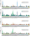
293T and QT-6 cells were transfected with plasmids expressing EG/D1 PB2, PB1, PA or NP with the indicated single mutations, a human or chicken polymerase I-driven plasmid expressing a vRNA-oriented or cRNA-oriented luciferase reporter gene, and a Renilla luciferase-expressing plasmid as an internal control. After 48 h incubation at 33 or 37°C, luciferase activities were measured and normalized to the internal Renilla luciferase activity. The data were expressed relative to the results for EG/D1 (wt). (A) Comparison of luciferase activity at 37 and 33°C in vRNA-oriented minigenome assays in 293T cells. (B) Comparison of polymerase activity at 37 and 33°C in cRNA-oriented minigenome assays in 293T cells. (C) Comparison of polymerase activity in 293T and QT6 cells in vRNA-oriented minigenome assays at 37°C. (D) Comparison of polymerase activity in 293T and QT6 cells in cRNA-oriented minigenome assays at 37°C. Each data point is the mean ± SD of three independent experiments. Colors on each x-axis highlight different virus gene segments. Single and double asterisks indicate a P value <0.05 and <0.01, respectively (ANOVA with Tukey’s multiple comparison test). Asterisks for mutations with negative effects on polymerase activity were omitted for clarity.
Fig 3
Fig 3. Effect of multiple mutations on polymerase activity.
293T and QT-6 cells were transfected with plasmids expressing EG/D1 PB2, PB1, PA and NP with the indicated multiple mutations, a human or chicken polymerase I-driven plasmid expressing a vRNA-oriented or cRNA-oriented luciferase reporter gene, and a Renilla luciferase-expressing plasmid as an internal control. Luciferase activities were assayed 48 h post-transfection and normalized to the internal Renilla luciferase activity. The data were expressed relative to the results for EG/D1 (wt). (A) Comparison of luciferase activity at 37 and 33°C in vRNA-oriented minigenome assays in 293T cells. (B) Comparison of polymerase activity at 37 and 33°C in cRNA-oriented minigenome assays in 293T cells. (C) Comparison of polymerase activity in 293T and QT6 cells in vRNA-oriented minigenome assays at 37°C. (D) Comparison of polymerase activity in 293T and QT6 cells in cRNA-oriented minigenome assays at 37°C. Each data point is the mean ± SD of three independent experiments. Colors on each x-axis highlight different virus gene segments. Single and double asterisks indicate a P value <0.05 and <0.01, respectively (ANOVA with Tukey’s multiple comparison test). Asterisks for mutations with negative effects on polymerase activity were omitted for clarity.
Fig 4
Fig 4. Effect of polymerase mutations on progeny vRNA production in primary human airway cells.
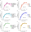
(A−D) SAE cells were infected with the indicated viruses at an MOI of 0.03 and incubated at 37°C (A and B) or 33°C (C and D). (E−F) CEFs were infected with the indicated viruses at an MOI of 0.005 and incubated at 37°C. The culture supernatants were harvested at the indicated times and assayed by quantitative real-time RT-PCR to determine the amount of progeny vRNA. Each data point is the mean ± SD of the log10 number of vRNA copies/ml from three separate experiments.
Fig 5
Fig 5. Relationship between polymerase activity and progeny vRNA production in primary human airway cells.
The vRNA-oriented luciferase activity data in Figs 2A and 3A were plotted against the progeny vRNA production data in S4A and S4B Fig at 37°C (A) and 33°C (B). The cRNA-oriented luciferase activity data in Figs 2B and 3B were plotted against the progeny vRNA production data in S4A and S4B Fig at 37°C (C) and 33°C (D). The data of the 29 mutations selected to investigate viral production (Figs 3 and S4) were expressed relative to the results for EG/D1 (wt). Pearson’s correlation coefficient (R) and a linear fit of the data were also calculated.
Fig 6
Fig 6. Effect of polymerase mutations on mortality and weight loss of infected mice.
Five-week-old BALB/c mice were inoculated intranasally with 2.5 × 10⁵ FFU of the indicated viruses. (A) Body weight of mice (5 mice per group) infected with the indicated viruses was monitored for 14 d post-infection. The mean ± SD of the percent body weight change for each group of infected mice is shown. (B) Survival of the infected mice (5 mice per group). Mortality was calculated including mice that were sacrificed after they had lost more than 30% of their body weight. (C) Virus titers in the lungs of infected mice (5 mice per group) at 3 d (left) and 6 d (right) post-infection. Each symbol marks the titer in an individual mouse. The asterisk indicates a P value <0.01 (ANOVA with Tukey’s multiple comparison test). (D) Photomicrographs of hematoxylin and eosin (H&E) stained (upper and middle panels) and immunohistochemically (IHC) stained (lower panel) lung sections from mice infected with the indicated viruses at 3 d post-infection. In IHC-stained tissues, the viral antigen is stained deep brown on a hematoxylin-stained background.
Fig 7
Fig 7. Structural model of the EG/D1 polymerase complex.
Structural model of the EG/D1 heterotrimeric polymerase complex bound to the vRNA promoter. (A) Surface view of the EG/D1 structure color-coded (as described below) according to the domain structure in which the mutations identified in this study were located. Left and right structures differ by 180° in orientation. (B) Surface view of the EG/D1 structure color-coded showing PB2 (light green), PB1 (pink), PA (light blue) and the mutations in this study (red). Left and right structures differ by 180° in orientation. (C) Transparent EG/D1 surface diagram showing the mutations identified in this study (red) located mainly on the PB1 β-ribbon (magenta) and β-hairpin structures (violet) sandwiching the vRNA promoter. Upper and lower structures differ by 90° in orientation.

References

    1. Cauldwell AV, Long JS, Moncorge O, Barclay WS (2014) Viral determinants of influenza A virus host range. J Gen Virol 95: 1193–1210. 10.1099/vir.0.062836-0 - DOI - PubMed
    1. Eisfeld AJ, Neumann G, Kawaoka Y (2015) At the centre: influenza A virus ribonucleoproteins. Nat Rev Microbiol 13: 28–41. 10.1038/nrmicro3367 - DOI - PMC - PubMed
    1. Imai M, Herfst S, Sorrell EM, Schrauwen EJ, Linster M, et al. (2013) Transmission of influenza A/H5N1 viruses in mammals. Virus Res 178: 15–20. 10.1016/j.virusres.2013.07.017 - DOI - PMC - PubMed
    1. Watanabe Y, Ibrahim MS, Suzuki Y, Ikuta K (2012) The changing nature of avian influenza A virus (H5N1). Trends Microbiol 20: 11–20. 10.1016/j.tim.2011.10.003 - DOI - PubMed
    1. Matrosovich M, Tuzikov A, Bovin N, Gambaryan A, Klimov A, et al. (2000) Early alterations of the receptor-binding properties of H1, H2, and H3 avian influenza virus hemagglutinins after their introduction into mammals. J Virol 74: 8502–8512. - PMC - PubMed

Publication types

Substances