. 2016 Jun 7;7(23):34229-39.
doi: 10.18632/oncotarget.8748.

XAF1 promotes neuroblastoma tumor suppression and is required for KIF1Bβ-mediated apoptosis

Affiliations

XAF1 promotes neuroblastoma tumor suppression and is required for KIF1Bβ-mediated apoptosis

Zhang'e Choo et al. Oncotarget. .

Abstract

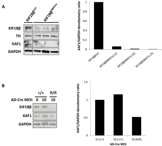
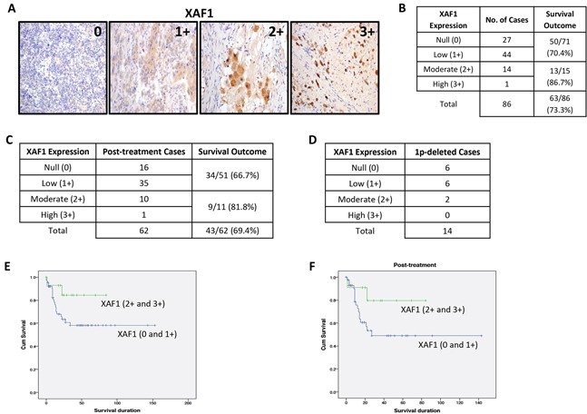
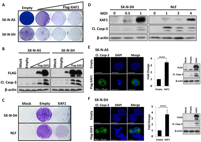
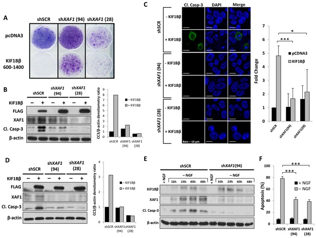
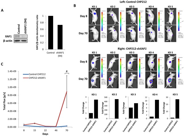
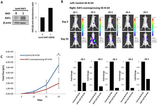
Neuroblastoma is an aggressive, relapse-prone childhood tumor of the sympathetic nervous system. Current treatment modalities do not fully exploit the genetic basis between the different molecular subtypes and little is known about the targets discovered in recent mutational and genetic studies. Neuroblastomas with poor prognosis are often characterized by 1p36 deletion, containing the kinesin gene KIF1B. Its beta isoform, KIF1Bβ, is required for NGF withdrawal-dependent apoptosis, mediated by the induction of XIAP-associated Factor 1 (XAF1). Here, we showed that XAF1 low expression correlates with poor survival and disease status. KIF1Bβ deletion results in loss of XAF1 expression, suggesting that XAF1 is indeed a downstream target of KIF1Bβ. XAF1 silencing protects from NGF withdrawal and from KIF1Bβ-mediated apoptosis. Overexpression of XAF1 impairs tumor progression whereas knockdown of XAF1 promotes tumor growth, suggesting that XAF1 may be a candidate tumor suppressor in neuroblastoma and its associated pathway may be important for developing future interventions.

Keywords: KIF1Bβ; XAF1; apoptosis; neuroblastoma.


Conflict of interest statement

FINANCIAL SUPPORT AND CONFLICTS OF INTEREST Zhang'e Choo and the project is supported by the National Medical Research Council, Singapore New Investigator Grant awarded to Zhi Xiong Chen. Susanne Schlisio is supported by grants from the Swedish Children Cancer Foundation and the Swedish Research Council. In addition, all authors declare that there is no conflict of interest.

Figures

Figure 1
Figure 1. XAF1 is poorly expressed in KIF1Bβ Knock-out mouse sympathetic neuroblasts
A. Immunoblot analysis of isolated primary sympathetic neuroblasts from superior cervical ganglia of wild type and conditional KIF1Bβ Knock-out (KO) mouse. Right – corresponding densitometry for XAF1 expression. B. Immunoblot analysis of isolated primary sympathetic neuroblasts deleted for KIF1Bβ ex vivo by treatment with Cre Recombinase adenovirus. Right – corresponding densitometry for XAF1 expression.
Figure 2
Figure 2. XAF1 is poorly expressed in neuroblastomas
A. XAF1 immunohistochemistry staining in a tissue microarray (TMA) (n=86). Images are acquired using a 20X objective. Staining intensity grade is indicated in the upper right corner of representative images shown; 0=null, 1+=low, 2+=moderate, 3+=high. Images of 2+ and 3+ XAF1 staining depict post-treatment neuroblastic tumors. B. Association between XAF1 expression and survival outcome for neuroblastoma patients. C. Association between XAF1 expression and survival outcome for post-treatment neuroblastoma patients. D. Distribution of XAF1 expression in 1p-deleted neuroblastoma patients. E. and F. Kaplan-Meier survival curves for patients with complete absence and low expression of XAF1 (0 and 1+) compared to moderate and high expression of XAF1 (2+ and 3+) – (E) Total number of cases and (F) post-treatment cases.
Figure 3
Figure 3. Overexpression of XAF1 induces apoptosis in neuroblastoma cells
A. Crystal violet staining to determine cell viability in SK-N-AS (top) and SK-N-SH (bottom) after transfection with an increasing dose of 1 μg, 2 μg and 4 μg Flag-XAF1 plasmid. Transfected cells were selected with 2 mg/ml G418 (SK-N-AS) and 500 μg/ml G418 (SK-N-SH) for several weeks. Empty vector (empty) served as negative control. B. Corresponding immunoblot analysis of SK-N-AS and SK-N-SH cells. C. Crystal violet staining of SK-N-SH (top) and NLF (bottom) cells that were transduced with lentivirus encoding XAF1 and selected with 1μg/ml puromycin for several weeks. Control virus (empty) served as negative control. D. Immunoblot analysis of SK-N-SH and NLF cell lines transduced with increasing MOI of lentivirus encoding XAF1. E. and F. Anti-cleaved caspase-3 (green) immunofluorescence staining of SK-N-AS (E) and SK-N-SH (F) 24 hours post-transfection with plasmid encoding Flag-XAF1 or empty vector. Cells were counterstained with DAPI to visualize nuclei (blue). Scale bar – 10 μm. Right – graphical representation showing fold change in number of cleaved caspase-3-positive cells transfected with Flag-XAF1 relative to empty vector-transfected cells (mean ± SD; n=3; ***, P<0.001) and corresponding immunoblot analysis.
Figure 4
Figure 4. XAF1 is required for KIF1Bβ-mediated and NGF withdrawal-dependent apoptosis
A. Crystal violet staining to determine cell viability in CHP212 cells after transduction with lentivirus encoding shRNAs targeting XAF1 (shXAF1 94 and 28) or non-targeting control virus (shSCR) and transfected with either Flag-KIF1Bβ(600-1400) or control empty pcDNA3 plasmids. Transfected cells were selected with 500 μg/ml G418 for several weeks. B. Corresponding immunoblot analysis of CHP212 cells. Right – densitometry for cleaved caspase-3 (CC3) expression. C. Anti-cleaved caspase-3 (green) immunofluorescence staining of CHP212 cells stably expressing shRNAs targeting XAF1 (shXAF1 94 and 28) or non-targeting control (shSCR) followed by transfection with Flag-KIF1Bβ(600-1400) or empty pcDNA3 plasmids as indicated. Cells were counterstained with DAPI to visualize nuclei (blue). Scale bar – 10 μm. Right -graphical representation showing fold change in number of cleaved caspase-3-positive CHP212 cells transduced with shRNAs targeting XAF1 (shXAF1 94 and 28) and transfected with either Flag-KIF1Bβ(600-1400) or empty pcDNA3 plasmids, relative to cells transduced with non-targeting control (shSCR) (mean ± SD; n=4; *, P<0.05; ***, P<0.001). D. Immunoblot analysis of NB1 cells stably transduced with shRNAs lentivirus targeting XAF1 (shXAF1 94 and 28) or control virus (shSCR), followed by transfection with either Flag-KIF1Bβ(600-1400) or control empty pcDNA3 plasmids. Right – corresponding densitometry for CC3 expression. E. Immunoblot analysis of differentiated PC12 cells before (+) and after (−) NGF withdrawal as indicated. Differentiated cells were transduced with lentivirus encoding shRNA targeting XAF1 (shXAF1 94) or non-targeting control virus (shSCR) prior to NGF withdrawal. F. Percentage of apoptosis in isolated primary rat sympathetic neuroblasts before (+) and after (−) NGF withdrawal. Neuroblasts were transduced with shRNAs lentivirus targeting XAF1 (shXAF1 94 and 28) or control virus (shSCR) prior to NGF withdrawal. Apoptosis was scored by apoptotic nuclei quantification in Tuj1-positive neuroblasts (mean ± SD; n=3; ***, P<0.001).
Figure 5
Figure 5. XAF1 knockdown promotes tumor progression in vivo
A. Anti-XAF1 immunoblot analysis of CHP212 cells with (shXAF1 94) or without (Control) stable XAF1 knockdown and corresponding densitometry. B. Bioluminescence imaging of five representative nude mice (KD 1-5) on Day 8 and 70. Each mouse was injected with control CHP212 and CHP212-shXAF1 neuroblastoma cells on the left and right flanks respectively. Bottom – corresponding graphical representation (KD 1-5) showing fold change in tumor growth generated by CHP212-shXAF1 cells relative to control CHP212 cells. C. Tumor growth analysis by quantifying bioluminescent signal in nude mice over a period of 70 days (mean ± SEM; n=5; #, P=0.05).
Figure 6
Figure 6. XAF1 overexpression delays tumor progression in vivo
A. Anti-XAF1 immunoblot analysis of SK-N-SH cells transduced with or without XAF1-overexpressing lentivirus and corresponding densitometry. B. Bioluminescence imaging of five representative nude mice (OE 1-5) on day 2 and 33. Each mouse was engrafted with control SK-N-SH and XAF1-overexpressing SK-N-SH neuroblastoma cells on the left and right flanks respectively. Bottom – corresponding graphical representation (OE 1-5) showing fold change in tumor growth generated by XAF1-overexpressing SK-N-SH cells relative to control SK-N-SH cells. C. Tumor growth analysis by quantifying bioluminescent signal in nude mice over a period of 33 days (mean ± SEM; n=5; *, P<0.05).

Similar articles

Cited by

References

    1. Maris JM, Hogarty MD, Bagatell R, Cohn SL. Neuroblastoma. Lancet. 2007;369:2106–20. doi: 10.1016/S0140-6736(07)60983-0. - DOI - PubMed
    1. Brodeur GM. Neuroblastoma: biological insights into a clinical enigma. Nature reviews Cancer. 2003;3:203–16. doi: 10.1038/nrc1014. - DOI - PubMed
    1. Lee S, Nakamura E, Yang H, Wei W, Linggi MS, Sajan MP, et al. Neuronal apoptosis linked to EglN3 prolyl hydroxylase and familial pheochromocytoma genes: developmental culling and cancer. Cancer Cell. 2005;8:155–67. doi: 10.1016/j.ccr.2005.06.015. Epub 2005/08/16. [pii] S1535-6108(05)00224-2. - DOI - PubMed
    1. Sommer L, Rao M. Neural stem cells and regulation of cell number. Progress in neurobiology. 2002;66:1–18. - PubMed
    1. Zhu Y, Parada LF. The molecular and genetic basis of neurological tumours. Nature reviews Cancer. 2002;2:616–26. doi: 10.1038/nrc866. - DOI - PubMed

MeSH terms