. 2016 Apr 20;11(4):e0153155.
doi: 10.1371/journal.pone.0153155. eCollection 2016.

Trisubstituted-Imidazoles Induce Apoptosis in Human Breast Cancer Cells by Targeting the Oncogenic PI3K/Akt/mTOR Signaling Pathway

Affiliations

Trisubstituted-Imidazoles Induce Apoptosis in Human Breast Cancer Cells by Targeting the Oncogenic PI3K/Akt/mTOR Signaling Pathway

Chakrabhavi Dhananjaya Mohan et al. PLoS One. .

Abstract

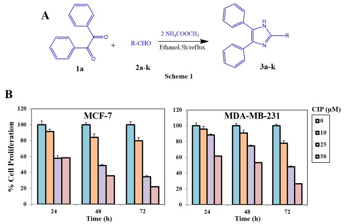
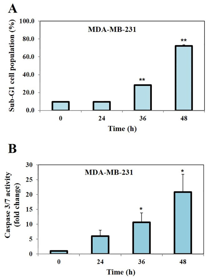
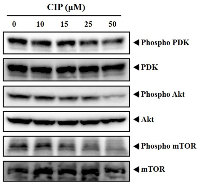
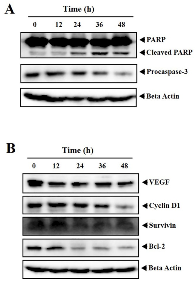
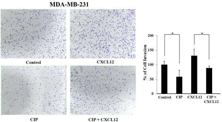
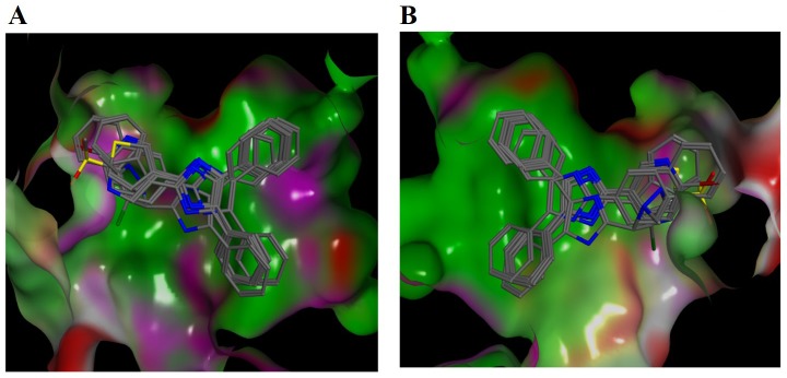
Overactivation of PI3K/Akt/mTOR is linked with carcinogenesis and serves a potential molecular therapeutic target in treatment of various cancers. Herein, we report the synthesis of trisubstituted-imidazoles and identified 2-chloro-3-(4, 5-diphenyl-1H-imidazol-2-yl) pyridine (CIP) as lead cytotoxic agent. Naïve Base classifier model of in silico target prediction revealed that CIP targets RAC-beta serine/threonine-protein kinase which comprises the Akt. Furthermore, CIP downregulated the phosphorylation of Akt, PDK and mTOR proteins and decreased expression of cyclin D1, Bcl-2, survivin, VEGF, procaspase-3 and increased cleavage of PARP. In addition, CIP significantly downregulated the CXCL12 induced motility of breast cancer cells and molecular docking calculations revealed that all compounds bind to Akt2 kinase with high docking scores compared to the library of previously reported Akt2 inhibitors. In summary, we report the synthesis and biological evaluation of imidazoles that induce apoptosis in breast cancer cells by negatively regulating PI3K/Akt/mTOR signaling pathway.


Conflict of interest statement

Competing Interests: The authors have declared that no competing interests exist.

Figures

Fig 1
Fig 1
A, Schematic representation for the synthesis of imidazole based small molecules. B, Breast cancer cells (2.5 X 10⁴/mL, MCF-7 & MDA-MB-231) were plated in triplicate, treated with indicated concentrations (0, 10, 25 and 50 μM) of CIP, and then subjected to MTT assay after 24, 48 and 72 h to analyse proliferation of cells. CIP suppresses the viability of various breast cancer cell lines in a dose- and time-dependent manner.
Fig 2
Fig 2
A, The analysis of cell cycle distribution following treatment with CIP was performed using flow cytometry. MDA-MB-231 cells were exposed to compound CIP (50 μM) for indicated time intervals (0, 24, 36 and 48 h), after which the cells were harvested and stained with PI. The cell distribution across the various phases of the cell cycle was analyzed with a flow cytometer. We observed that CIP induced significant apoptosis in a time dependent manner as evidenced by increased accumulation of cells in Sub-G1 phase of the cell cycle. B, MDA-MB-231 cells were exposed to compound CIP (50 μM) at indicated time intervals (0, 24, 36 and 48 h), after which they were harvested and caspase3/7 activity was measured using Caspase-Glo® 3/7 assay kit. We found that treatment of MDA-MB-231 cells with CIP caused the significant increase in the caspases-3/7 activity. * for p<0.05, ** for p<0.005.
Fig 3
Fig 3. MDA-MB-231 cells were treated with CIP at indicated concentrations (0, 10, 15, 25 and 50 μM) for 8 h and levels of phospho-PDK, phospho-Akt, phospho-mTOR and PDK, Akt, mTOR was analysed using western blotting.
We found that, CIP downregulated the phosphorylation of Akt, PDK and mTOR in a dose-dependent manner without any change in the expression levels of Akt, PDK and mTOR proteins.
Fig 4
Fig 4. A and B, MDA-MB-231 cells were treated with CIP (25 μM) for indicated time intervals (0, 12, 24, 36 and 48 h) and expression of apoptotic markers (PARP, Procaspase-3), antiapoptotic proteins (Bcl-2, Survivin, VEGF), cell cycle regulator (Cyclin D1) was profiled using western blot analysis.
We observed the significant decline in the expression of PARP, procaspase-3, cyclin D1, VEGF, survivin and Bcl-2 proteins in the time-dependent manner without alteration in the levels of beta actin.
Fig 5
Fig 5. MDA-MB-231 cells were suspended in serum-free DMEM and seeded into the Matrigel transwell chambers and were incubated with CIP (25 μM) for 8 h.
The invading cells were fixed and stained with crystal violet solution and invaded cells were counted in five randomly selected areas. CIP significantly suppressed the cell invasion both in the presence and absence of CXCL12 suggesting that CIP interferes with invasive potential of breast cancer cells.
Fig 6
Fig 6. In silico molecular docking studies of trisubstituted imidazoles against kinase domain of Akt2:
Common binding poses of trisubstituted imidazoles towards the Akt2 kinase domain. The molecular surface of the protein is represented based on the surface polarity; green, pink and red colours show hydrophobic, polar and solvent exposed regions, respectively. For the sake of better visualisation of the binding pocket surface of Akt2, molecular surface was rendered in two panels (A and B) in which ligands were rotated 180 degrees.

References

    1. Ludwig JA, Lamhamedi-Cherradi S-E, Lee H-Y, Naing A, Benjamin R. Dual Targeting of the Insulin-Like Growth Factor and Collateral Pathways in Cancer: Combating Drug Resistance. Cancers. 2011;3(3):3029–54. 10.3390/cancers3033029. PMC3759185. - DOI - PMC - PubMed
    1. Arcaro A, Guerreiro AS. The Phosphoinositide 3-Kinase Pathway in Human Cancer: Genetic Alterations and Therapeutic Implications. Current Genomics. 2007;8(5):271–306. 10.2174/138920207782446160. PMC2652403. - DOI - PMC - PubMed
    1. Makinoshima H, Takita M, Saruwatari K, Umemura S, Obata Y, Ishii G, et al. Signaling through the Phosphatidylinositol 3-Kinase (PI3K)/Mammalian Target of Rapamycin (mTOR) Axis Is Responsible for Aerobic Glycolysis mediated by Glucose Transporter in Epidermal Growth Factor Receptor (EGFR)-mutated Lung Adenocarcinoma. Journal of Biological Chemistry. 2015;290(28):17495–504. 10.1074/jbc.M115.660498 - DOI - PMC - PubMed
    1. Keane KN, Cruzat VF, Carlessi R, de Bittencourt PIH, Newsholme P. Molecular Events Linking Oxidative Stress and Inflammation to Insulin Resistance and β-Cell Dysfunction. Oxidative Medicine and Cellular Longevity. 2015;2015:15 10.1155/2015/181643 - DOI - PMC - PubMed
    1. LoPiccolo J, Blumenthal GM, Bernstein WB, Dennis PA. Targeting the PI3K/Akt/mTOR pathway: effective combinations and clinical considerations. Drug resistance updates: reviews and commentaries in antimicrobial and anticancer chemotherapy. 2008;11(1–2):32–50. Epub 2008/01/02. 10.1016/j.drup.2007.11.003 ; PubMed Central PMCID: PMCPmc2442829. - DOI - PMC - PubMed

Publication types

MeSH terms