. 2016 Apr 20;11(4):e0152877.
doi: 10.1371/journal.pone.0152877. eCollection 2016.

Spaceflight Activates Lipotoxic Pathways in Mouse Liver

Affiliations

Spaceflight Activates Lipotoxic Pathways in Mouse Liver

Karen R Jonscher et al. PLoS One. .

Erratum in

  • Correction: Spaceflight Activates Lipotoxic Pathways in Mouse Liver.
    Jonscher KR, Alfonso-Garcia A, Suhalim JL, Orlicky DJ, Potma EO, Ferguson VL, Bouxsein ML, Bateman TA, Stodieck LS, Levi M, Friedman JE, Gridley DS, Pecaut MJ. Jonscher KR, et al. PLoS One. 2016 May 4;11(5):e0155282. doi: 10.1371/journal.pone.0155282. eCollection 2016. PLoS One. 2016. PMID: 27145222 Free PMC article.

Abstract

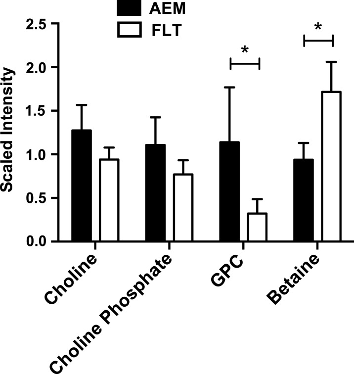
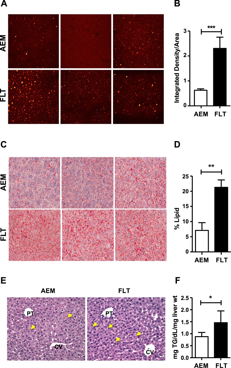
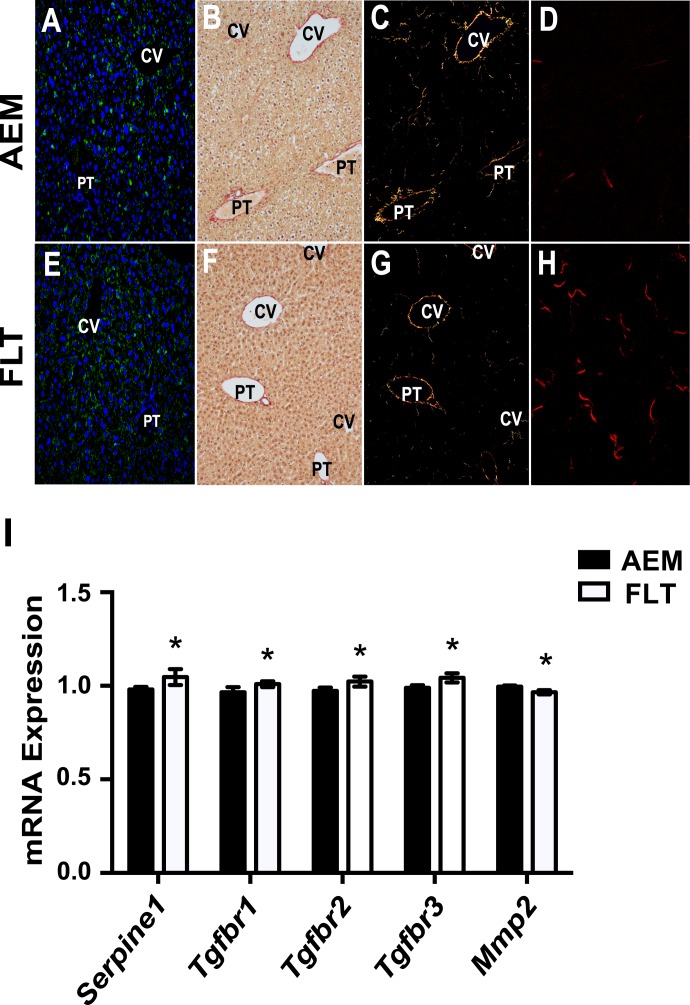
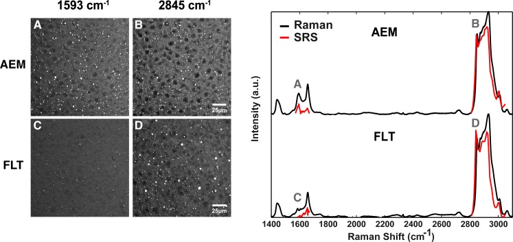
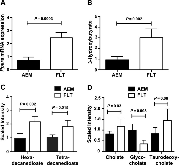
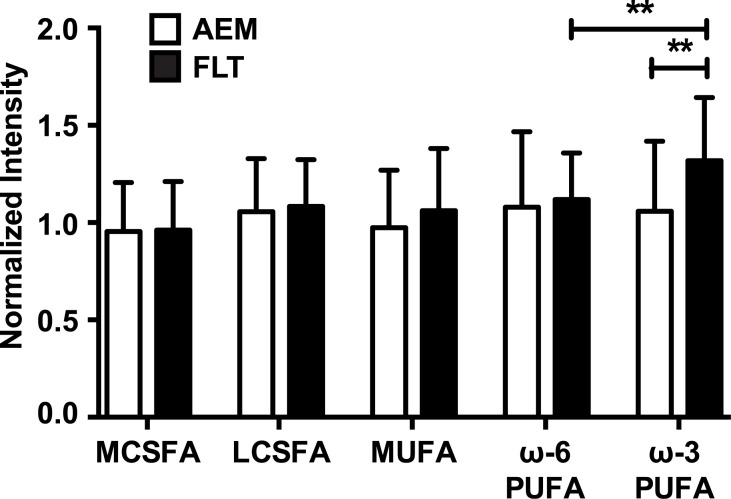
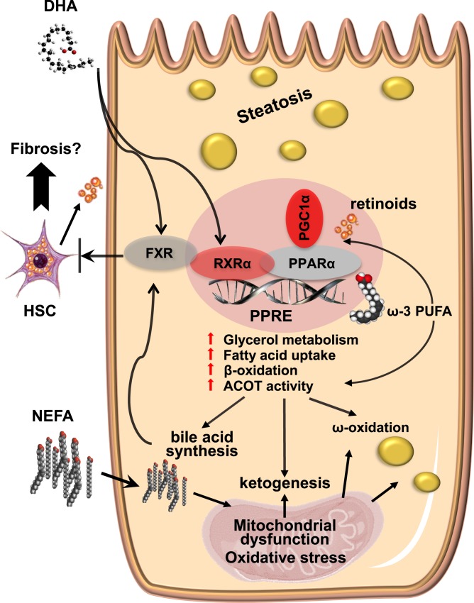
Spaceflight affects numerous organ systems in the body, leading to metabolic dysfunction that may have long-term consequences. Microgravity-induced alterations in liver metabolism, particularly with respect to lipids, remain largely unexplored. Here we utilize a novel systems biology approach, combining metabolomics and transcriptomics with advanced Raman microscopy, to investigate altered hepatic lipid metabolism in mice following short duration spaceflight. Mice flown aboard Space Transportation System -135, the last Shuttle mission, lose weight but redistribute lipids, particularly to the liver. Intriguingly, spaceflight mice lose retinol from lipid droplets. Both mRNA and metabolite changes suggest the retinol loss is linked to activation of PPARα-mediated pathways and potentially to hepatic stellate cell activation, both of which may be coincident with increased bile acids and early signs of liver injury. Although the 13-day flight duration is too short for frank fibrosis to develop, the retinol loss plus changes in markers of extracellular matrix remodeling raise the concern that longer duration exposure to the space environment may result in progressive liver damage, increasing the risk for nonalcoholic fatty liver disease.


Conflict of interest statement

Competing Interests: The authors have declared that no competing interests exist.

Figures

Fig 1
Fig 1. Betaine levels are increased in FLT mouse livers.
Mass spectrometry-based metabolomics was performed on n = 6 mice per group as described in Methods and Materials. Mass spectra corresponding to choline and related metabolites were identified and extracted chromatographic peaks used for relative quantification. Mann-Whitney analysis of results showed a significant increase in levels of betaine in FLT mice as compared with AEM controls, with a concomitant decrease in concentrations of metabolic precursors. *P < 0.05; data are mean ± SEM.
Fig 2
Fig 2. Spaceflight mice have increased accumulation of hepatic lipid droplets.
A) Frozen liver tissue was cryosectioned using OCT solution and sections imaged by CARS at a magnification of 60×. Representative images are shown from 3 different animals in each group. Images from AEM ground controls appear on the top panel and FLT mice on the bottom panel. B) Multiple regions were imaged from 2 cryosections taken at different tissue depths per animal (n = 5/group). Images were processed using ImageJ and integrated pixel intensity measured for each unit area. A Mann-Whitney test was used to compare integrated intensity values between groups. ***P < 0.001. C) Cryosectioned samples were stained with Oil Red O and imaged at 40× using an Aperio scanner. Sections from AEM and FLT mice were analyzed. Representative images from 3 mice per group are shown. D) Following color deconvolution, where dark red staining was used as an indicator of positive lipid signal, the percent of positive lipid signal was calculated per unit area for samples from n = 5 mice per group. A Mann-Whitney test was used to compare between groups and the mean ± SEM is shown. ** P < 0.01. E) H&E staining was performed on fixed liver sections from n = 4–5 mice/group to investigate liver histology. Inspection of the H&E stained sections revealed that the AEM ground control mice (left panel) had predominantly small zone 2 cytoplasmic lipid droplets (CLD) whereas the FLT mice (right panel) had an increase in slightly larger CLD in a panlobular pattern. Multiple lipid droplets are indicated using yellow arrows in representative images from each group. F) Total triglycerides were measured from n = 6 mice/group using a colorimetric assay (540 nm). A Mann-Whitney test was used to compare the calculated concentration values between groups. Data shown are mean ± SEM. * P < 0.05.
Fig 3
Fig 3. Lipid droplets in spaceflight mouse liver have reduced retinol content.
Stimulated Raman scattering (SRS) images of liver sections (left panel) revealed a decreased intensity of embedded lipid droplets at ~1593 cm-1 for spaceflight mice (FLT, C) with respect to ground mice (AEM, A), and showed no difference at 2845 cm-1 (B, D), the C-H2 symmetric stretching band characteristic of lipids. Hyperspectral SRS imaging (right panel) around the two frequencies of interest unveil quasi-identical spectra of the lipid droplets of the two mouse groups, except for the peak at ~1593 cm-1 (red curves). Spontaneous Raman spectra (black curves) agree with the SRS results. Raman spectra were acquired using n = 3 mice per group, 2 sections per mouse, and 2–3 droplets per section. Technical replicates were also performed for several samples. SRS imaging was used to confirm the Raman results and was performed on sections from one mouse in each group.
Fig 4
Fig 4. Remodeling of the ECM is increased in livers from spaceflight mice.
Fixed tissues were stained for PLIN2 (green) and ACTA2 (red) and nuclei were stained with DAPI (blue) to visualize lipid droplets and markers of activation in stellate cells (A, E). Mouse liver tissues were either fixed as described in Materials and Methods and stained with Picrosirius Red (B, C, F, G) or flash frozen and cryosectioned (D, H). Collagen staining was observed in Picrosirius Red stained sections (B, F) primarily near the portal triad (PT) and central vein (CV) areas. Using cross-polarized light, signal was more readily observed (C, G). SHG imaging was performed on cryosectioned samples (D, H) and increased collagen signal in interstitial regions was observed in livers from FLT mice as compared with AEM controls. Representative images are shown from each group (n = 3–5 mice/group). Magnification is 200× for stained samples and 60× for SHG images. Transcriptomic analysis reveals that expression levels of several important modulators of ECM remodeling are changed in spaceflight (I). Values are mean ± SEM; * P < 0.05. n = 5–6 / group.
Fig 5
Fig 5. Products of PPARα-mediated thioesterase activity are upregulated in spaceflight mice.
A) Hepatic PPARα mRNA expression was quantified using qRT-PCR and normalized to 18S rRNA levels for both FLT mice and AEM controls. An unpaired t-test with Welch’s correction was used for comparison. PPARα targets include acyl-coA thioesterases, which act in different cellular compartments to mediate lipid metabolism. Mass spectrometry-based metabolomics analysis of liver compounds showed increased concentrations of products of B) ACOT2, C) ACOT1 and D) ACOT8 activity. Data (mean ± SEM) were plotted for n = 3–5 mice/group.
Fig 6
Fig 6. Increased ω-3 polyunsaturated fatty acid concentration following spaceflight.
Mass spectrometry-based metabolomics was performed and spectra corresponding to both saturated and unsaturated fatty acids were identified and chromatographic peaks quantified. Mann-Whitney analysis of results from n = 5 mice per group showed a significant increase in levels of ω-3 PUFAs in spaceflight mice as compared with AEM controls. **P < 0.01; data are mean ± SEM.
Fig 7
Fig 7. Spaceflight induces activation of PPARα pathways maintained by a feedback loop involving hepatic thioesterase activity and mediated by DHA.
Elements of the space environment such as microgravity, oxidative stress and radiation may lead to activation of the PPARα-RXRα heterodimer by ω-3 fatty acids (including DHA), PGC-1α and retinoids from activated HSCs, increasing thioesterase activity. Hepatic steatosis, as well as synthesis of bile acids, ketone bodies and dicarboxylic acids, results from activation of downstream pathways. Fibrosis may also ensue from transformation of the activated HSCs. DHA and bile acids are ligands for FXR, which may be activated in a compensatory manner and help protect from HSC-induced remodeling of the ECM. PPRE, peroxisome proliferator response element.

References

    1. Da Silva MS, Zimmerman PM, Meguid MM, Nandi J, Ohinata K, Xu Y, et al. Anorexia in space and possible etiologies: an overview. Nutrition. 2002;18(10):805–13. Epub 2002/10/04. . - PubMed
    1. Lane HW, Gretebeck RJ, Smith SM. Nutrition, endocrinology, and body composition during space flight. Nutr Res. 1998;18(11):1923–34. Epub 2001/09/07. . - PubMed
    1. Tobin BW, Uchakin PN, Leeper-Woodford SK . Insulin secretion and sensitivity in space flight: diabetogenic effects. Nutrition. 2002;18(10):842–8. Epub 2002/10/04. . - PubMed
    1. Baqai FP, Gridley DS, Slater JM, Luo-Owen X, Stodieck LS, Ferguson V, et al. Effects of spaceflight on innate immune function and antioxidant gene expression. Journal of Applied Physiology. 2009;106(6):1935–42. 10.1152/japplphysiol.91361.2008 - DOI - PMC - PubMed
    1. Armstrong MJ, Adams LA, Canbay A, Syn WK. Extrahepatic complications of nonalcoholic fatty liver disease. Hepatology. 2014;59(3):1174–97. Epub 2013/09/05. 10.1002/hep.26717 . - DOI - PubMed

Publication types