

Breast Cancers Detected at Screening MR Imaging and Mammography in Patients at High Risk: Method of Detection Reflects Tumor Histopathologic Results
- PMID: 27097237
- PMCID: PMC5006733
- DOI: 10.1148/radiol.2016151419
Breast Cancers Detected at Screening MR Imaging and Mammography in Patients at High Risk: Method of Detection Reflects Tumor Histopathologic Results
Abstract
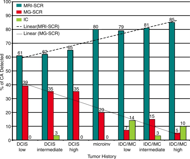
Purpose To compare the clinical, imaging, and histopathologic features of breast cancers detected at screening magnetic resonance (MR) imaging, screening mammography, and those detected between screening examinations (interval cancers) in women at high risk. Materials and Methods This retrospective institutional review board-approved, HIPAA-compliant review of 7519 women at high risk for breast cancer who underwent screening with MR imaging and mammography between January 2005 and December 2010 was performed to determine the number of screening-detected and interval cancers diagnosed. The need for informed consent was waived. Medical records were reviewed for age, risk factors (family or personal history of breast cancer, BRCA mutation status, history of high-risk lesion or mantle radiation), tumor histopathologic results, and time between diagnosis of interval cancer and most recent screening examination. The χ(2) test and logistic regression methods were used to compare the features of screening MR imaging, screening mammography, and interval cancers. The Wilcoxon signed-rank test was used to calculate P values. Results A total of 18 064 screening MR imaging examinations and 26 866 screening mammographic examinations were performed. Two hundred twenty-two cancers were diagnosed in 219 women, 167 (75%) at MR imaging, 43 (19%) at mammography, and 12 (5%) interval cancers. Median age at diagnosis was 52 years. No risk factors were associated with screening MR imaging, screening mammography, or interval cancer (P > .06). Cancers found at screening MR imaging were more likely to be invasive cancer (118 of 167 [71%]; P < .0001). Of the 43 cancers found at screening mammography, 38 (88%) manifested as calcifications and 28 (65%) were ductal carcinoma in situ. Interval cancers were associated with nodal involvement (P = .005) and the triple-negative subtype (P = .03). Conclusion In women at high risk for breast cancer who underwent screening with mammography and MR imaging, invasive cancers were more likely to be detected at MR imaging, whereas most cancers detected at screening mammography were ductal carcinoma in situ. Interval cancers were found infrequently and were more likely to be node positive and of the triple-negative subtype. (©) RSNA, 2016.
Figures
Similar articles
-
Screening breast MR imaging in women with a history of lobular carcinoma in situ.Radiology. 2011 Nov;261(2):414-20. doi: 10.1148/radiol.11110091. Epub 2011 Sep 7. Radiology. 2011. PMID: 21900617
-
Breast MR imaging screening in women with a history of breast conservation therapy.Radiology. 2014 Aug;272(2):366-73. doi: 10.1148/radiol.14131893. Epub 2014 Mar 17. Radiology. 2014. PMID: 24635678
-
Screening breast MR imaging in women with a history of chest irradiation.Radiology. 2011 Apr;259(1):65-71. doi: 10.1148/radiol.10100991. Epub 2011 Feb 15. Radiology. 2011. PMID: 21325032
-
Overview of Breast Cancer Screening and Diagnosis.PET Clin. 2018 Jul;13(3):301-323. doi: 10.1016/j.cpet.2018.02.001. PET Clin. 2018. PMID: 30100072 Review.
-
Abbreviated Magnetic Resonance Imaging (MRI) for Breast Cancer Screening: Rationale, Concept, and Transfer to Clinical Practice.Annu Rev Med. 2019 Jan 27;70:501-519. doi: 10.1146/annurev-med-121417-100403. Annu Rev Med. 2019. PMID: 30691370 Review.
Cited by
-
Differential diagnosis of plasma cell mastitis and invasive ductal carcinoma using multiparametric MRI.Gland Surg. 2020 Apr;9(2):278-290. doi: 10.21037/gs.2020.03.30. Gland Surg. 2020. PMID: 32420252 Free PMC article.
-
Sizing It Up: Concordance between Breast Imaging and Pathologically Determined Tumor Measurement.Ann Surg Oncol. 2025 Jun 16. doi: 10.1245/s10434-025-17663-5. Online ahead of print. Ann Surg Oncol. 2025. PMID: 40522576
-
Emerging nanoparticle-based x-ray imaging contrast agents for breast cancer screening.Nanomedicine (Lond). 2025 May;20(10):1149-1166. doi: 10.1080/17435889.2025.2496129. Epub 2025 Apr 22. Nanomedicine (Lond). 2025. PMID: 40261216 Review.
-
Feasibility of Velocity-Selective Arterial Spin Labeling in Breast Cancer Patients for Noncontrast-Enhanced Perfusion Imaging.J Magn Reson Imaging. 2021 Oct;54(4):1282-1291. doi: 10.1002/jmri.27781. Epub 2021 Jun 13. J Magn Reson Imaging. 2021. PMID: 34121250 Free PMC article.
-
Imaging surveillance for the detection of ipsilateral local tumor recurrence in patients who underwent oncoplastic breast-conserving surgery with acellular dermal matrix: abbreviated MRI versus conventional mammography and ultrasonography.World J Surg Oncol. 2021 Sep 27;19(1):290. doi: 10.1186/s12957-021-02403-2. World J Surg Oncol. 2021. PMID: 34579740 Free PMC article.
References
-
- Smith RA, Duffy SW, Gabe R, Tabar L, Yen AM, Chen TH. The randomized trials of breast cancer screening: what have we learned? Radiol Clin North Am 2004;42(5):793–806, v. - PubMed
-
- Tabár L, Vitak B, Chen TH, et al. . Swedish two-county trial: impact of mammographic screening on breast cancer mortality during 3 decades. Radiology 2011;260(3):658–663. - PubMed
-
- Humphrey LL, Helfand M, Chan BK, Woolf SH. Breast cancer screening: a summary of the evidence for the U.S. Preventive Services Task Force. Ann Intern Med 2002;137(5 Part 1):347–360. - PubMed
-
- Tilanus-Linthorst M, Verhoog L, Obdeijn IM, et al. . A BRCA1/2 mutation, high breast density and prominent pushing margins of a tumor independently contribute to a frequent false-negative mammography. Int J Cancer 2002;102(1):91–95. - PubMed
-
- Brekelmans CT, Seynaeve C, Bartels CC, et al. . Effectiveness of breast cancer surveillance in BRCA1/2 gene mutation carriers and women with high familial risk. J Clin Oncol 2001;19(4):924–930. - PubMed
Publication types
MeSH terms
Substances
Grants and funding
LinkOut - more resources
Full Text Sources
Other Literature Sources
Medical
