. 2016 Apr 21:7:11291.
doi: 10.1038/ncomms11291.

Virtual memory T cells develop and mediate bystander protective immunity in an IL-15-dependent manner

Affiliations

Virtual memory T cells develop and mediate bystander protective immunity in an IL-15-dependent manner

Jason T White et al. Nat Commun. .

Abstract

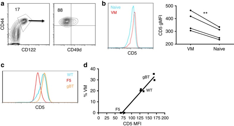
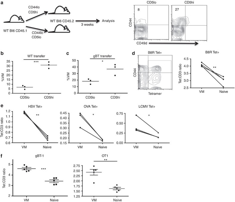
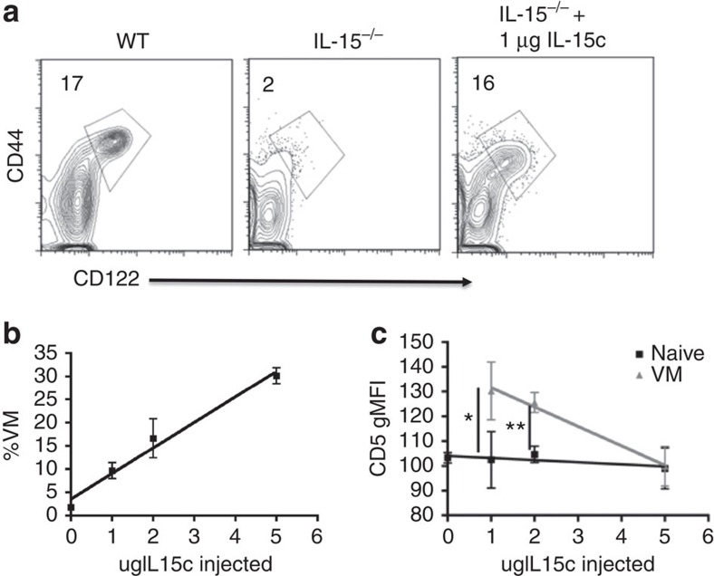
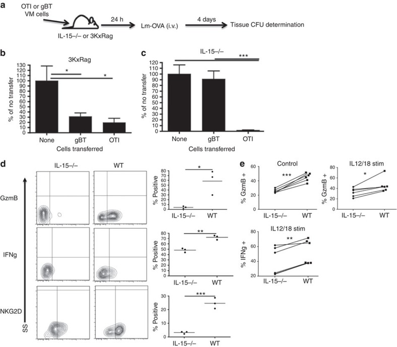
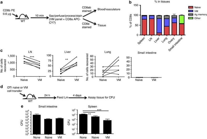
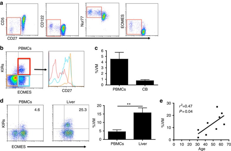
Virtual memory cells (VM) are an antigen-specific, memory phenotype CD8 T-cell subset found in lymphoreplete, unchallenged mice. Previous studies indicated that VM cells were the result of homeostatic proliferation (HP) resembling the proliferation observed in a lymphopenic environment. Here we demonstrate that HP is ongoing in lymphoreplete mice, the degree of which is dictated by the number of naive CD8 T cells with a sufficiently high affinity for self-antigen interacting with peripheral IL-15. VM cell transcriptional profiles suggest a capacity to mediate protective immunity via antigen non-specific bystander killing, a function we show is dependent on IL-15. Finally, we show a VM-like population of human cells that accumulate with age and traffic to the liver, displaying phenotypic and functional attributes consistent with the bystander protective functions of VM cells identified in the mouse. These data identify developmental and functional attributes of VM cells, including their likely role in protective immunity.


Figures

Figure 1
Figure 1. VM cells resemble cells that have undergone HP in both integrin expression and affinity.
(a) Flow cytometry on splenocytes isolated from unmanipulated WT mice. Gating on CD44hi/CD122hi CD8 T cells (left, percentage of CD8 above gate). Gated population visualized by CD44/CD49d (right, percentage of VM cells [CD44hi, CD49dlo] above population). (b) Naive (CD44lo) and VM (CD44hi, CD49dlo) CD8 T cells from the same mouse stained for CD5. Representative histogram overlay of the two populations (left), gMFI comparison (right; **P<0.01, paired t-test, representative of two experiments of n=4 per group and n=3 per group). (c,d) Naive (CD44lo) CD8 T-cell populations from polyclonal and WT mice were stained for CD5. (c) Representative flow histogram overlay of naive CD5 distributions for WT (blue), F5xRag−/− (red) and gBTxRag−/− (orange) (one mouse per histogram; representative of three experiments). (d) gMFI of naive populations graphed against per cent VM cells found in each mouse (linear regression, r2: 0.97, P<0.0001; representative of two experiments with at least n=2 per group).
Figure 2
Figure 2. CD5 expression is a predictor of a naive cell's propensity to become VM.
(a) Naive (CD44lo), CD5hi (the 20% highest CD5 expressers) or CD5lo (the 20% lowest CD5 expressers) CD8 T cells were sorted out of unmanipulated WT mice, and 4.0 × 10⁵ of each population were adoptively transferred into congenic WT recipients, which were left undisturbed for 3 weeks (left). Recipients were then killed and splenocytes were analysed for transferred cells changing phenotype (right, per cent VM in quadrant). (b,c) Comparisons between WT mice receiving WT (b) or gBTxRag−/− (c) transferred cells (*P<0.05, ***P<0.001, t-test; n=3 per group). (d,e) Tetramer staining and pulldown of CD8s specific for foreign cognate antigen in WT mice. Representative contour flow plot of CD44/tetramer staining for B8R tetramer (left), overlay of pseudocolour dot plots for CD3 and tetramer staining in naive (blue) and VM (red) CD8 T cells. Comparison within mice of tetramer:CD3 gMFI staining on individual tetramer+ (Tet+) cells (d, B8R [TSYKFESV]; e, HSV [SSIEFARL], OVA [SIINFEKL], LCMV [KAVYNFATC]; *P<0.05, **P<0.01, paired t-test, representative of 2–4 experiments with n=3 per group). (f) PBMCs from gBT-1 and OTI TCR transgenic mice were stained with the HSV and OVA MHCI tetramers, respectively, as described above. VM and naive populations were compared within mice for Tet:CD3 ratios (**P<0.01, ***P<0.001, paired t-test; n=5 per group, error bars represent s.e.m.).
Figure 3
Figure 3. Availability of IL-15 dictates both the size and CD5 composition of the VM population.
(a) Percentage of VM (CD44hi, CD122hi) cells found in WT (left), IL-15−/− (middle) and IL-15−/− 4 days after IL-15 complex injections (right, per cent VM above gate). (b,c) IL-15 complexes were injected intravenously and given 4 days to interact with the hematopoietic system. Splenocytes were then analysed for their percentage of VM cells as a function of IL-15 complexes injected (b, linear regression, r2=0.98, P=0.01; representative of two experiments with n=3 per group), and the gMFI of CD5 in the VM and naive pools (c, *P<0.05, **P<0.01, t-test, representative of three experiments with n=3 per group, error bars represent s.e.m.).
Figure 4
Figure 4. RNAseq highlights differences in cytokine responsiveness between CD8 T naive CD5hi/lo and VM cells.
(a) The T-cell population of a WT mouse was stained for eomesodermin expression. The representative histogram overlay of the different populations is given (left), as well as a comparison between naive (CD44lo) CD5lo/CD5hi cells (**P<0.01, paired t-test, representative of two experiments with n=3 per group). (b) Naive CD8 T cells were also stained for IL-2/15Rβ (CD122) expression (*P<0.05, paired t-test, representative of two experiments with n=3 per group).
Figure 5
Figure 5. VM cells, being similar in phenotype to previously reported bystander cells, mediate their effects in a bystander-like manner.
(a) OTI and gBTxRag−/− mice were sorted for VM cells and subsequently injected into 3KxRag−/− (b) or IL-15−/− (c) mice (2.0 × 10⁵ cells per mouse). One day after transfer, mice were challenged with Lm-OVA via tail vein (1.0 × 10⁵ CFU per mouse for 3KxRag−/− and 2.5 × 10³ CFU per mouse for IL-15KO). Four days later, mice were killed and spleens were processed for CFU (b,c, *P<0.05, ***P<0.001, one-way analysis of variance, data combined from two experiments with n=3 per group, error bars represent s.e.m.). (d) Mice from the gBTxRag−/− transfer conditions (into WT or IL-15−/− mice) were injected with Brefeldin A before killing, and the transferred cells were subsequently stained for effector molecule expression (*P<0.05, **P<0.01, ***P<0.001, t-test, representative of two experiments with n=3 per group). (e) Congenically marked WT T cells were incubated with WT or IL-15−/− splenocytes for 24 hours with (IL12/18 stim) or without (control) IL12/18 stimulation, then stained for effector molecule expression (*P<0.05, **P<0.01, ***P<0.001, paired t-test, data combined from two experiments with n=3 per group).
Figure 6
Figure 6. VM cells show a tropism for certain tissues under steady-state and trafficking conditions.
(a,b) WT mice were injected with CD8b antibody that was allowed to briefly circulate. Mice were then killed and stained with an antibody panel to determine naive, VM and antigen-experienced CD8 T cells, and whether the cells were in the vasculature or tissues (a). (b) Representative bar graphs of T-cell distribution (pooled data from two experiments with n=3 per experiment, error bars represent s.e.m.). (c) WT splenocytes were sorted into CD8 naive and VM populations, stained with different tracking dyes and co-adoptively transferred into recipient WT congenic mice. Recipients were killed 48 h later and tissues were processed as in a, and transferred cell populations were compared (**P<0.01, ***P<0.001, paired t-test, data combined from two experiments with n=3 per group). (d) OTI splenocytes were sorted into naive and VM populations and adoptively transferred into WT mice. One day later, mice were orally challenged with Lm expressing mutant internalin-A, and killed 4 days subsequent to challenge. (e) Tissues of mice orally infected were assayed for Lm CFUs (**P<0.01, ***P<0.001, one-way analysis of variance, representative of two experiments with n=3 per group, error bars represent s.e.m.).
Figure 7
Figure 7. Human subset with VM characteristics.
(a) Human PBMCs were stained for markers denoting CD8 T-cell interaction with self or IL-15. Representative flow plots of CD8+, CD45RA+ cells are shown. (b) Putative human VM memory cells are analysed for their corresponding CD27 levels. (c,d) Percentage of CD3+/CD8+/CD45RA+/KIR+/EOMES+ (VM) cells are compared between adult PBMC samples and cord blood samples (c) or between healthy adult PBMCs (n=9, 6 males and 3 females, age range 29–72 (mean 50) years) and healthy liver (n=5, 4 males and 1 female, age range 23–56 (mean 40) years; error bars represent s.e.m.; (d) **P<0.01, t-test). (e) Normal splenocytes banked from surgeries (n=7 benign pancreatic mass and n=2 non-functioning neuroendocrine tumour) were analysed for their populations of VM cells (CD3+/CD8+/CD45RA+/KIR+/EOMES+) and correlated with age (linear regression, r2=0.47, P=0.04).

References

    1. Cheroutre H., Lambolez F. & Mucida D. The light and dark sides of intestinal intraepithelial lymphocytes. Nat. Rev. Immunol. 11, 445–456 (2011) . - PMC - PubMed
    1. Weinreich M. A., Odumade O. A., Jameson S. C. & Hogquist K. A. T cells expressing the transcription factor PLZF regulate the development of memory-like CD8+ T cells. Nat. Immunol. 11, 709–716 (2010) . - PMC - PubMed
    1. Lee Y. J., Jameson S. C. & Hogquist K. A. Alternative memory in the CD8 T cell lineage. Trends Immunol. 32, 50–56 (2011) . - PMC - PubMed
    1. Huang T., Wei B., Velazquez P., Borneman J. & Braun J. Commensal microbiota alter the abundance and TCR responsiveness of splenic naive CD4+ T lymphocytes. Clin. Immunol. 117, 221–230 (2005) . - PubMed
    1. Haluszczak C. et al.. The antigen-specific CD8+ T cell repertoire in unimmunized mice includes memory phenotype cells bearing markers of homeostatic expansion. J. Exp. Med. 206, 435–448 (2009) . - PMC - PubMed

MeSH terms