

Bilateral Synchronous Ectopic Ethmoid Sinus Olfactory Neuroblastoma: A Case Report
- PMID: 27097989
- PMCID: PMC4841357
- DOI: 10.12659/ajcr.897623
Bilateral Synchronous Ectopic Ethmoid Sinus Olfactory Neuroblastoma: A Case Report
Abstract
Background: Olfactory neuroblastoma (ONB), also known as esthesioneuroblastoma, is a rare malignant head and neck cancer thought to originate from the olfactory epithelium. It typically invades contiguous structures at presentation. We report a very rare case of multifocal and ectopic ONB.
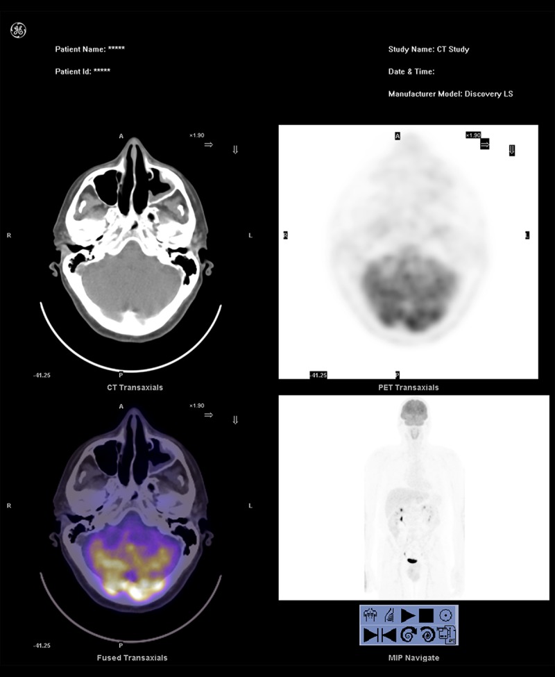
Case report: A 41-year-old man presented with left nasal obstruction and occasional left epistaxis associated with headache. Endoscopic examination of the nasal cavities and computed tomography suggested bilateral polypoid masses. Histopathological diagnosis after endoscopic resection established bilateral olfactory neuroblastoma of the ethmoid sinuses. The patient received postoperative radiotherapy. He remains free of disease 4 years after treatment.
Conclusions: To the best of our knowledge this is the second documented case of multifocal ectopic olfactory neuroblastoma. Clinicians should consider ONB in the differential diagnosis of bilateral synchronous nasal and paranasal masses to avoid delayed diagnosis. Endoscopic resection of ONB could be an option in selected cases.
Figures


Similar articles
-
Ectopic primary olfactory neuroblastoma of the maxillary sinus.Ann Diagn Pathol. 2016 Jun;22:45-8. doi: 10.1016/j.anndiagpath.2016.04.001. Epub 2016 Apr 11. Ann Diagn Pathol. 2016. PMID: 27180059 Review.
-
[Clinicopathologic study of sinonasal teratocarcinosarcoma and its contrast with olfactory neuroblastoma].Zhonghua Bing Li Xue Za Zhi. 2008 Jul;37(7):458-64. Zhonghua Bing Li Xue Za Zhi. 2008. PMID: 19035117 Chinese.
-
Huge sphenoid sinus olfactory neuroblastoma: a case report.Kaohsiung J Med Sci. 2009 Feb;25(2):87-92. doi: 10.1016/S1607-551X(09)70046-4. Kaohsiung J Med Sci. 2009. PMID: 19321412 Free PMC article. Review.
-
[Ethmoid esthesioneuroblastoma presenting with ophthalmologic manifestations].J Fr Ophtalmol. 2014 Jun;37(6):e87-9. doi: 10.1016/j.jfo.2013.09.013. Epub 2014 Apr 16. J Fr Ophtalmol. 2014. PMID: 24743035 French. No abstract available.
-
Sinonasal teratocarcinosarcoma ("mixed olfactory neuroblastoma-craniopharyngioma") presenting with syndrome of inappropriate secretion of antidiuretic hormone.Clin Neuropathol. 2000 Mar-Apr;19(2):63-9. Clin Neuropathol. 2000. PMID: 10749286
Cited by
-
Ectopic olfactory neuroblastoma is associated with increased frequency of syndrome of inappropriate antidiuretic hormone secretion and reduced disease control: Case series with systematic review and pooled analysis.Int Forum Allergy Rhinol. 2025 Jan;15(1):45-67. doi: 10.1002/alr.23502. Epub 2024 Dec 11. Int Forum Allergy Rhinol. 2025. PMID: 39661032 Free PMC article.
-
Ectopic Olfactory Neuroblastoma: Systematic Review of a Rare Clinical Entity among Sinonasal Tumors.J Neurol Surg B Skull Base. 2023 Jan 5;85(2):109-118. doi: 10.1055/a-1993-7790. eCollection 2024 Apr. J Neurol Surg B Skull Base. 2023. PMID: 38463937 Free PMC article.
-
Intranasal Esthesioneuroblastoma: CT Patterns Aid in Preventing Routine Nasal Polypectomy.AJNR Am J Neuroradiol. 2018 Feb;39(2):344-349. doi: 10.3174/ajnr.A5464. Epub 2017 Dec 7. AJNR Am J Neuroradiol. 2018. PMID: 29217745 Free PMC article.
-
Olfactory Neuroblastoma Is Not Always Located at the Roof of the Nasal Cavity.J Belg Soc Radiol. 2024 Apr 12;108(1):39. doi: 10.5334/jbsr.3562. eCollection 2024. J Belg Soc Radiol. 2024. PMID: 38826683 Free PMC article.
-
Endoscopic surgery versus various open approaches in esthesioneuroblastoma: a systematic review of the literature.Front Oncol. 2025 May 28;15:1512771. doi: 10.3389/fonc.2025.1512771. eCollection 2025. Front Oncol. 2025. PMID: 40502638 Free PMC article.
References
-
- Broich G, Pagliari A, Ottaviani F. Esthesioneuroblastoma: A general review of the cases published since the discovery of the tumour in 1924. Anticancer Res. 1997;17(4A):2683–706. - PubMed
-
- Dulguerov P, Allal AS, Calcaterra TC. Esthesioneuroblastoma: A meta-analysis and review. Lancet Oncol. 2001;2(11):683–90. - PubMed
-
- Morita A, Ebersold MJ, Olsen KD, et al. Esthesioneuroblastoma: Prognosis and management. Neurosurgery. 1993;32(5):706–14. discussion 714–15. - PubMed
-
- Dulguerov P, Calcaterra T. Esthesioneuroblastoma: the UCLA experience 1970–1990. Laryngoscope. 1992;102(8):843–49. - PubMed
-
- Koka VN, Julieron M, Bourhis J, et al. Aesthesioneuroblastoma. J Laryngol Otol. 1998;112(7):628–33. - PubMed
Publication types
MeSH terms
LinkOut - more resources
Full Text Sources
