Review
. 2016 Apr 21:9:43.
doi: 10.1186/s13041-016-0224-4.

Control of adult neurogenesis by programmed cell death in the mammalian brain

Affiliations
Review

Control of adult neurogenesis by programmed cell death in the mammalian brain

Jae Ryun Ryu et al. Mol Brain. .

Abstract

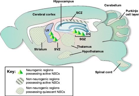
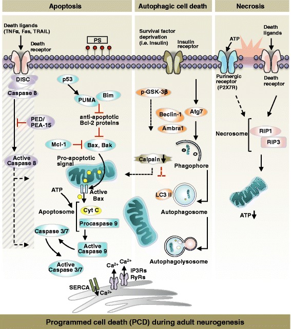
The presence of neural stem cells (NSCs) and the production of new neurons in the adult brain have received great attention from scientists and the public because of implications to brain plasticity and their potential use for treating currently incurable brain diseases. Adult neurogenesis is controlled at multiple levels, including proliferation, differentiation, migration, and programmed cell death (PCD). Among these, PCD is the last and most prominent process for regulating the final number of mature neurons integrated into neural circuits. PCD can be classified into apoptosis, necrosis, and autophagic cell death and emerging evidence suggests that all three may be important modes of cell death in neural stem/progenitor cells. However, the molecular mechanisms that regulate PCD and thereby impact the intricate balance between self-renewal, proliferation, and differentiation during adult neurogenesis are not well understood. In this comprehensive review, we focus on the extent, mechanism, and biological significance of PCD for the control of adult neurogenesis in the mammalian brain. The role of intrinsic and extrinsic factors in the regulation of PCD at the molecular and systems levels is also discussed. Adult neurogenesis is a dynamic process, and the signals for differentiation, proliferation, and death of neural progenitor/stem cells are closely interrelated. A better understanding of how adult neurogenesis is influenced by PCD will help lead to important insights relevant to brain health and diseases.

Keywords: Adult neurogenesis; Apoptosis; Autophagy; Necrosis; Neural stem cells; Neuroblasts; Programmed cell death.


Figures

Fig. 1
Fig. 1
Adult neurogenesis in spontaneous neurogenic regions and non-neurogenic regions. Neurogenic regions possessing active neural stem cells (NSCs): In the adult brain, neurogenesis in the subgranular zone (SGZ) of the hippocampal dentate gyrus (DG) and the subventricular zone (SVZ) of the lateral ventricle actively supplies newly generated cells. SGZ and SVZ have been identified as spontaneous neurogenic regions possessing self-renewing neural stem cells (NSCs) and neural progenitor cells (NPCs), respectively. Non-neurogenic regions possessing active NSCs: In addition to these two discrete regions, subcallosal zone (SCZ) is the sources for continuously generating multi-potent NSCs. Non-neurogenic regions possessing quiescent NSCs: Recent reports have suggested that NSCs may be widely distributed in the adult brain. The existence of NSCs is proposed by in vitro neurosphere culture and BrdU+ labeling in many regions which were previously believed to be non-neurogenic, such as striatum, thalamus, hypothalamus, spinal cord, and Purkinje cell layer of the cerebellum. One of the difficulties for identifying NSCs in the non-neurogenic regions is possibly due to the mitotic quiescence of the NSCs, which has inducible capacity for self-renewal and multi-potency under pathological conditions
Fig. 2
Fig. 2
The key players in the regulation of PCD during adult neurogenesis. Apoptosis: Pro-apoptotic signals induce Bax translocation to the outer membrane of mitochondria. Bax-mediated pore formation leads to the release of apoptogenic cytochrome c (Cyt c) into the cytosol and apoptosome formation. The apoptosome activates procaspase-9 and catalytically active caspase-9 induces activation of downstream effector caspases, caspase-3 and -7 (Caspase 3/7). Phosphatidylserine (PS) exposure at the cell surface is required for the clearance of apoptotic cells. In the adult neural stem cells (NSCs), p53, Bim and PUMA have been implicated in activating apoptosis. In addition, the pro-apoptotic proteins, Bax and Bak are the key regulators. Mcl-1 antagonizes the pro-apoptotic proteins, and therefore, considered as a critical anti-apoptotic protein for the survival of the NSCs. In extrinsic apoptosis, a death ligand (TNFα, Fas, or TRAIL) binding activates death receptor and induces DISC complex formation near the receptor. Upon DISC complex-mediated activation of caspase-8, intrinsic and extrinsic apoptosis converge at the level of the executioner caspase cascade. In adult NSCs, PED/PEA-15 represses the activation of caspase-8. Release of Ca2+ from the ER and subsequent transfer to the mitochondria promtes the commitment of NSCs to cell death. Autophagic cell death (ACD): Autophagy is induced when cells are starved of nutrients or survival factors. Atg7 regulates the maturation of autophagosome and initiates the lipidation of LC3 (also called LC3 II). Cargos subjected to degradation are degraded in the autophagolysosome. AMBRA1 and Beclin-1-induced autophagy is inversely correlated with apoptosis in adult NSCs. Under insulin-deprived condition, the adult hippocampal neural stem (HCN) cells succumb to ACD wherein the cell fate is under the control of GSK-3β activation. Inhibition of GSK-3β phosphorylation (p-GSK-3β) induces ACD. The negative regulator of ACD is calpain, which also mediates the crosstalk between apoptosis and ACD. Necrosis: Extracellular ATP or death receptor activation rapidly induces RIP1/RIP3 necrosome formation. Necrotic cell death results from the depletion of cytoplasmic ATP due to mitochondrial dysfunction. A purinergic P2X7 receptor-mediated necrosis induction has been reported in adult NSCs. However, regulator of necrosis in adult NSCs has not been identified to date

References

    1. Buss RR, Sun W, Oppenheim RW. Adaptive roles of programmed cell death during nervous system development. Annu Rev Neurosci. 2006;29:1–35. doi: 10.1146/annurev.neuro.29.051605.112800. - DOI - PubMed
    1. Oppenheim RW. Viktor Hamburger (1900–2001). Journey of a neuroembryologist to the end of the millennium and beyond. Neuron. 2001;31:179–190. doi: 10.1016/S0896-6273(01)00366-X. - DOI - PubMed
    1. Oppenheim RW. Cell death of motoneurons in the chick embryo spinal cord. V. Evidence on the role of cell death and neuromuscular function in the formation of specific peripheral connections. J Neurosci. 1981;1:141–151. - PMC - PubMed
    1. Oppenheim RW, Chu-wang I. Spontaneous cell death of spinal motoneurons following peripheral innervation in the chick embryo. Brain Res. 1977;125:154–160. doi: 10.1016/0006-8993(77)90367-5. - DOI - PubMed
    1. Oppenheim RW, Prevette D, Yin QW, Collins F, MacDonald J. Control of embryonic motoneuron survival in vivo by ciliary neurotrophic factor. Science. 1991;251:1616–1618. doi: 10.1126/science.2011743. - DOI - PubMed

Publication types