. 2016 Apr 21:6:24774.
doi: 10.1038/srep24774.

Yeast V-ATPase Proteolipid Ring Acts as a Large-conductance Transmembrane Protein Pore

Affiliations

Yeast V-ATPase Proteolipid Ring Acts as a Large-conductance Transmembrane Protein Pore

Sergio Couoh-Cardel et al. Sci Rep. .

Abstract

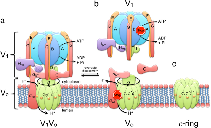
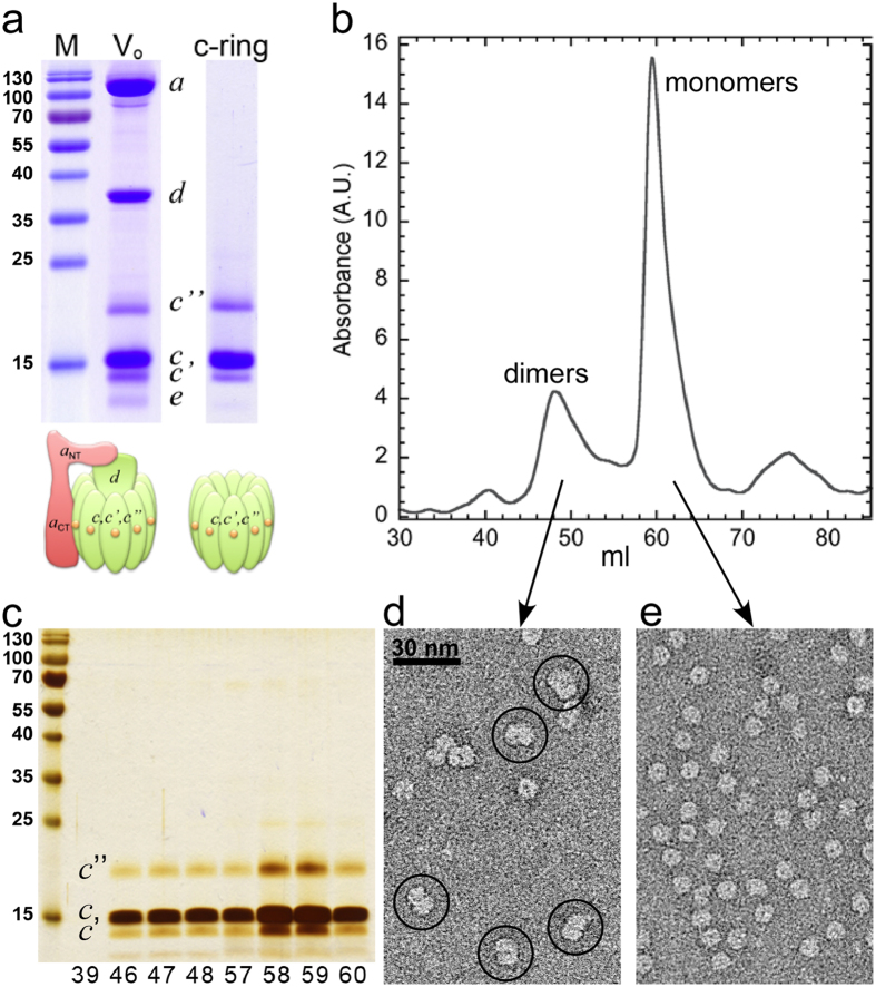
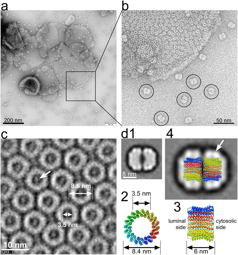
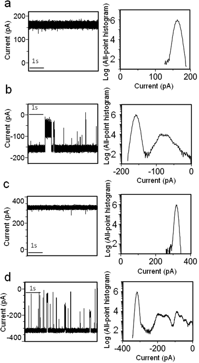
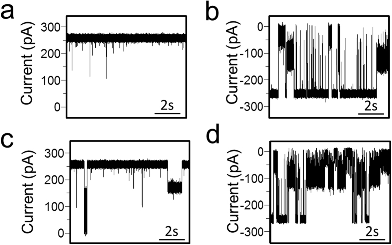
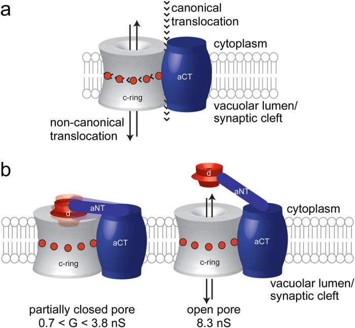
The vacuolar H(+)-ATPase (V-ATPase) is a rotary motor enzyme that acidifies intracellular organelles and the extracellular milieu in some tissues. Besides its canonical proton-pumping function, V-ATPase's membrane sector, Vo, has been implicated in non-canonical functions including membrane fusion and neurotransmitter release. Here, we report purification and biophysical characterization of yeast V-ATPase c subunit ring (c-ring) using electron microscopy and single-molecule electrophysiology. We find that yeast c-ring forms dimers mediated by the c subunits' cytoplasmic loops. Electrophysiology measurements of the c-ring reconstituted into a planar lipid bilayer revealed a large unitary conductance of ~8.3 nS. Thus, the data support a role of V-ATPase c-ring in membrane fusion and neuronal communication.


Figures

Figure 1
Figure 1. Subunit architecture and regulation of yeast V-ATPase.
(a) Schematic of yeast V-ATPase subunit architecture. Rotor subunits (DFdc8cc”) are in green, the stator complex ((EG)3CHa) is in orange and the catalytic hexamer (A3B3) is in blue (adapted from reference30). Yeast V-ATPase c subunit ring (c-ring) contains three proteolipid isoforms, c, c’ and c”. While c and c’ each contain four, c” is predicted to contain five transmembrane segments with the N-terminal α helix that is not found in c and c’. This α helix is likely located inside the central pore of the c-ring. While c” is conserved across species, the c’ isoform has as of yet not been identified in the mammalian enzyme. (b) Regulation of V-ATPase’s ATP hydrolysis-driven proton pumping activity by reversible dissociation into inactive V1 and Vo sectors. (c) Yeast V-ATPase c-ring.
Figure 2
Figure 2. Purification and structural characterization of yeast V-ATPase c-ring.
(a) SDS-PAGE of purified Vo (10 μg) as well as dialyzed and concentrated c-ring (5 μg). The gels were stained with Coomassie blue. (b) Size-exclusion chromatography (Superdex 200, 16 × 500 mm2) of c-ring (1 mg) in 0.1% DDM containing buffer. (c) Peak fractions (10 μl each) were analyzed by SDS-PAGE and silver staining. (d,e) Negative stain EM of fraction 48 and 58, respectively. Dimeric c-ring complexes are highlighted by circles in (d).
Figure 3
Figure 3. 2-D crystallization and single-particle image analysis of c-ring.
For 2-D crystallization, c-ring (1.5 mg/ml) was mixed with DOPC (0.5 mg/ml) and detergent was removed by the addition of BioBeads. (a) Negative stain TEM of DOPC reconstituted c-ring after detergent removal for 7 days. At this stage, para-crystalline arrays of c-rings were visible, but reconstitution was incomplete with numerous dimeric c-ring complexes visible next to lipid vesicles (see molecules highlighted by circles in (b)). (c) Reconstituted c-rings after complete detergent removal were imaged by cryo-EM and crystalline areas were analyzed by correlation averaging. (d1) Averaged projection (367 images) of dimeric c-ring. A dataset of 4337 images of dimeric c-rings from images, as shown in (b) was analyzed by reference free alignment and classification, as implemented in EMAN1.9. (d2,3) Top- and side-view of a bacterial V-ATPase c-ring (K10 ring from E. hirae; 2bl2.pdb). (d4) Model of the dimeric yeast V-ATPase c-ring superimposed on the EM average. The stain-excluding detergent belt is indicated by the arrow.
Figure 4
Figure 4. Representative single-channel electrical traces along with their corresponding semi-logarithmic all-point, current-amplitude histograms.
The traces were acquired with the c-ring at various applied transmembrane potentials. (a) +20 mV. (b) −20 mV. (c) +40 mV. (d) −40 mV. All single-channel traces were low-pass Bessel filtered at a frequency of 10 kHz. These electrical traces are typical among n = 8 distinct single-channel electrical recordings.
Figure 5
Figure 5. Interaction of the d subunit with the c-ring produces current blockades of varying amplitude.
In panels (a,b) single-channel electrical signature of the c-ring is illustrated at +30 and −30 mV, respectively. In (c,d) the single-channel traces are (a,b) in the presence of 0.3 μM d subunit, respectively, which was added to the cis side. These electrical traces are typical among n = 3 distinct single-channel electrical recordings.
Figure 6
Figure 6. Canonical and non-canonical ion translocation across the Vo complex/c-ring.
(a) Schematic model of the aCT-c-ring complex highlighting canonical and non-canonical ion translocation pathways. (b) Dispersity in the unitary conductance of the Vo complex was likely caused by structural fluctuations or different conformations of the aNT-d complex and its interaction with the c-ring cytoplasmic domains.

References

    1. Forgac M. Vacuolar ATPases: rotary proton pumps in physiology and pathophysiology. Nat Rev Mol Cell Biol 8, 917–29 (2007). - PubMed
    1. Sun-Wada G. H. & Wada Y. Role of vacuolar-type proton ATPase in signal transduction. Biochim Biophys Acta 1847, 1166–1172 (2015). - PubMed
    1. Cotter K., Stransky L., McGuire C. & Forgac M. Recent Insights into the Structure, Regulation, and Function of the V-ATPases. Trends Biochem Sci 40, 611–22 (2015). - PMC - PubMed
    1. Zhang Z. et al. Structure of the yeast vacuolar ATPase. J Biol Chem 283, 35983–95 (2008). - PMC - PubMed
    1. Rawson S. et al. Structure of the Vacuolar H(+)-ATPase Rotary Motor Reveals New Mechanistic Insights. Structure 23, 461–71 (2015). - PMC - PubMed

Publication types

MeSH terms

Substances

LinkOut - more resources