

doi: 10.1038/srep24818.
Optogenetic control of human neurons in organotypic brain cultures
Affiliations
- PMID: 27098488
- PMCID: PMC4838935
- DOI: 10.1038/srep24818
Item in Clipboard
Optogenetic control of human neurons in organotypic brain cultures
Sci Rep.
.
Abstract
Optogenetics is one of the most powerful tools in neuroscience, allowing for selective control of specific neuronal populations in the brain of experimental animals, including mammals. We report, for the first time, the application of optogenetic tools to human brain tissue providing a proof-of-concept for the use of optogenetics in neuromodulation of human cortical and hippocampal neurons as a possible tool to explore network mechanisms and develop future therapeutic strategies.
Figures
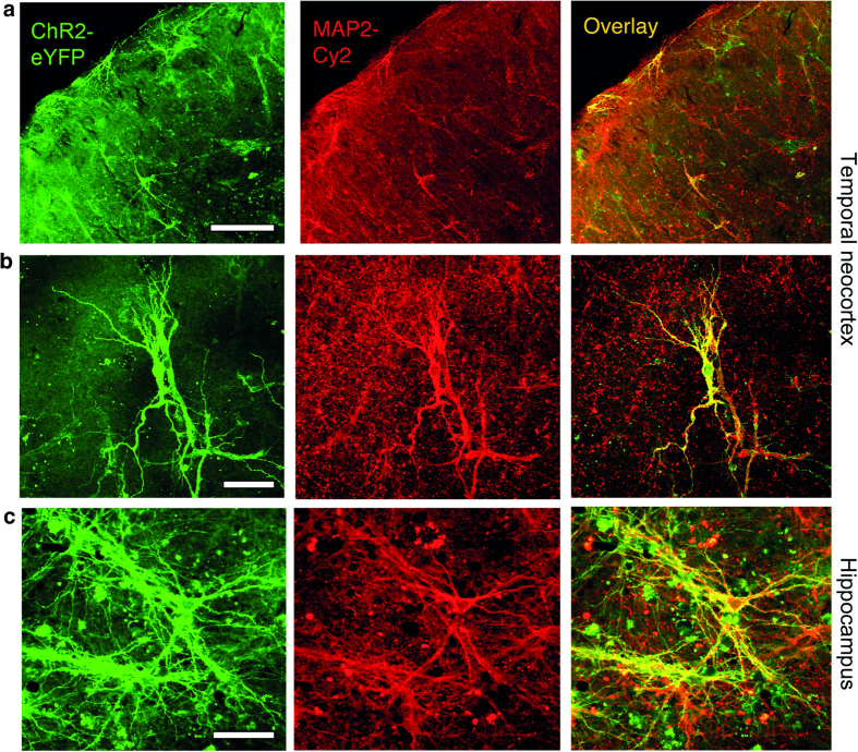
Representative confocal images of enhanced yellow fluorescent protein expression (eYFP, green), neuron-specific microtubule associated protein-2 (MAP2, red) and the overlay of both channels. (a) Left, middle and right image from a cortical organotypic tissue culture (scalebar 200 μm) with a higher magnification image in (b) (scalebar 50 μm). (c) Left, middle and right image from a hippocampal organotypic tissue culture (scalebar 50 μm).
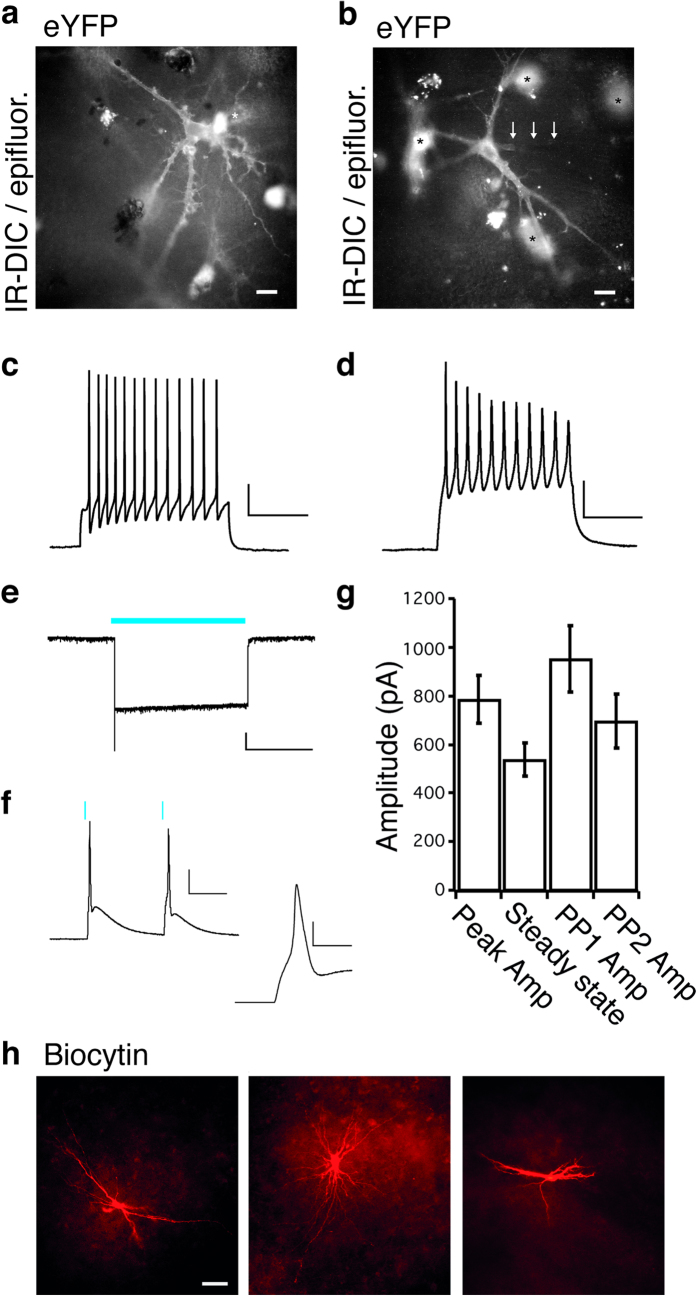
ChR2-expressing cells were identified for whole-cell patch-clamp recordings with the expression of the reporter EYFP in infrared-differential interference contrast (IR-DIC) microscopy combined with epifluorescence exemplified in (a) a cultured temporal neocortical neuron and in (b) a cultured hippocampal neuron (scale bars 20 μm). Neurons in both preparations, temporal neocortical neuron (c) and hippocampal neuron (d) fired action potentials in response to a depolarising current step (scale bars 20 mV, 200 ms). (e) A 10-s continuous blue light-pulse induced an inward current in voltage clamp (scale bar 50 pA, 5 s) and in (f) paired 1 ms light pulses elicited action potentials in current-clamp (scale bar 20 mV, 50 ms and 5 ms in inset). (g) Average current obtained from hippocampal and temporal neocortical neurons induced by blue light application, measured as peak amplitude (peak amp) or steady state (2 s after peak) evoked by a 5 s light pulse (n = 31) or peak amplitude in first response (PP1) or second response (PP2) after paired 1 ms light pulses, 100 ms interval (n = 32). (h) Biocytin staining showing three representative patterns of dendritic morphology from whole-cell recorded neurons (scale bar 50 μm).
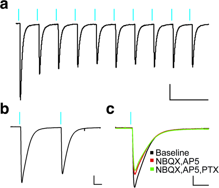
(a) A 10-pulse train (1 ms at 50 Hz) of blue light consistently elicited inward currents in voltage-clamp mode. (b) Paired-pulse light stimulation with a 100 ms interval (scale bar 200 pA, 20 ms). (c) Representative traces showing that a large part of the light-induced currents remained after AMPA (NBQX), NMDA-(AP5) and GABAA-(PTX)-receptor blockade (scale bar 200 pA, 20 ms).
References
-
- Ramirez S. et al. Creating a false memory in the hippocampus. Science 341, 387–91 (2013). - PubMed
Publication types
MeSH terms
Substances
LinkOut - more resources
Full Text Sources
Other Literature Sources
