. 2016 Apr 21:6:24823.
doi: 10.1038/srep24823.

Generation of a monoclonal antibody recognizing the CEACAM glycan structure and inhibiting adhesion using cancer tissue-originated spheroid as an antigen

Affiliations

Generation of a monoclonal antibody recognizing the CEACAM glycan structure and inhibiting adhesion using cancer tissue-originated spheroid as an antigen

Yumi Sato et al. Sci Rep. .

Erratum in

Abstract

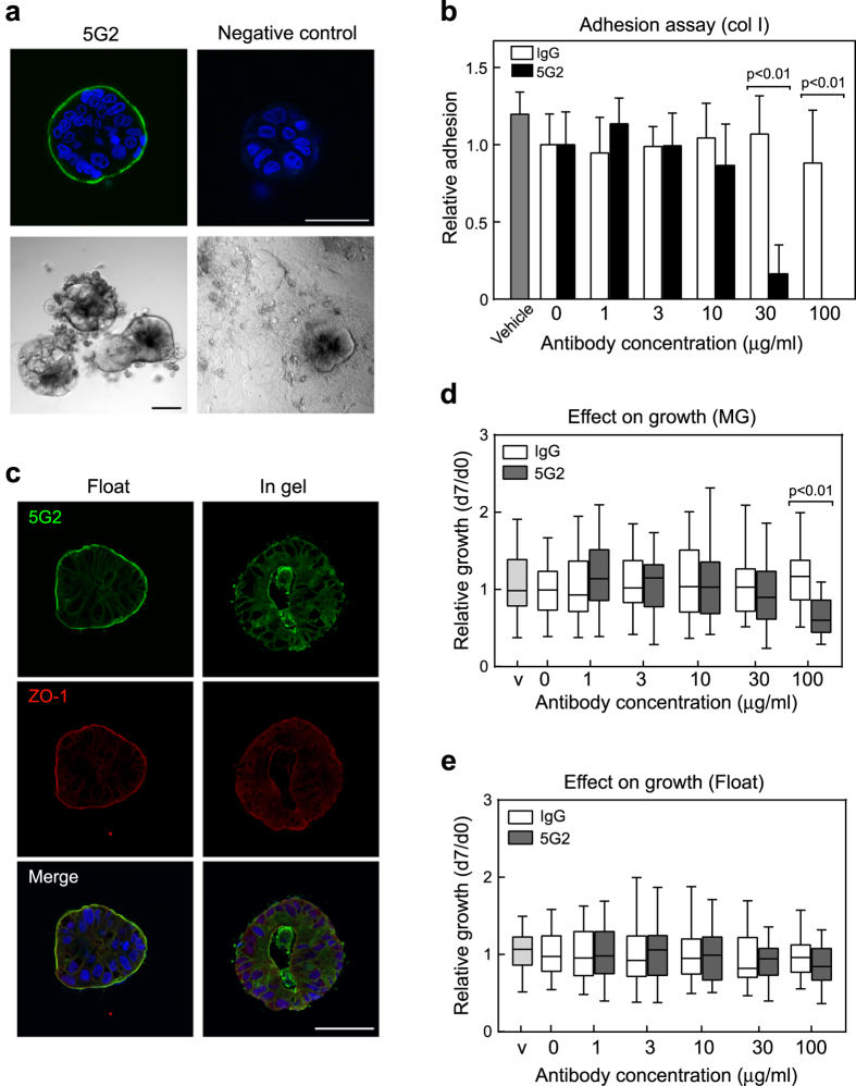
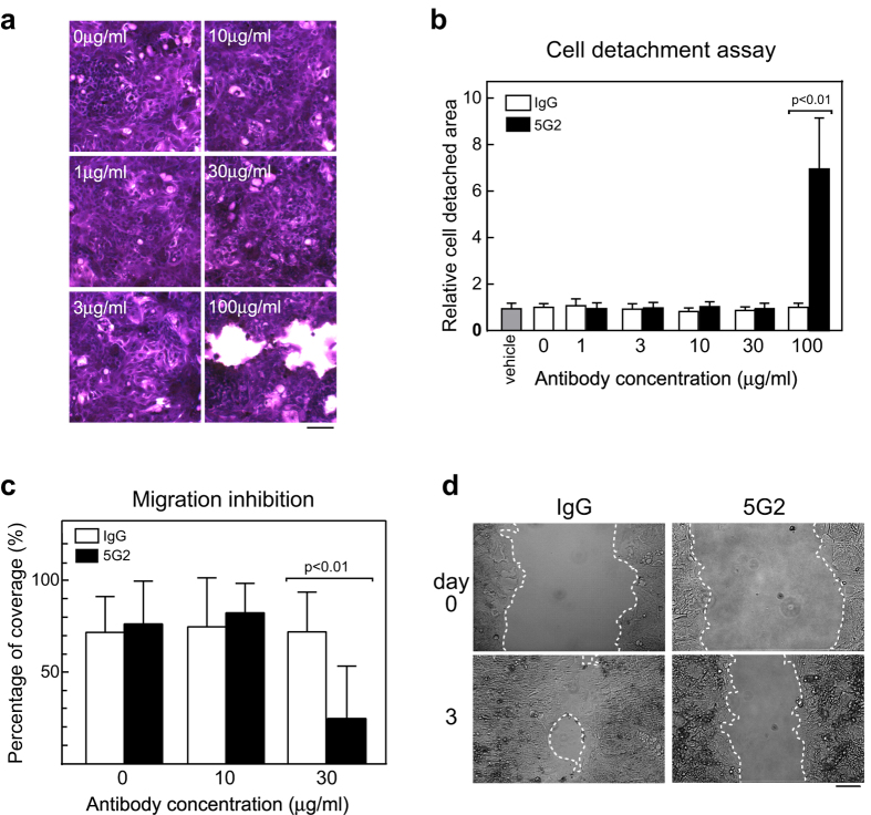
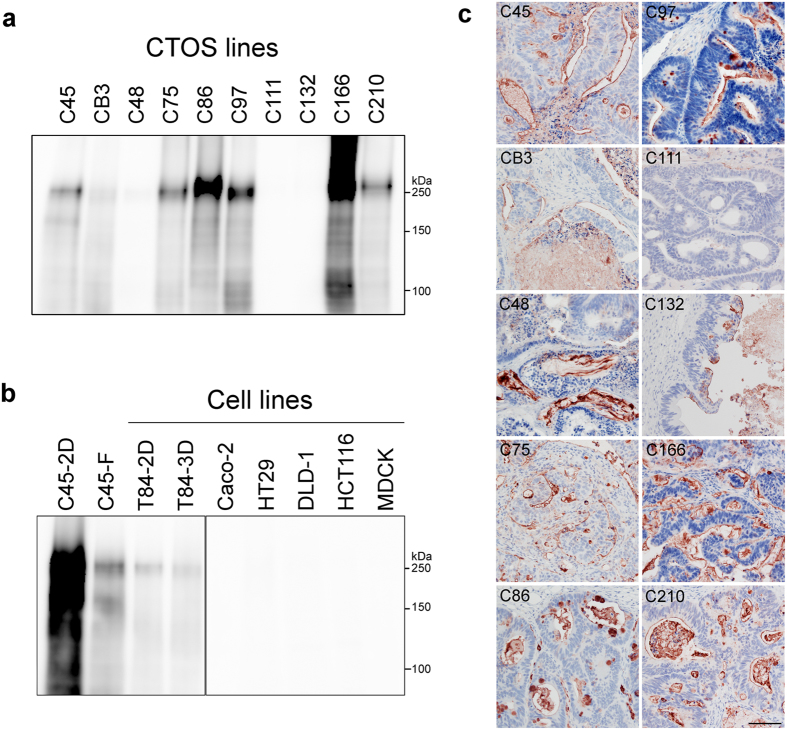
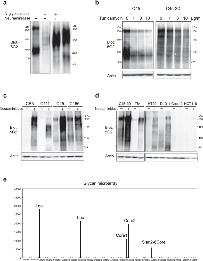
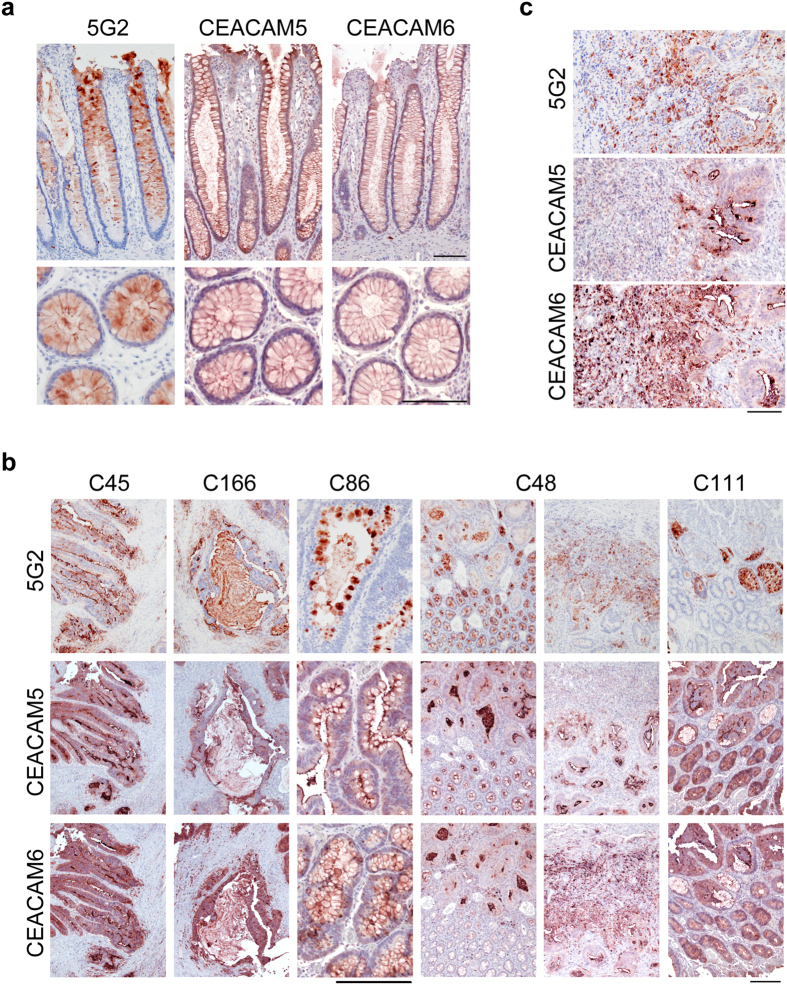
Spheroids cultured directly from tumours can better reflect in vivo tumour characteristics than two-dimensional monolayer culture or three-dimensional culture of established cell lines. In this study, we generated antibodies by directly immunizing mice with primary-cultured living spheroids from human colorectal cancer. We performed phenotypic screening via recognition of the surface of the spheroids and inhibition of their adhesion to extracellular matrices to identify a monoclonal antibody, clone 5G2. The antibody inhibited cell migration in two-dimensional culture and promoted cell detachment. Western blotting and immunohistochemistry detected the 5G2 signal in many colorectal cancer spheroids, as well as patient tumours, but failed to detect in various cell lines examined. We found that 5G2 recognized the Le(a) and Le(c) on N-glycan, and their major carrier proteins were CEACAM5 and CEACAM6. Pre-incubation of the spheroids with 5G2 impaired translocation of integrin β4 from the lateral membrane to the contact interface between the extracellular matrix when embedded in it. As we successfully obtained a functional antibody, which antigen was glycan structures and lost in cell lines, cancer tissue-originated spheroids can be a useful antigen for generating novel anti-cancer antibodies.


Figures

Figure 1
Figure 1. Generation of monoclonal antibody against the CTOS surface.
(a) Top, whole mount immunocytochemistry (ICC) without permeabilization of live CTOSs with 5G2 mAb. CTOSs were incubated with 1 μg/ml purified 5G2 mAb or mouse IgG3κ isotype control and observed by confocal microscopy. Scale bar, 50 μm. Bottom, phase contrast images of an adhesion assay. CTOSs were cultured for 5 days with or without 5G2 hybridoma culture media on a type I collagen-coated dish. Scale bar, 100 μm. (b) Quantification of adhesion to type I collagen-coated plates based on the percentage of adhered CTOSs. Data indicate mean ± SD from three independent experiments. Each condition was performed in triplicate and data are shown relative to the negative control (non-treated) in each experiment. Vehicle: 10% glycerol-PBS. Differences between 5G2 and control IgG were analysed using ANOVA. (c) Immunohistochemistry of sectioned CTOSs with 5G2 mAb (green) and ZO-1 (red). CTOSs were cultured in floating or gel embedded conditions. Gel embedded CTOSs were cultured for 3 days in type I collagen gel. Scale bar, 50 μm. (d,e) Effect of pre-incubation with 5G2 mAb on CTOS growth under Matrigel embedded (d) or floating (e) culture conditions. Growth of CTOSs was evaluated by the day 7 to day 0 size ratio and the data normalized to a mean of 0 μg/ml for each experiment. Values are relative to the negative control (non-treated). Statistical analysis was performed by the Kruskal-Wallis test. Horizontal bar: median; boxes: 25th and 75th percentiles; bars: 10th and 90th percentiles. Data are from more than three independent experiments. V, vehicle, 10% glycerol-PBS.
Figure 2
Figure 2. 5G2 mAb promoted C45-2D cell detachment and inhibited cell migration on collagen-coated dishes.
(a) Cresyl violet staining of C45-2D cells. Confluent C45-2D cells were incubated with 5G2 mAb for 3 days. Scale bar, 100 μm. (b) Quantification of cell detachment areas when treated with the indicated antibodies. Data indicate mean ± SD from three independent experiments. Differences were analysed by the Kruskal-Wallis test. (c) Quantification of cell migration. C45-2D cell migration was assessed by the percentage of closure after 3 days in culture after removal of the culture insert and antibody addition. Data indicate mean ± SD from four independent experiments. Differences were analysed by the Kruskal-Wallis test. (d) Representative images of the cell migration assay in (c). The antibody doses were 30 μg/ml. The white dotted lines indicate the edge of the space. Scale bar, 100 μm.
Figure 3
Figure 3. Expression of 5G2 mAb antigen in colorectal cancer-derived CTOSs and cell lines.
(a,b) Western blot of lysate from colorectal cancer-derived CTOSs (a) and cell lines (b) using 5G2 mAb. (c) Immunohistochemical analysis of CTOS-derived xenotumour with 5G2 mAb. Scale bar, 100 μm.
Figure 4
Figure 4. 5G2 mAb recognized asialo-type N-glycan structure.
(a) Western blot of glycosidase digested membrane fractions of C45 CTOSs using 5G2 mAb. (b) Western blot of lysate from tunicamycin-treated cells using 5G2 mAb. C45 CTOSs and C45-2D cells were cultured with the indicated doses of tunicamycin for 2 days. (c,d) Western blot of sialic acid-digested cell lysate using 5G2 mAb. Protein lysates from CTOSs (c) and cell lines (d) were digested by neuraminidase. Actin is shown as a loading control. (e) Glycan microarray for determination of the glycan structure recognized by 5G2 mAb. The names of glycan structures with high affinity are indicated.
Figure 5
Figure 5. CEACAM5 and CEACAM6 were major carriers of 5G2 antigens.
(a) Western blot of lysate from siRNA-transduced C45 CTOSs using the indicated antibodies. Actin is shown as a loading control. (b) Western blot of lysate from T84 cells in which HA-tagged CEACAM5 and CEACAM6 were forcibly expressed. Cell lysates were immunoprecipitated by HA antibody and detected by HA, CECAM5, CEACAM6, or 5G2 antibodies. The two major bands are indicated by arrowheads. (c) Western blot of lysate from colorectal cancer CTOS lines with CEACAM5, CEACAM6, and 5G2 antibodies. Actin is shown as a loading control.
Figure 6
Figure 6. Localization of 5G2 antigen is consistent with CEACAM5 and CEACAM6 in patient tissues.
(a) Immunohistochemical analysis of human normal colon and (b,c) tumour sections from patient tumours stained with CEACAM5, CEACAM6, and 5G2 antibodies as indicated. (c) High magnifications of the invasive front region of C48. Scale bar, 100 μm (a,c), 200 μm (b).
Figure 7
Figure 7. Pre-incubation with 5G2 mAb inhibited translocation of integrin β4 in CTOSs.
(a) Immunocytochemical analysis of CTOSs using integrin β4 antibody. C45 CTOSs under floating conditions (i.e., suspension culture) were pre-incubated with the indicated antibody and cultured for 20 h in Matrigel. (b) CTOS surface coverage with the integrin β4 signal. C45 CTOSs were treated the same as in (a). Differences were analysed by the Mann Whitney U-test. IgG, n = 111; 5G2, n = 139. Horizontal bar, median; boxes, 25th and 75th percentiles; bars, 10th and 90th percentiles. (c) Integrin β4 signal intensity on lateral membranes in CTOSs. C45 CTOSs were treated the same as in (a). Data indicate mean ± SD. Differences were analysed by the Mann Whitney U-test. IgG, n = 1512; 5G2, n = 2066. (d) Western blot using phosphor-FAK (pFAK) or total FAK (tFAK) antibody. C45 CTOSs in floating conditions (F) were pre-incubated with the indicated antibody and cultured for 6 h in Matrigel (G). (e) Immunoprecipitation analysis of floating cultured C45 CTOSs. Antibodies used for immunoprecipitation (IP) and detection (Blot) are shown.

Similar articles

Cited by

References

    1. Breslin S. & O’Driscoll L. Three-dimensional cell culture: the missing link in drug discovery. Drug Discov. Today 18, 240–249, doi: 10.1016/j.drudis.2012.10.003 (2013). - DOI - PubMed
    1. Fennema E., Rivron N., Rouwkema J., van Blitterswijk C. & de Boer J. Spheroid culture as a tool for creating 3D complex tissues. Trends Biotechnol. 31, 108–115, doi: 10.1016/j.tibtech.2012.12.003 (2013). - DOI - PubMed
    1. Hongisto V. et al. High-throughput 3D screening reveals differences in drug sensitivities between culture models of JIMT1 breast cancer cells. Plos One 8, e77232, doi: 10.1371/journal.pone.0077232 (2013). - DOI - PMC - PubMed
    1. Weigelt B., Lo A. T., Park C. C., Gray J. W. & Bissell M. J. HER2 signaling pathway activation and response of breast cancer cells to HER2-targeting agents is dependent strongly on the 3D microenvironment. Breast Cancer Res. Treat. 122, 35–43, doi: 10.1007/s10549-009-0502-2 (2010). - DOI - PMC - PubMed
    1. Luca A. C. et al. Impact of the 3D microenvironment on phenotype, gene expression, and EGFR inhibition of colorectal cancer cell lines. Plos One 8, e59689, doi: 10.1371/journal.pone.0059689 (2013). - DOI - PMC - PubMed

Publication types